Copyright © 2019 The Korean Society for Bone and Mineral Research
This is an Open Access article distributed under the terms of the Creative Commons Attribution Non-Commercial Li- cense (http://creativecommons.org/licenses/by-nc/4.0/) which permits unrestricted non-commercial use, distribu- tion, and reproduction in any medium, provided the original work is properly cited.
Position Statement on the Use of Bone Turnover Markers for Osteoporosis Treatment
So Young Park1, Seong Hee Ahn2, Jun-Il Yoo3, Youn-Jee Chung4, Yun Kyung Jeon5, Byung-Ho Yoon6, Ha Young Kim7, Seung Hun Lee8, Jehoon Lee9, Seongbin Hong2
1Division of Endocrinology and Metabolism, Department of Internal Medicine, Kyung Hee University Hospital, Seoul;
2Division of Endocrinology and Metabolism, Department of Internal Medicine, Inha University Hospital, Inha University School of Medicine, Incheon;
3Department of Orthopaedic Surgery, Gyeongsang National University Hospital, Jinju;
4Department of Obstetrics and Gynecology, Seoul St. Mary's Hospital, College of Medicine, The Catholic University of Korea, Seoul;
5Division of Endocrinology and Metabolism, Department of Internal Medicine, Pusan National University Hospital, Busan;
6Department of Orthopaedic Surgery, Seoul Paik Hospital, Inje University College of Medicine, Seoul;
7Division of Endocrinology, Department of Internal Medicine, Wonkwang University Sanbon Hospital, Wonkwang University School of Medicine, Gunpo;
8Division of Endocrinology and Metabolism, Asan Medical Center, University of Ulsan College of Medicine, Seoul;
9Department of Laboratory Medicine, College of Medicine, The Catholic University of Korea, Seoul, Korea
Current evidences continue to support the clinical application of bone turnover markers (BTMs) in the management of postmenopausal osteoporosis. The limitations of bone mineral density measured by dual energy X-ray absorptiomet especially emphasize the beneficial roles of BTMs, such as serum C-terminal telopeptide of type I collagen and se- rum procollagen type I N-propeptide, as monitoring tools to assess the responses to treatment. Therefore, the proper application and assessment of BTM in clinical practice is very important. However, their use in Korea is still insufficient. Therefore, the BTM com- mittee has set up by the Korean Society for Bone and Mineral Research have been con- stituted and provided a position statement which will suggest on the clinical application of BTM for the management of postmenopausal osteoporosis in Korea.
Key Words: Bone remodeling · Osteoporosis · Republic of Korea
INTRODUCTION
Osteoporosis is a major health burden and its impact is expected to rise through- out the world. Osteoporosis is defined as a disease characterized by low bone mass and deteriorated bone quality, which leads to decreased bone strength and sub- sequent increase in the risk of fracture.[1] Bone mass is mainly expressed by bone mineral density (BMD) and bone quality is composed of microarchitecture, bone turnover rate, mineralization, and microdamage accumulation.[2] The BMD mea- surement using dual energy X-ray absorptiomet (DXA) is the most commonly used tool for the diagnosis of osteoporosis.[3] Although BMD is used for the determina- tion of treatment strategy and the evaluation of bone loss rate or treatment re- sponse, it still does not completely capture the risk of osteoporotic fracture. More- over, serial BMD measurements as a tool for treatment response require a long in- Corresponding author
Seongbin Hong
Division of Endocrinology and Metabolism, Department of Internal Medicine, Inha University Hospital, Inha University School of Medicine, 27 Inhang-ro, Jung-gu, Incheon 22332, Korea
Tel: +82-32-890-3597 Fax: +82-32-882-6578 E-mail: sbhongmd@inha.ac.kr Received: November 1, 2019 Revised: November 22, 2019 Accepted: November 23, 2019
Review Article
pISSN 2287-6375 eISSN 2287-7029
terval of more than a year.
Bone turnover, which is the process of removal of old bones by bone resorption and followed by replacement of new bones by bone formation, is continuously occurring.
[4] The change of bone turnover rate could affect the bone quality. Bone turnover marker (BTM) is an index reflecting the rate of bone turnover and BTM can be measured with urine and blood non-invasively. Considering the limitations of BMD and the characteristic of BTMs reflecting bone qual- ity, there has been growing interest in the potential role of BTMs in predicting fracture risk and to monitoring the treat- ment response in clinical practice.
There is emerging evidence on clinical use of BTMs to predict bone loss and fracture risk and to monitor the re- sponse to osteoporosis treatment.[5-7] Also, the measure- ment of BTMs will gives us a better understanding of the pathogenesis of osteoporosis. However, the value of the BTMs can be influenced by several physiological and path- ological factors, and, in some cases, by multiple methodol- ogies used for the same analyst. Among various BTMs, se- rum C-terminal telopeptide of type I collagen (CTX-I) and serum propeptide of type I collagen (PINP) are recently rec- ommended as a monitoring targets for osteoporosis treat- ment by several osteoporosis guidelines including the In- ternational Osteoporosis Foundation (IOF), the American Association of Clinical Endocrinologists/American College of Endocrinology (AACE/ACE), and the National Osteoporo- sis Foundation (NOF) and Japan Osteoporosis Society.[8-10]
Despite this current attention in the clinical implication of BTMs for the management of postmenopausal osteopo- rosis, the use of BTMs is still insufficient in Korea. Therefore, the Korean Society for Bone Mineral Research organized the BTM committee to provide recommendations on their use to clinicians in Korea.
STANDARDIZATION OF BTMs 1. What are available assay methods for
measurement of BTMs in Korea?
BTMs are classified as either bone formation markers or bone resorption makers.
1) Bone formation markers
Bone formation markers include osteocalcin, bone spe- cific alkaline phosphatase (BSALP), carboxyterminal pro-
peptide of type I procollagen (PICP), and PINP. BSALP and osteocalcin are released by osteoblasts and play a major role in bone mineralization. PICP and PINP are cleaved from procollagen type I during collagen synthesis. In Korea, os- teocalcin and BSALP are most commonly used bone for- mation markers.[11] Osteocalcin is measured by immune- radiometric assay (DIAsource Immunoassays S.A., Nivelles, Belgium) and an electrochemiluminescence assay (ECLIA;
Roche Diagnostics, Mannheim, Germany). BSALP is mea- sured by chemiluminescence assay (Beckman Coulter Inc., Sacramento, CA, USA), enzyme immunoassay (Quidel Cor- poration, San Diego, CA, USA), and electrophoresis assay (Helena Laboratories, Beaumont, TX, USA). Although PINP has not been widely used yet in Korea, it can be measured by the ECLIA (Roche Diagnostics) and covered by insurance in osteoporotic patients recently.
2) Bone resorption markers
Bone resorption markers include CTX-I, N-terminal telo- peptide of collagen type I (NTX-1), free and total pyridino- line (PYD), and free and total deoxyPYD.
In Korea, CTX-I is the most commonly used as a bone re- sorption marker and has been mainly measured by the ECLIA using β-CrossLaps kit (Roche Diagnostics).
2. Patient sample collection procedure standardization
1) Importance of standardized patient sample collection procedure
The primary challenge to the adoption of many BTMs in routine practice has been the poor reproducibility within- subject and between-laboratory. For clinical utility of BTMs, the pre-analytical sources of variability, along with the un- derlying disease process, must be identified, minimized, and controlled through carefully standardized patient prep- aration and sample handling procedures.[12,13]
Because serum PINP and CTX-I have recently been rec- ommended as a reference BTM,[14] we focused on the proper sample collection procedures for both CTX-I and PINP in this position statement.
2) Sample collection and processing
Although either serum or plasma sample may be used for the measurements of CTX-I and PINP, ethylenediaminetet- raacetic acid plasma has the advantage over the serum as it
has superior sample stability.[15-17] The same sample type should be used consistently when monitoring a patient. CTX- I exhibits a circadian rhythm in blood. CTX-I level peaks dur- ing the early morning hours (2-5 a.m.) and reaches a nadir be- tween 11 a.m. and 2 p.m. The only known modulator having a major effect on this circadian pattern is food intake. Overnight fasting markedly reduced the circadian variation of CTX-I.
Therefore, blood samples for CTX-I measurement must be collected following an overnight fast during the morning be- tween 7:30 a.m. and 10:00 a.m.[18-20] Other BTMs have min- imal circadian rhythm and are minimally affected by food in- take. Sample processing is described previously.[11]
3) Sources of pre-analytical variability: Patient related factors
The patient related factors can be divided into controlla- ble and uncontrollable factors (Table 1). Controllable fac- tors include the menstrual cycle, seasonal variation, and physical activities. The optimal time to collect samples in pre-menopausal women is the early-mid-follicular phase.
[21] There is a minor but detectable seasonal variation for CTX-I in older adults and those with severe vitamin D defi- cit.[22,23] Intensive physical training (e.g., elite soccer play- ers) moderately increases serum CTX-I and slightly decreas- es PINP. Vigorous exercise should be avoided the day prior
to sampling.[24] Uncontrollable factors include age, sex, pregnancy, geography, renal function, and specific diseas- es and medications.[14]
3. Interpretation of BTMs concentrations - The role of reference intervals
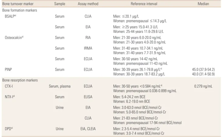
The reference intervals of BTMs are useful for interpreting the results in patients with osteoporosis. Several prospec- tive studies have reported that the presence of incre ased BTMs have an additive effect on the increase risk of fracture in women.[7] The very high BTM values (>3 standard devia- tion above the mean of the reference values) during the ini- tial assessment suggests other metabolic bone disease than osteoporosis. However, sufficient consensus has not been achieved for determining a cut-off point of BTMs that pre- dicts an increased fracture risk or assesses response of treat- ment. It is necessary to establish reference intervals of BTMs for different geographic areas and ethnicities. Although ref- erence intervals of PINP in Korean population were previ- ously reported [12] median level was not suggested accord- ing to menopausal state. Median serum CTX-I level was 0.279 ng/mL (range, 0.036-0.899 ng/mL) in 321 healthy pre- menopausal Korean women.[25] For the other BTMs includ- ing osteocalcin and NTX-1, the only data on reference inter- vals are given by manufactures (Table 2).
CLINICAL UTILITY OF BTMs 1. Can BTMs predict fractures?
Elevated levels of BTM can predict more rapid rates of bone loss and higher fracture risk
Several BTMs were introduced as having an additive ef- fect on fracture risk in women with a low BMD.[26] Two large population-based studies found that increased levels of serum BSALP, urinary and serum CTX-I are significantly associated with an increased risk of osteoporotic fracture (hip and vertebra) with a relative risk of 2.3 to 4.8.[5,27]
The predictive value of BTMs for the risk of fracture was slightly weakened, but persisted after adjustment for BMD.
Additionally, the patients with both increased bone resorp- tion markers and decreased bone formation makers were at higher risk of fracture than the patient with either one of 2 predictors.[7,27-29] These studies suggested that the index of BTMs provides the information on the risk of frac- ture independently of BMD, therefore, the risk of fracture Table 1. Factors determining pre-analytical variability of bone turn-
over markers Uncontrolled
factors - Age
- Menopausal status - Gender
- Fracture
- Pregnancy and lactation - Bed rest/Immobility - Geography and ethnicity - Day to day variation
- Drugs (corticosteroids, anticonvulsants, heparin, GnRH agonists, oral contraceptives)
- Diseases (thyrotoxicosis, diabetes, renal impair- ment, liver disease)
Controlled factors - Circadian rhythm - Fasting status - Exercise - Menstrual cycle - Season
- Lifestyle factors (smoking, alcohol, diet) GnRH, gonadotropin-releasing hormone.
can be assessed and supplemented with BTMs if DXA is not available.[28]
Nonetheless, uncertainties over their clinical use still re- mains, and the routine use of BTMs to assess the fracture risk is not recommended. Despite with the association be- tween BTMs and the risk of fracture, the previous studies have shown inconsistent findings due to their large vari- ability.[30,31] The difference in the predictive value of the various BTMs were noted in many studies and the reasons for this discrepancy are not clear. Another critical flaw is that a common approach to statistically analyze the results of BTMs in the prediction of osteoporotic fracture is odds ratios of fractures per standard deviation of increase in BTMs (e.g., the risk of fracture in patients with higher values is compared with that with lower values).[28] The use of odds ratios is not ideal for clinical decision-making in predicting fractures, so it is not easy to apply the results to the gener- al population.[32]
Therefore, the measurement of absolute risks, such as
10-year probabilities, should be evaluated in a further study with large population. Another possible avenue remains the development of international reference standards and standardization of their measurement that may help mini- mize or eliminate old problems.[33,34]
2. Can BTM monitor therapeutic efficacy?
BTMs can reflect the therapeutic responses to anti-osteoporosis therapies earlier than BMD 1) Anti-resorptive drugs
Inhibition of bone resorption by anti-resorptive drugs results in a decrease in bone resorption markers followed by a plateau. Changes in BTMs during anti-resorptive therapy depend on the mechanism of action of the drug, degree of inhibition of bone resorption, and the route of administra- tion. This inhibition of bone resorption secondarily causes a decrease in bone formation markers, due to physiologic mechanisms linking osteoclast and osteoblast activity.
Table 2. The reference intervals and median value of available bone turnover markers in Korea
Bone turnover marker Sample Assay method Reference interval Median
Bone formation markers
BSALPa) Serum CLIA Men: ≤20.1 μg/L
Women: premenopausal ≤14.3 μg/L
Serum EIA Men: ≥25 years 15.0-41.3 U/L
Women: 25-44 years 11.6-29.6 U/L
Osteocalcina) Serum RIA Men: 21-30 years 6.0-20.0 ng/mL
Women: 21-30 years 4.0-20.0 ng/mL
Serum IRMA Men: 31-40 years 10.7-34.1 ng/mL
Women: 31-40 years 7.7-31.9 ng/mL
Serum ECLIA Men: 30-50 years 14-42 ng/mL
Women: premenopausal 11-43 ng/mL
PINP Serum ECLIA Men: 30-39 years 26.1-79.8 μg/La)
Women: 30-39 years 18.7-83.2 μg/L 45.0 (37.9-54.2) 40.0 (31.4-50.9) Bone resorption markers
CTX-I Serum, plasma ECLIA Men: 30-50 years <0.584 ng/mLa)
Women: premenopausal 0.036-0.899 ng/mL 0.279 ng/mL
NTX-Ia) Serum ELISA Men: 5.4-24.2 nm BCE
Women: 6.2-19.0 nm BCE
Urine EIA Men: 3.0-63.0 nmol BCE/mmol·Cr
Women: 5.0-65.0 nmol BCE/mmol·Cr CLIA Men: 21-83 nmol BCE/mmol·Cr
Women: premenopausal 17-94 nmol BCE/mmol
DPDa) Urine EIA, CLEIA Men: 2.3-5.4 nmol BCE/mmol·Cr
Women: 3.0-7.4 nmol BCE/mmol·Cr
a)Described in kit manufacturer’s package insert or manufacturer’s in-house data.
BSALP, bone specific alkaline phosphatase; PINP, propeptide of type I collagen; CTX-I, C-terminal telopeptide of type I collagen; NTX-I, N-terminal telopep- tide of collagen type I; DPD, deoxypyridinoline; CLIA, chemiluminescence assay; EIA, enzyme immunoassay; RIA, radioimmunoassay; IRMA, immunora- diometric assay; ECLIA, electrochemiluminescence assay; ELISA, enzyme-linked immunosorbent assay; CLEIA, chemiluminescence enzyme immunoassay.
(1) Bisphosphonate (BP)
BPs are the most commonly used drugs for treatment of osteoporosis. Bone resorption markers are maximally sup- pressed after 8 weeks of treatment and bone formation markers are maximally suppressed after 26 weeks of treat- ment. The oral BPs, such as alendronate, ibandronate, and risedronate, have been compared in the TRIO study [35] to evaluate the clinical utility of BTMs to assess treatment re- sponse. Alendronate and ibandronate decreased BTM (CTX- I, NTX-1) more than risedronate. In this study, more than 80% of patients responded to treatment as defined by a decrease more than the least significant change (LSC) for CTX-I (56%) and PINP (38%) after 3 months of treatment.
Response can also be defined as a reduction to a level be- low the median found in healthy young women.[35] BPs administrated intravenously inhibit bone resorption and decrease levels of bone resorption markers faster than BPs administrated orally. Zoledronic acid is given by annual in- travenous infusion, thus avoiding concerns about poor ab- sor ption. It results in a reduction in CTX-I by 2 weeks and when it is given for 6 years as in the Horizon Study, the sup- pression of CTX-I and PINP is maintained.[36]
(2) Denosumab
Denosumab, a fully human monoclonal antibody to re- ceptor activator of nuclear factor-κB ligand, is administrat- ed subcutaneously inhibited bone resorption 12 hr after administration.[13] Bone resorption markers (such as CTX- I) decrease within 24 hr of treatment. Denosumab results in a greater inhibition of bone resorption than zoledronic acid.[37] PINP decreases over several months to a lesser extent than the bone resorption markers and remains sup- pressed with continued dosing for up to 10 years.[38] Once the drug is stopped, the BTM overshoot so that their levels are increased compared to baseline. These elevation of BTMs results are associated with accelerated bone loss, and there are recent reports of multiple vertebral fractures associated with this high BTM.[39]
(3) Selective estrogen receptor modulator (SERM)
SERM such as raloxifene have a weaker effect on the change of bone turnover than BPs and denosumab. In 60% to 65% of women with osteopenia, a significant response could be demonstrated using the LSC approach with CTX-I or PINP.[40]
2) Anabolic drugs
The linkage between osteoclasts and osteoblasts can also function in the opposite direction, with anabolic drugs compared to anti-resorptive drugs.
(1) Recombinant human parathyroid hormone (PTH) 1-34 (teriparatide)
Teriparatide is typically characterized as an anabolic agent, but results in increases in both bone formation and resorp- tion markers.[41,42] Bone formation markers increase with- in a few days of starting treatment,[42] peaking by 3 months.
PINP has been proven to be the most responsive BTM to this treatment.
For monitoring of early response to teriparatide therapy, PINP is measured prior to the initiation of teriparatide, and then after 1 to 3 months of therapy. Because patients who were pre- treated with alendronate then switched to teriparatide showed PINP response rates of 79% at 1 month and 97% at 3 months, follow up assessment of PINP at 3 months may be more helpful than earlier assessments in this group of patients.[43]
The increase in serum PINP concentration by >10 pg/mL may be predictive of a greater increase in BMD.[43]
(2) Abaloparatide
Abaloparatide, recombinant human PTH related peptide (1-34), is a newly licensed anabolic therapy for osteoporo- sis.[44] It works through the PTH receptor as does with terip- aratide. This drug stimulates rapid bone formation and re- sorption and works in a dose dependent manner in wom- en with postmenopausal osteoporosis, but less than terip- aratide, leading to a greater increase in BMD compared with teriparatide.[45] The clinical utility of BTMs for monitoring abaloparatide therapy has not yet been fully reported.
(3) Romosozumab
Romosozumab, anti-sclerostin monoclonal antibody, also works in a dose-dependent manner and increases bone formation markers. But, unlike teriparatide, there is an early but transient increase in PINP level and a decrease in serum CTX-I.[46] PINP level begin to increase from 1 week after drug administration and reaches a peak in 1 month, then slowly return to pretreatment values within 6 months.
CTX-I initially decreased and remained below the baseline value at 12 month.[46] These BTM changes are associated with a rapid increase in BMD.
3. Can BTM monitor compliance?
In postmenopausal women receiving a treatment, BTMs can be used to monitor the individual compliance
Monitoring of BTM at an individual level may improve the compliance of patients on anti-osteoporotic treatment.[47]
The IOF has proposed that a BTM such as PINP or CTX-I mea- sured within 3 months of starting therapy would help to identify those with poor adherence which occur commonly with osteoporosis therapy, especially oral BPs.[48] The ab- sence of change in the BTMs concentration during anti-os- teoporotic therapy might reflect poor compliance (e.g., BP not taken at all, or not taken in the fasting state, or taken with milk), poor adherence, inappropriate technique of teripara- tide injection, a medical problem (e.g., recent vertebral frac- ture), secondary osteoporosis, or real absence of response.
According to recently published Consensus Statement in Asia-Pacific region,[49] in patients who are receiving anti- resorptive therapies, serum CTX-I and/or PINP can be used to monitor compliance and drug response with measure- ments at baseline, 3, 6, and 12 months after starting treat- ment. In patients who are receiving anabolic therapies, se- rum PINP can be used to monitor compliance and drug re- sponse, with measurements at baseline, 1 to 3, 6, and 12 months after starting treatment.
However, further studies are necessary to define an ade- quate response through the change in BTM concentration that is associated with a greater reduction in fracture inci- dence. The reference range of BTMs concentration for the adequate response could be helpful when used as target for anti-osteoporotic treatment.
4. Can BTMs be useful for drug holiday?
Monitoring BTMs during drug holiday can be helpful to decide to resume treatment, but the evidence was insufficient yet
BPs are considered as a first-line treatment for osteopo- rosis. It is effective in reduction of fracture risk over 3 to 5 years of treatment. However, long-term use of BPs is asso- ciated with the rare but serious adverse effects such as os- teonecrosis of the jaw (ONJ) and atypical femoral fractures (AFF). Therefore, the concept of a ‘drug holiday’ from BPs has emerged as being potentially beneficial in avoiding these adverse effects. The effect of BP such as anti-resorp- tion and fracture prevention persist for a long period after cessation of drugs because of long-term deposition of BPs
in bones. However, despite BP has long skeletal retention time, fracture risk will increase after discontinuation of it.
Therefore, continuous monitoring during drug holiday will be needed for the assessment of fracture risk and to restart treatment if needed. There are limited clinical practice gui- delines on BTM monitoring during drug holiday. There are few studies about an assessment of fracture risk using BTMs during drug holiday. A recent retrospective study showed that those in newly developed fracture group during drug holiday was older and had lower BMD than non-fracture group. Other factors associated with fracture such as BTMs, vitamin D level, body mass index, and PTH were not signifi- cantly different between 2 groups.[50] Another study pre- sented that BMD by DXA, BTMs (CTX-I, PINP), and fracture- risk assessment tool score may help to guide when to re- sume treatment during drug holiday.[51] Some research- ers suggested that empirical approaches such as monitor- ing BMD and BTMs are necessary during drug holiday.[52]
Several meta-analyses presented that increased bone turn- over is a significant determinant of fracture susceptibility.
[53-55] In one guideline, they suggested that the increase in bone resorption markers compared to the levels before treatment might be a signal to stop the “holiday”.[56] Dur- ing drug holiday, a significant decrease in BMD or a signifi- cant increase in BTMs suggests that the residual effect of BP therapy may be diminishing and that it may be time to resume active therapy.[52,57,58] Roberts and colleagues [51] recommended that if BTMs rise over 30% after drug holiday, treatment should be resumed.
5. Could BTMs predict side effects such as ONJ and AFF?
Although there is still controversy, BTMs can be used carefully as a predictor of long-term side effects of BPs such as ONJ and AFF
Prolonged BP therapy has been related with rare adverse events such as ONJ and AFF. ONJ is a rare oral disease in which the jaw bone’s healing power is impaired because of use of BP or other antiresorptive agents. Patients may be considered to have ONJ if they had current or previous treatment with antiresorptive or antiangiogenic agents and as if there is exposed bone in the jaw that has persist- ed for more than 8 weeks but without history of radiation therapy or obvious metastatic disease.[59] A number of case reports have suggested that long-term treatment
with BPs may be related to the occurrence of fractures of the femur below the lesser trochanter, which was regarded as an atypical based on their unusual site and radiographic appearance in patients with osteoporosis.[60] These rare but serious side effects were supposed to be related to the severely suppression of bone turnover. Therefore, several biochemical markers have been used as effective evalua- tion methods during the bone-remodeling period. In one cross-sectional study, 4 BTMs including serum CTX-I, os- teocalcin, PTH, and BSALP were compared between medi- cation-related ONJ patients and control groups. Serum CTX-I, osteocalcin, and BSALP levels were lower and serum PTH level was higher in ONJ patients than in control groups.
[59] Another study showed that high CTX-I level was asso- ciated with a low risk of ONJ in patients who was treated with BPs and had a tooth extraction.[61] BSALP level over 10 μg/L denoted faster healing and indicated a better prog- nosis in ONJ patients.[62] Some dentists have suggested CTX-I measurement for the prediction of ONJ. If patients’
CTX-I level is lower than 0.100 to 0.150 ng/mL, the risk of ONJ is high, therefore, drug holiday or having surgery after recovery of CTX-I level could be recommended.[63,64] One prospective cross-sectional study suggested that the levels of BTMs including PINP, tartrate-resistant acid phosphatase 5b (TRACP-5b) and undercarboxylated osteocalcin were significantly lower in patients with AFF than in patients with typical osteoporotic femoral fracture. They suggested that severe suppression of bone turnover was associated with the pathogenesis of AFF.[65] Systematic review of case report and case series on AFF presented that bone formation marker was decreased in 14%, normal in 79%, and increased in 7.0% in AFF patients. And bone resorp- tion marker was decreased 18.2%, normal 69.7%, and in- creased 12.1% in AFF patients.[60]
6. How should BTM be used in patient with chronic kidney disease (CKD)?
The various serum and urine BTMs are affected by renal dysfunction. The measurement of BASLP and PTH is recommended as a BTM in patients with CKD
Because of urinary and some serum BTMs are excreted into urine by kidney, the serum level of those are affected by renal dysfunction. BSALP, intact PINP, and TRACP-5b are not affected by renal impairment.
1) BSALP
Its serum concentration seems to be independent of glomerular filtration rate. Combination of a low PTH (<150 pg/mL) and a low BSALP (<27 IU/L) improved the specific- ity of diagnosing adynamic bone disease in 103 dialysis patients with bone biopsy. In the newer automated Ostase BSALP assay, the cut-off <20 IU/L is used.[66]
2) Osteocalcin
Since osteocalcin is cleared by kidney, its use in CKD pa- tients is limited. The combination of osteocalcin (<41 ng/L) and BSALP (<23 U/L) improved the positive predictive val- ue for diagnosing adynamic bone disease to 77% in a CKD- 5 cohort.[66]
3) PINP
PINP is present in 2 major forms, an intact trimeric form and a monomeric one. Some assays recognize both forms (‘Total PINP’; Roche Elecsys, Mannheim, Germany) while other assays recognize the trimeric form only (‘Intact PINP’, Orion Diagnostica and IDS iSYS). The proportion of the mo- nomeric form is elevated in patients with CKD, wher eas the apparent concentration of intact PINP is unaffected by glo- merular filtration rate in kidney disease patients.[67] PINP monomers are not cleared by conventional dialysis ses- sions and the LSC is of 32% for the intact assay.[68]
4) CTX-I
CTX-I is renally excreted and accumulates in CKD patients.
CTX-I is cleared by dialysis and therefore pre-dialysis sam- pling is required for longitudinal monitoring.[66]
5) TRACP-5b
TRACP-5b is an enzyme released by osteoclasts to break- down bone matrix. High serum levels of TRACP-5b there- fore reflect increased osteoclastic activity and resorption.
Levels of TRACP-5b correlate with PTH and ALP and are un- affected by renal function. However, its use is limited by availability of automated assays.[66]
6) The Kidney Disease: Improving Global Outcomes (KDIGO) guidelines for CKD-mineral and bone disorder (CKD-MBD)
The KDIGO guidelines recommended that the bone de- rived markers of collagen synthesis and breakdown, includ-
ing CTX-I, should not be routinely measured in patients with CKD stages 3 to 5D.[69] The primary rationale for this recommendation was that the levels of such markers did not appear to be more effective at predicting clinical out- comes or bone histology than serum PTH or BASLP.[70]
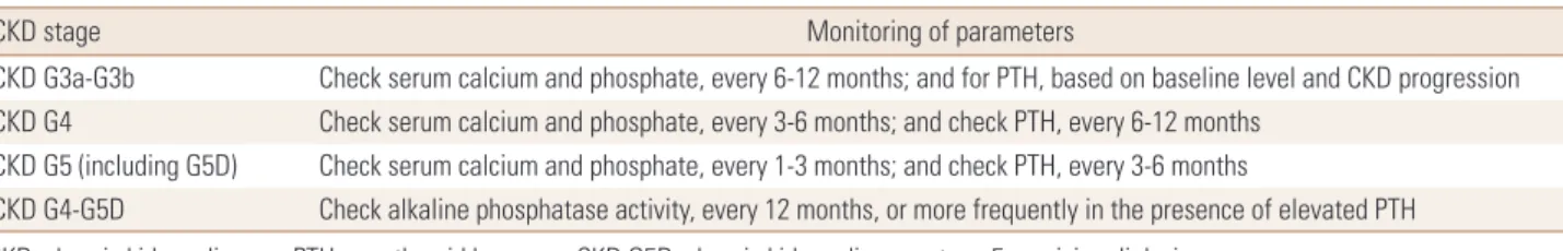
However, they suggest that measurements of serum PTH or BASLP can be used to evaluate bone disease because markedly high or low values predict underlying bone turn- over in patients with CKD G3a-G5D.[69] They also recom- mend the interval of monitoring serum calcium, phosphate, and PTH on the presence and magnitude of abnormalities, based on rate of progression of CKD (Table 3).[69]
CONCLUSION
The appropriate use of BTMs in treatment of osteoporo- sis could be helpful to predict fracture risk and monitor treatment response and patient compliance. But, BTMs have not been used widely in clinical practice due to their poor within-subject and between-lab reproducibility. Re- searchers are constantly trying to automate and standard- ize the measurement of bone markers to minimize vari- ability of BTMs. In addition, insurance coverage for BTMs have recently been possible in Korea. Thus, BTMs could be used easily as a dynamic index reflecting bone quality, com- plementary to BMD. More studies about the efficacy of BTMs are needed in the future.
Table 3. The interval of monitoring serum calcium, phosphate, and parathyroid hormone based on rate of progression of chronic kidney disease
CKD stage Monitoring of parameters
CKD G3a-G3b Check serum calcium and phosphate, every 6-12 months; and for PTH, based on baseline level and CKD progression CKD G4 Check serum calcium and phosphate, every 3-6 months; and check PTH, every 6-12 months
CKD G5 (including G5D) Check serum calcium and phosphate, every 1-3 months; and check PTH, every 3-6 months
CKD G4-G5D Check alkaline phosphatase activity, every 12 months, or more frequently in the presence of elevated PTH CKD, chronic kidney disease; PTH, parathyroid hormone; CKD G5D, chronic kidney disease stage 5 receiving dialysis.
Fig. 1. The algorithm of bone turnover marker use in osteoporosis treatment. PINP, propeptide of type I collagen; BSALP, bone specific alkaline phosphatase; CTX-1, C-terminal telopeptide of type I collagen; NTX-1, N-terminal telopeptide of collagen type I; DPD, deoxypyridinoline; PYD, pyridinoline; BP, bisphosphonate; SERM, selective estrogen receptor modulator; LSC, least significant changes.
Definite diagnosis of osteoporosis Exclude the causes of secondary osteoporosis
Before initiating drug treatment for osteoporosis, measure bone turnover markers 1) Bone formation markers: PINP, Osteocalcin, BSALP
2) Bone resorption markers: CTX-I, NTX, DPD, PYD
Anti-resorptive agent
(BPs, Denosumab, SERMs) Anabolic agent
(abaloparatide, romosozumab, teriparatide) Follow-up
Bone resorption marker after 3-6 months Bone formation marker after 6 months
Follow-up
Bone formation marker after 1-3 months Bone resorption marker after 3-6 months
Continue current treatment
Yes No
Reassess treatment: Poor compliance, Other issue?
If no cause is present, consider switching to another drug One of two
1) Exceeds LSC
2) Within reference range of premenopausal women
SUMMARIES OF CONSENSUS STATEMENT (Fig. 1)
- Consider using BTMs in the initial evaluation and follow- up of patients with osteoporosis.
- Elevated levels of BTMs can predict more rapid rates of bone loss and higher fracture risk.
- BTMs can reflect the therapeutic responses to anti-os- teoporosis therapies earlier than BMD and help in se- lecting osteoporosis treatment and in assessing response to therapies.
- CTX-I and/or PINP can be used to evaluate patient ad- herence and response to anti-resorptive drugs with mea- surement at baseline, 3 to 6 months after starting treat- ment.
- PINP can be used to evaluate patient adherence and responses to anabolic agents with measurement at base- line, 1 to 3 months after starting treatment.
- For patients taking anti-resorptive agents, target range for successful treatment is to have levels of BTMs in the reference range for premenopausal women. Also, rela- tive change of BTMs from baselines above LSC can be also used.
- Monitoring BTMs during drug holiday can be helpful to decide to resume treatment, but the evidence was in- sufficient yet.
- Although there is still controversy, BTMs can be used carefully as a predictor of long-term side effects of BPs such as ONJ and AFF.
- The various serum and urine BTMs are affected by renal dysfunction. The measurement of BASLP and PTH is rec- ommended as a BTM in patients with CKD.
DECLARATIONS
Ethics approval and consent to participate
Not applicable.Conflict of interest
No potential conflict of interest relevant to this article was reported.
ORCID
So Young Park https://orcid.org/0000-0002-4820-9415 Yun Kyung Jeon https://orcid.org/0000-0002-4319-5181
Byung-Ho Yoon https://orcid.org/0000-0001-8518-6331 Ha Young Kim https://orcid.org/0000-0002-0651-2213 Seongbin Hong https://orcid.org/0000-0002-8189-395X
REFERENCES
1. Peck WA, Burckhardt P, Christiansen C, et al. Consensus development conference: diagnosis, prophylaxis, and treat- ment of osteoporosis. Am J Med 1993;94:646-50.
2. Weinstein RS. True strength. J Bone Miner Res 2000;15:621-5.
3. Blake GM, Fogelman I. Role of dual-energy X-ray absorpti- ometry in the diagnosis and treatment of osteoporosis. J Clin Densitom 2007;10:102-10.
4. Feng X, McDonald JM. Disorders of bone remodeling. Annu Rev Pathol 2011;6:121-45.
5. Garnero P, Hausherr E, Chapuy MC, et al. Markers of bone resorption predict hip fracture in elderly women: the EPI- DOS Prospective Study. J Bone Miner Res 1996;11:1531-8.
6. Garnero P, Sornay-Rendu E, Claustrat B, et al. Biochemical markers of bone turnover, endogenous hormones and the risk of fractures in postmenopausal women: the OFE- LY study. J Bone Miner Res 2000;15:1526-36.
7. Ross PD, Kress BC, Parson RE, et al. Serum bone alkaline phosphatase and calcaneus bone density predict fractures:
a prospective study. Osteoporos Int 2000;11:76-82.
8. Cosman F, de Beur SJ, LeBoff MS, et al. Clinician’s guide to prevention and treatment of osteoporosis. Osteoporos Int 2014;25:2359-81.
9. Orimo H, Nakamura T, Hosoi T, et al. Japanese 2011 guide- lines for prevention and treatment of osteoporosis--exec- utive summary. Arch Osteoporos 2012;7:3-20.
10. Vasikaran S, Eastell R, Bruyere O, et al. Markers of bone turnover for the prediction of fracture risk and monitoring of osteoporosis treatment: a need for international refer- ence standards. Osteoporos Int 2011;22:391-420.
11. Park SY, Ahn SH, Yoo JI, et al. Clinical application of bone turnover markers in osteoporosis in Korea. J Bone Metab 2019;26:19-24.
12. Nishizawa Y, Ohta H, Miura M, et al. Guidelines for the use of bone metabolic markers in the diagnosis and treatment of osteoporosis (2012 edition). J Bone Miner Metab 2013;
31:1-15.
13. Szulc P. The role of bone turnover markers in monitoring treatment in postmenopausal osteoporosis. Clin Biochem 2012;45:907-19.
14. Szulc P, Naylor K, Hoyle NR, et al. Use of CTX-I and PINP as bone turnover markers: National Bone Health Alliance recommendations to standardize sample handling and patient preparation to reduce pre-analytical variability.
Osteoporos Int 2017;28:2541-56.
15. Christgau S, Rosenquist C, Alexandersen P, et al. Clinical evaluation of the Serum CrossLaps One Step ELISA, a new assay measuring the serum concentration of bone-de- rived degradation products of type I collagen C-telopep- tides. Clin Chem 1998;44:2290-300.
16. Garnero P, Borel O, Delmas PD. Evaluation of a fully auto- mated serum assay for C-terminal cross-linking telopep- tide of type I collagen in osteoporosis. Clin Chem 2001;47:
694-702.
17. Morovat A, Catchpole A, Meurisse A, et al. IDS iSYS auto- mated intact procollagen-1-N-terminus pro-peptide as- say: method evaluation and reference intervals in adults and children. Clin Chem Lab Med 2013;51:2009-18.
18. Qvist P, Christgau S, Pedersen BJ, et al. Circadian variation in the serum concentration of C-terminal telopeptide of type I collagen (serum CTx): effects of gender, age, meno- pausal status, posture, daylight, serum cortisol, and fast- ing. Bone 2002;31:57-61.
19. Redmond J, Fulford AJ, Jarjou L, et al. Diurnal rhythms of bone turnover markers in three ethnic groups. J Clin En- docrinol Metab 2016;101:3222-30.
20. Clowes JA, Hannon RA, Yap TS, et al. Effect of feeding on bone turnover markers and its impact on biological vari- ability of measurements. Bone 2002;30:886-90.
21. Gass ML, Kagan R, Kohles JD, et al. Bone turnover marker profile in relation to the menstrual cycle of premenopaus- al healthy women. Menopause 2008;15:667-75.
22. Bhattoa HP, Nagy E, More C, et al. Prevalence and seasonal variation of hypovitaminosis D and its relationship to bone metabolism in healthy Hungarian men over 50 years of age: the HunMen Study. Osteoporos Int 2013;24:179-86.
23. Pasco JA, Henry MJ, Kotowicz MA, et al. Seasonal periodic- ity of serum vitamin D and parathyroid hormone, bone resorption, and fractures: the Geelong Osteoporosis Study.
J Bone Miner Res 2004;19:752-8.
24. Weiler R, Keen R, Wolman R. Changes in bone turnover markers during the close season in elite football (soccer) players. J Sci Med Sport 2012;15:255-8.
25. Bae SJ, Kim BJ, Lim KH, et al. Efficacy of intravenously ad- ministered ibandronate in postmenopausal Korean wom-
en with insufficient response to orally administered bisphos- phonates. J Bone Miner Metab 2012;30:588-95.
26. Johnell O, Odén A, De Laet C, et al. Biochemical indices of bone turnover and the assessment of fracture probability.
Osteoporos Int 2002;13:523-6.
27. Vergnaud P, Garnero P, Meunier PJ, et al. Undercarboxylat- ed osteocalcin measured with a specific immunoassay predicts hip fracture in elderly women: the EPIDOS Study.
J Clin Endocrinol Metab 1997;82:719-24.
28. Garnero P, Cloos P, Sornay-Rendu E, et al. Type I collagen racemization and isomerization and the risk of fracture in postmenopausal women: the OFELY prospective study. J Bone Miner Res 2002;17:826-33.
29. Gerdhem P, Ivaska KK, Alatalo SL, et al. Biochemical mark- ers of bone metabolism and prediction of fracture in el- derly women. J Bone Miner Res 2004;19:386-93.
30. Marques EA, Gudnason V, Lang T, et al. Association of bone turnover markers with volumetric bone loss, periosteal apposition, and fracture risk in older men and women:
the AGES-Reykjavik longitudinal study. Osteoporos Int 2016;27:3485-94.
31. Melton LJ 3rd, Crowson CS, O'Fallon WM, et al. Relative contributions of bone density, bone turnover, and clinical risk factors to long-term fracture prediction. J Bone Miner Res 2003;18:312-8.
32. Leiper JM, Paterson KR, Lunan CB, et al. A comparison of biosynthetic human insulin with porcine insulin in the blood glucose control of diabetic pregnancy. Diabet Med 1986;3:49-51.
33. Ulivieri FM, Piodi LP, Grossi E, et al. The role of carboxy-ter- minal cross-linking telopeptide of type I collagen, dual x- ray absorptiometry bone strain and Romberg test in a new osteoporotic fracture risk evaluation: A proposal from an observational study. PLoS One 2018;13:e0190477.
34. Vasikaran S. Assessment of bone turnover in osteoporosis:
harmonization of the total testing process. Clin Chem Lab Med 2018;56:1603-7.
35. Naylor KE, Jacques RM, Paggiosi M, et al. Response of bone turnover markers to three oral bisphosphonate therapies in postmenopausal osteoporosis: the TRIO study. Osteo- poros Int 2016;27:21-31.
36. Black DM, Reid IR, Cauley JA, et al. The effect of 6 versus 9 years of zoledronic acid treatment in osteoporosis: a ran- domized second extension to the HORIZON-Pivotal Frac- ture Trial (PFT). J Bone Miner Res 2015;30:934-44.
37. Miller PD, Pannacciulli N, Brown JP, et al. Denosumab or zoledronic acid in postmenopausal women with osteopo- rosis previously treated with oral bisphosphonates. J Clin Endocrinol Metab 2016;101:3163-70.
38. Bone HG, Wagman RB, Brandi ML, et al. 10 years of deno- sumab treatment in postmenopausal women with osteo- porosis: results from the phase 3 randomised FREEDOM trial and open-label extension. Lancet Diabetes Endocri- nol 2017;5:513-23.
39. Lamy O, Gonzalez-Rodriguez E, Stoll D, et al. Severe re- bound-associated vertebral fractures after denosumab discontinuation: 9 clinical cases report. J Clin Endocrinol Metab 2017;102:354-8.
40. Naylor KE, Jacques RM, Peel NF, et al. Response of bone turnover markers to raloxifene treatment in postmeno- pausal women with osteopenia. Osteoporos Int 2016;27:
2585-92.
41. Finkelstein JS, Leder BZ, Burnett SM, et al. Effects of teripa- ratide, alendronate, or both on bone turnover in osteopo- rotic men. J Clin Endocrinol Metab 2006;91:2882-7.
42. Glover SJ, Eastell R, McCloskey EV, et al. Rapid and robust response of biochemical markers of bone formation to teriparatide therapy. Bone 2009;45:1053-8.
43. Eastell R, Krege JH, Chen P, et al. Development of an algo- rithm for using PINP to monitor treatment of patients with teriparatide. Curr Med Res Opin 2006;22:61-6.
44. Shirley M. Abaloparatide: First global approval. Drugs 2017;
77:1363-8.
45. Miller PD, Hattersley G, Riis BJ, et al. Effect of Abalopara- tide vs placebo on new vertebral fractures in postmeno- pausal women with osteoporosis: A randomized clinical trial. JAMA 2016;316:722-33.
46. McClung MR, Grauer A, Boonen S, et al. Romosozumab in postmenopausal women with low bone mineral density.
N Engl J Med 2014;370:412-20.
47. Delmas PD, Vrijens B, Eastell R, et al. Effect of monitoring bone turnover markers on persistence with risedronate treatment of postmenopausal osteoporosis. J Clin Endo- crinol Metab 2007;92:1296-304.
48. Diez-Perez A, Naylor KE, Abrahamsen B, et al. International osteoporosis foundation and European calcified tissue so- ciety working group. Recommendations for the screening of adherence to oral bisphosphonates. Osteoporos Int 2017;28:767-74.
49. Sun V, Raz DJ, Erhunmwunsee L, et al. Improving family
caregiver and patient outcomes in lung cancer surgery:
Study protocol for a randomized trial of the multimedia self-management (MSM) intervention. Contemp Clin Tri- als 2019;83:88-96.
50. Bindon B, Adams W, Balasubramanian N, et al. Osteopo- rotic fractures during bisphosphonate drug holiday. En- docr Pract 2018;24:163-9.
51. Roberts J, Castro C, Moore AE, et al. Changes in bone min- eral density and bone turnover in patients on ‘drug holi- day’ following bisphosphonate therapy: real-life clinic set- ting. Clin Endocrinol (Oxf) 2016;84:509-15.
52. McClung M, Harris ST, Miller PD, et al. Bisphosphonate therapy for osteoporosis: benefits, risks, and drug holiday.
Am J Med 2013;126:13-20.
53. Bonnick SL, Shulman L. Monitoring osteoporosis therapy:
bone mineral density, bone turnover markers, or both?
Am J Med 2006;119:S25-31.
54. Wasnich RD, Miller PD. Antifracture efficacy of antiresorp- tive agents are related to changes in bone density. J Clin Endocrinol Metab 2000;85:231-6.
55. Cummings SR, Karpf DB, Harris F, et al. Improvement in spine bone density and reduction in risk of vertebral frac- tures during treatment with antiresorptive drugs. Am J Med 2002;112:281-9.
56. Camacho PM, Petak SM, Binkley N, et al. American associ- ation of clinical endocrinologists and American college of endocrinology clinical practice guidelines for the diagno- sis and treatment of postmenopausal osteoporosis - 2016.
Endocr Pract 2016;22:1-42.
57. Lee SH, Gong HS, Kim TH, et al. Position statement: Drug holiday in osteoporosis treatment with bisphosphonates in South Korea. J Bone Metab 2015;22:167-74.
58. Anagnostis P, Stevenson JC. Bisphosphonate drug holi- days-when, why and for how long? Climacteric 2015;18 Suppl 2:32-8.
59. Peisker A, Raschke GF, Fahmy MD, et al. Cross-sectional study of four serological bone turnover markers for the risk assessment of medication-related osteonecrosis of the jaw. J Craniofac Surg 2018;29:e137-e40.
60. Giusti A, Hamdy NA, Papapoulos SE. Atypical fractures of the femur and bisphosphonate therapy: A systematic re- view of case/case series studies. Bone 2010;47:169-80.
61. Friedlander AH, Chang TI, Hazboun RC, et al. High C-ter- minal cross-linking telopeptide levels are associated with a minimal risk of osteonecrosis of the jaws in patients tak-
ing oral bisphosphonates and having exodontia. J Oral Maxillofac Surg 2015;73:1735-40.
62. Lee JJ, Cheng SJ, Wang JJ, et al. Factors predicting the prog- nosis of oral alendronate-related osteonecrosis of the jaws:
a 4-year cohort study. Head Neck 2013;35:1787-95.
63. Lee CY, Suzuki JB. CTX biochemical marker of bone me- tabolism. Is it a reliable predictor of bisphosphonate-asso- ciated osteonecrosis of the jaws after surgery? Part II: a prospective clinical study. Implant Dent 2010;19:29-38.
64. Lee CY, Suzuki JB. CTX biochemical marker of bone me- tabolism. Is it a reliable predictor of bisphosphonate-asso- ciated osteonecrosis of the jaws after surgery? Part I: bio- logical concepts with a review of the literature. Implant Dent 2009;18:492-500.
65. Iizuka Y, Iizuka H, Kaneko T, et al. Bone turnover markers and the factors associated with atypical femur fractures among Japanese patients. Injury 2016;47:2484-9.
66. Chiang C. The use of bone turnover markers in chronic kidney disease-mineral and bone disorders. Nephrology
(Carlton) 2017;22 Suppl 2:11-3.
67. Ueda M, Inaba M, Okuno S, et al. Clinical usefulness of the serum N-terminal propeptide of type I collagen as a mark- er of bone formation in hemodialysis patients. Am J Kid- ney Dis 2002;40:802-9.
68. Cavalier E, Delanaye P, Moranne O. Variability of new bone mineral metabolism markers in patients treated with main- tenance hemodialysis: implications for clinical decision making. Am J Kidney Dis 2013;61:847-8.
69. Isakova T, Nickolas TL, Denburg M, et al. KDOQI US com- mentary on the 2017 KDIGO clinical practice guideline update for the diagnosis, evaluation, prevention, and treat- ment of chronic kidney disease-mineral and bone disor- der (CKD-MBD). Am J Kidney Dis 2017;70:737-51.
70. Delanaye P, Souberbielle JC, Lafage-Proust MH, et al. Can we use circulating biomarkers to monitor bone turnover in CKD haemodialysis patients? Hypotheses and facts. Nep- hrol Dial Transplant 2014;29:997-1004.