Highlights
• A significant correlation was evident between hemiplegic shoulder pain (HSP) and range of motion (ROM) of the shoulder.
• HSP did not correlate with magnetic resonance (MR) arthrography findings such as rotator cuff tendon tear except rotator cuff atrophy.
Brain Neurorehabil. 2018 Mar;11(1):e6 https://doi.org/10.12786/bn.2018.11.e6 pISSN 1976-8753·eISSN 2383-9910
Clinical Research
Received: Sep 9, 2017 Revised: Feb 19, 2018 Accepted: Mar 23, 2018 Correspondence to Deog Young Kim
Department and Research Institute of Rehabilitation Medicine, Yonsei University College of Medicine, 50 Yonsei-ro, Seodaemun-gu, Seoul 03722, Korea.
E-mail: kimdy@yuhs.ac
Sang Chul Lee, Ae Ryoung Kim,Won Hyuk Chang, Ju-sun Kim,Deog Young Kim
Hemiplegic Shoulder Pain in Shoulder Subluxation after Stroke: Associated with Range of Motion Limitation
Brain & NeuroRehabilitation
Copyright © 2018. Korea Society for Neurorehabilitation i
ABSTRACT
We grouped hemiplegic patients with shoulder subluxation according to the presence of hemiplegic shoulder pain (HSP) and aimed to compare the difference in clinical and magnetic resonance (MR) arthrography findings between 2 groups in the effort to find factors possibly related to HSP. We performed a retrospective, cross-sectional analysis of all eligible inpatients treated at one university rehabilitation hospital. Clinical findings including passive range of motion (ROM), motor power, and spasticity and MR arthrography findings in the affected shoulder were compared between the 2 groups. Of 59 patients who met the criteria, 21 (35.6%) and 38 (64.4%) were classified as HSP group and no-HSP group, respectively. Limitation of ROM in flexion and rotator cuff atrophy significantly related with HSP (R2 = 0.449, p < 0.05). Limitation of ROM in flexion and rotator cuff atrophy in MR arthrography was 1.1 and 6.4 times more likely associated with HSP, respectively.
Additionally, spasticity in flexion correlated with limitation of ROM in flexion (p = 0.049), external rotation (p = 0.034), and with total limitation of ROM (p = 0.033). A significant correlation was evident between HSP and ROM of the shoulder. However, HSP did not correlate with MR arthrography findings, except rotator cuff atrophy.
Keywords: Magnetic Resonance Imaging; Shoulder Pain; Shoulder Dislocation; Stroke
INTRODUCTION
Shoulder subluxation, one possible reason of hemiplegic shoulder pain (HSP), refers to a non-traumatic, partial or total change of relation between the scapula and the humerus in all directions and planes as compared to the non-affected shoulder [1]. Shoulder subluxation in hemiplegic patients after stroke varies commonly in 17% to 64% [2]. The development of shoulder subluxation occurs mostly during the first 3 weeks of hemiplegia [3].
HSP is a general term used to describe pain in the paralyzed shoulder in stroke patients. HSP impacts on upper limb functional recovery, activities of daily living performance, and quality of life and is associated with a higher incidence of depression both during and after rehabilitation [4]. In a hemiplegic shoulder, the humeral head is displaced inferiorly and anteriorly by a loss of normal shoulder muscle activity, particularly the supraspinatus and posterior deltoid muscles,
Clinical Research
Received: Sep 9, 2017 Revised: Feb 19, 2018 Accepted: Mar 23, 2018 Correspondence to Deog Young Kim
Department and Research Institute of Rehabilitation Medicine, Yonsei University College of Medicine, 50 Yonsei-ro, Seodaemun-gu, Seoul 03722, Korea.
E-mail: kimdy@yuhs.ac
Copyright © 2018. Korea Society for Neurorehabilitation
This is an Open Access article distributed under the terms of the Creative Commons Attribution Non-Commercial License (https://
creativecommons.org/licenses/by-nc/4.0) which permits unrestricted non-commercial use, distribution, and reproduction in any medium, provided the original work is properly cited.
ORCID iDs Sang Chul Lee
https://orcid.org/0000-0002-6241-7392 Ae Ryoung Kim
https://orcid.org/0000-0003-3765-3024 Won Hyuk Chang
https://orcid.org/0000-0002-4969-7895 Ju-sun Kim
https://orcid.org/0000-0003-1178-7884 Deog Young Kim
https://orcid.org/0000-0001-7622-6311 Conflict of Interest
The authors have no potential conflicts of interest to disclose.
Sang Chul Lee ,1 Ae Ryoung Kim ,2 Won Hyuk Chang ,3 Ju-sun Kim ,1 Deog Young Kim 1
1 Department and Research Institute of Rehabilitation Medicine, Yonsei University College of Medicine, Seoul, Korea
2Department of Rehabilitation Medicine, Kyungpook National University College of Medicine, Daegu, Korea
3 Department of Physical and Rehabilitation Medicine, Samsung Medical Center, Sungkyunkwan University School of Medicine, Seoul, Korea
Hemiplegic Shoulder Pain in Shoulder
Subluxation after Stroke: Associated
with Range of Motion Limitation
and the weight of the upper limb stretches the joint capsule, muscles, tendons, and ligaments which in turn leads to painful ischemia in the peri-articular tissues of shoulder joint [5]. Latent shoulder pain after shoulder subluxation occurs due to fibrous changes or injury in these peri- articular tissues due to the improper alignment between the humerus and scapula. However, the relationship between the subluxation and HSP is controversial so far.
Various interventions for subluxation includes positioning, slings, strapping and electrical stimulation [1,6]. Among these treatments, strapping and slings have the shortcomings of holding the upper extremity in a bad position that seems to cause peri-articular tissue contracture and have a disadvantageous effect on body image, balance, and symmetry. One purpose of sling is to prevent HSP from shoulder subluxation, but sling can result in rather deteriorating HSP.
A recent and relatively large study reported that HSP was significantly more frequent in subjects with shoulder subluxation [7]. As in previous studies, simple radiography is not able to reveal a correlation between the subluxation and HSP. Compared with simple radiography, magnetic resonance (MR) arthrography may clarify the relationship between soft-tissue injuries and HSP. Therefore, we hypothesized that there were more abnormal findings on MR arthrography in patients with HSP than those without HSP. In the current study, we intended to group hemiplegic patients with shoulder subluxation according to the presence of HSP and to compare the difference in clinical and MR arthrography findings between groups in the effort to find factors possibly related to HSP, with the eventual aim of evaluating and managing HSP properly in hemiplegic patients with shoulder subluxation.
MATERIALS AND METHODS
Setting and participants
After approval from the Institutional Review Board (approval number: 4-2015-0327), we reviewed medical records of stroke survivors who admitted to a university rehabilitation institute for the rehabilitation of the sequelae of an ischemic or hemorrhagic stroke between March 2013 and February 2015 retrospectively to identify all eligible patients.
Inclusion criteria included 1) stroke occurring ≤ 3 months before MR arthrography evaluation;
2) hemiplegia; 3) shoulder subluxation; 4) MR arthrography and all clinical evaluation conducted within 7 days; and 5) > 24 on the Mini-Mental Status Examination scale for pain assessment [8]. Shoulder subluxation was diagnosed when there was a gap of more than one fingerbreadth between the acromion and the head of the humeral bone by palpation [9]. In addition, we confirmed all shoulder subluxation by anteroposterior radiographs centered on the glenohumeral joint. During the shoulder X-ray examination, patients were instructed to sit with an upright posture with the arm in a neutral position hanging down under gravity. Exclusion criteria included 1) prestroke shoulder pain; 2) previous stroke; 3) analgesics and muscle relaxants from 7 days before MR and clinical evaluation; 4) botulinum toxin at any time before MR and clinical evaluation; 5) hemineglect; 6) complex regional pain syndrome; 7) cervical originated pain;
8) central and/or other neuropathic pain; and 9) intensive trauma of the shoulder.
Data collection
Collected specific characteristics included age, gender, time from stroke onset to MR arthrography evaluation, duration of poststroke shoulder pain, stroke type (hemorrhagic
2/9 https://doi.org/10.12786/bn.2018.11.e6
Pain in Shoulder Subluxation Brain & NeuroRehabilitation
https://e-bnr.org
versus ischemic), side of hemiplegia, and severity of HSP rated with a 10-cm horizontal visual analog scale (VAS). VAS score 0 was defined as no pain, 1 to 3 as mild pain, and 4 to 10 as moderate–severe pain [10]. Finding of physical examination also were collected.
Motor status was documented based on the medical research council scale. Passive range of motions (ROM) of shoulder was measured using a goniometer with the patient sitting on a stool. Abduction, forward flexion and external rotation were measured. Muscle spasticity was measured by the modified Ashworth scale (MAS; range 0–5, with 0 representing no spasticity and 5 representing extreme spasticity) [11]. Subjects were divided into 2 groups according to presence of HSP. HSP was diagnosed in patients with a VAS value ≥ 4 [12].
MR arthrography assessment
All patients underwent shoulder MR arthrography. All MR examinations were performed on 3.0-T scanners (Horizon, GE Healthcare, Milwaukee, WI, USA; or Gyroscan Intera, Philips Healthcare, Bothell, WA, USA; or Magnetom Trio, Siemens, Elrangen, Germany) with an 8-channel shoulder coil (Invivo, Gainesville, FL, USA). Patients received an intra-articular injection of 15–18 mL of diluted dimeglumine gadopentetate solution with a concentration of 0.08 mL of Magnevist (Schering, Berlin, Germany) per 18 mL of normal saline and 2 mL of iodine contrast into the glenohumeral joint performed under fluoroscopic guidance before MR arthroscopy. The following scanning sequences were performed: axial, sagittal, and coronal fat-suppressed T1-weighted spin-echo sequences. All MR arthroscopy images were interpreted or confirmed by one radiologist with more than 8 years of experience in musculoskeletal MR. The following requirements were requested when prescribing MR arthrography such as frozen shoulder including inferior recess thickening; rotator cuff interval measurements; rotator cuff injury (including biceps longhead tendon); and presence/absence of subdeltoid-subacromial bursitis.
Statistical analyses
SPSS version 21.0 (SPSS Inc., Chicago, IL, USA) was used for all statistical analyses. Patients whose VAS scores with a clinically meaningful amount (≥ 4 points) were classified as HSP group, whereas patients who did not were classified as no-HSP group. Independent t-test and χ2 test were used to compare differences between the 2 groups. In addition, logistic regression analyses were used to identify the relating factors for HSP. HSP group was coded as 1, and the no-HSP group was coded as 0. The significant relating factors with a significance level of p < 0.20, identified by univariate logistic analyses, were entered into the forward multivariate logistic regression models. A p value < 0.05 was considered to be statistically significant.
Additional analysis was done to evaluate whether ROM and spasticity of the shoulder were correlated irrespective of findings of adhesive capsulitis in MR arthrography. Since ROM and spasticity of the shoulder did not show normal distribution respectively, Spearman correlation coefficients were calculated to investigate the association between them.
RESULTS
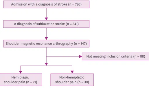
A total of 726 stroke patients were admitted to the university rehabilitation institute and assessed for study eligibility between March 2013 and February 2015. Of these, 59 patients (mean age 65.0
± 11.0 years); mean body weight 65.4 ± 14.1 kg; mean body height 162.6 ± 8.4 cm; 56% male; 74%
ischemic stroke) were eligible for the criteria. The flowchart of the study is presented in Fig. 1.
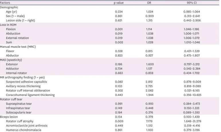
Twenty-one (35.6%) and 38 (64.4%) patients were classified as the HSP and no-HSP groups, respectively (Table 1). Table 2 reports the results of univariate logistic regression on the HSP group.
4/9 https://doi.org/10.12786/bn.2018.11.e6
Pain in Shoulder Subluxation Brain & NeuroRehabilitation
https://e-bnr.org
Admission with a diagnosis of stroke (n = 726)
A diagnosis of subluxation stroke (n = 341)
Shoulder magnetic resonance arthrography (n = 147)
Not meeting inclusion criteria (n = 88)
Hemiplegic
shoulder pain (n = 21) Non-hemiplegic shoulder pain (n = 38) Fig. 1. Flowchart of the study.
Table 1. Comparison of the pain and the no-pain groups
Factors HSP group (n = 21) No-HSP group (n = 38) p value*
Demographic
Age (yr) 60.5 ± 15.2 56.0 ± 13.3 0.237
Sex (male:female) 11:10 19:19 0.861
Lesion side (right:left) 8:13 17:21 0.621
Time from stroke onset to MR arthrography evaluation (day) 66.8 ± 10.0 62.8 ± 8.3 0.111
Duration of poststroke shoulder pain (mon) 40.1 ± 7.3 - -
VAS 6.1 ± 1.5 1.7 ± 1.2 < 0.001
Loss in ROM (°)
Flexion 21.4 ± 15.9 6.2 ± 1.5 < 0.001
Abduction 24.3 ± 19.6 11.1 ± 17.7 0.010
External rotation 23.8 ± 22.6 10.9 ± 15.1 0.026
Sum 69.5 ± 44.3 28.2 ± 33.7 < 0.001
Manual muscle test (MRC)
Flexor 1.7 ± 0.9 1.8 ± 0.9 0.534
Abductor 1.7 ± 0.9 1.8 ± 0.8 0.827
MAS (spasticity)
Extensor 0.57 ± 0.87 0.29 ± 0.69 0.178
Adductor 0.38 ± 0.70 0.32 ± 0.74 0.739
Internal rotator 0.33 ± 0.66 0.43 ± 0.93 0.668
MR arthrography finding (yes:no)
Suspected adhesive capsulitis 9:12 8:30 0.132
Axillary recess thickening 8:13 7:31 0.124
Rotator cuff interval obliteration 5:16 5:33 0.244
Coracohumeral ligament thickening 3:18 3:35 0.656
Supraspinatus tear 5:16 9:29 0.991
Infraspinatus tear 13:8 16:22 0.145
Subscapularis tear 5:16 4:34 0.174
Biceps lesion 6:15 5:33 0.146
Rotator cuff atrophy 8:13* 3:35 0.004
Acromioclavicle joint arthrosis 11:10 22:16 0.448
Humerus chondromalacia 10:11 19:19 0.861
HSP, hemiplegic shoulder pain; MR, magnetic resonance; VAS, visual analog scale; ROM, range of motion; MRC, medical research council scale.
*Compared with HSP group.
The results from multivariate logistic regression analysis are summarized in Table 3. Of particular note, ROM of flexion and rotator cuff atrophy were significantly related to HSP in stroke patients (Nagelkerke R2 = 0.449, p < 0.05).
The final logistic regression equation showed that ROM of flexion was 1.1 times more likely associated with HSP in patients with stroke. Similarly, HSP in the stroke patients with the rotator cuff atrophy apparent in MR arthrography was 6.4 times more likely than in those without the rotator cuff atrophy in MR arthrography. However, the ROM of abduction, external rotation, and total ROM of shoulder were not significantly related with HSP. Muscle power and spasticity were also not related with HSP. Of MR arthrography findings, those compatible with adhesive capsulitis were not significantly related with HSP. Findings compatible with Table 2. Univariate logistic regression analysis to identify relating factors associated with the hemiplegic shoulder pain
Factors p value OR 95% CI
Demographic
Age (yr) 0.234 1.024 0.985–1.064
Sex (1 = male) 0.861 0.909 0.313–2.641
Lesion side (1 = right) 0.621 1.315 0.443–3.906
Loss in ROM
Flexion 0.001 1.114 1.046–1.186
Abduction 0.019 1.038 1.006–1.071
External rotation 0.019 1.038 1.006–1.072
Sum 0.002 1.028 1.010–1.046
Manual muscle test (MRC)
Flexor 0.528 0.815 0.431–1.539
Abductor 0.823 0.927 0.475–1.807
MAS (spasticity)
Extensor 0.186 1.600 0.797–3.212
Adductor 0.734 1.137 0.542–2.384
Internal rotator 0.663 0.859 0.434–1.702
MR arthrography finding (1 = yes)
Suspected adhesive capsulitis 0.082 2.812 0.878–9.009
Axillary recess thickening 0.103 2.725 0.818–9.080
Rotator cuff interval obliteration 0.302 2.062 0.521–8.165
Coracohumeral ligament thickening 0.443 1.944 0.356–10.625
Rotator cuff tear
Supraspinatus tear 0.991 0.993 0.284–3.473
Infraspinatus tear 0.149 0.448 0.150–1.333
Subscapularis tear 0.184 0.376 0.089–1.593
Biceps lesion 0.154 0.379 0.100–1.439
Rotator cuff atrophy 0.009 7.179 1.648–31.279
Acromioclavicle joint arthrosis 0.449 1.512 0.518–4.416
Humerus chondromalacia 0.861 1.100 0.379–3.196
OR, odds ratio; CI, confidence interval; ROM, range of motion; MRC, medical research council scale; MAS, modified Ashworth scale; MR, magnetic resonance.
Table 3. Multivariate logistic regression analysis to identify relating factors associated with the hemiplegic shoulder pain
Factors p value OR 95% CI
Constant < 0.001 0.111
ROM of flexion 0.002 1.102 1.037–1.172
Rotator cuff atrophy (1 = yes) 0.028 6.394 1.222–33.443
Nagelkerke R2 0.449
−2 log likelihood 53.49
Hosmer-Lemeshow goodness-of-fit
χ2 (df) 7.304 (4)
p value 0.121
OR, odds ratio; CI, confidence interval; ROM, range of motion.
adhesive capsulitis included axillary recess thickening, rotator cuff interval obliteration, and coracohumeral ligament thickening. Rotator cuff tear, biceps lesion, acromioclavicle joint arthrosis, and humerus chondromalacia were also not significantly correlated with HSP.
From additional analysis, MAS of flexion correlated with ROM in flexion (coefficient
= 0.257, p = 0.049), external rotation (coefficient = 0.276, p = 0.034), and total ROM (coefficient=0.278, p = 0.033).
DISCUSSION
The purpose of this study was to group hemiplegic patients with shoulder subluxation according to the presence of HSP and to compare the difference in clinical and MR
arthrography findings between 2 groups in the effort to find factors possibly related to HSP.
To our knowledge, this is the first study to evaluate MR arthrography findings in hemiplegic shoulders with subluxation. A significant correlation between HSP and restricted ROM of the shoulder was evident. An unexpected finding was that HSP did not relate with MR arthrography findings, except for rotator cuff muscle atrophy.
Previous studies of MR findings suggestive of adhesive capsulitis between stoke survivors with and without HSP reported that synovial capsule thickening, synovial capsule
enhancement, and enhancement in the rotator cuff interval were more prominent in those with HSP [13,14]. Unlike these studies, our study conducting on shoulder subluxation found no more MR arthrography findings suggesting adhesive capsulitis in the HSP group.
Although many stroke survivors suffer from restricted ROM, it is not fully established whether the pathologic biomechanics are same with adhesive capsulitis. Another potential reason of restricted ROM in hemiplegic shoulder, besides the adhesive capsulitis-like process, might be spasticity around the shoulder joint. It was reported that hemiplegic shoulder had less stiff glenohumeral joint capsules than those with adhesive capsulitis, who had similar ROM limitations [15]. In this context, restricted ROM of the hemiplegic shoulder could result from the combination of adhesive capsulitis and spasticity rather than a pure adhesive capsulitis since spasticity were correlated with restricted ROM of the shoulder in the current study. Therefore, prevention and aggressive management of spasticity and contracture in hemiplegic patients with shoulder subluxation are needed to reduce HSP.
Arm slings have been used for shoulder subluxation. Slings might be theoretically useful while patients ambulate to support the dependent extremity against the effects of gravity and prevent further subluxation and/or decrease pain. A 2009 Cochrane review, however, found insufficient evidence to determine whether slings prevent subluxation, decrease pain, or increase function after stroke [16]. Furthermore, in case of hemislings, positioning of the arm in adduction and internal rotation can predispose these patients to adhesive capsulitis and can promote contractures as the arm is kept in a flexor synergy pattern [17]. If the arm is incorrectly positioned, or if use of a sling is not alternated with therapeutic exercise, soft- tissue contractures can occur. Such contractures can contribute to the very pain the sling is intended to prevent. Since HSP in patients with shoulder subluxation are directly affected by restricted ROM of the shoulder, more active ROM exercise of the shoulder is required regardless of applying slings. Also, the present findings suggest that pathologic lesions like rotator cuff tear may not be related with HSP in patients with shoulder subluxation. Further studies are needed.
6/9 https://doi.org/10.12786/bn.2018.11.e6
Pain in Shoulder Subluxation Brain & NeuroRehabilitation
https://e-bnr.org
Previous studies demonstrated increased prevalence of the rotator cuff tear in hemiplegia [18-20], contrary to other reports that suggested no association between them [21-23].
Occurrence of rotator cuff tear in hemiplegia depends on the severity of motor weakness [24]. A subluxed shoulder can have altered biomechanics and may predispose the patients to rotator cuff injury. Although this study was not designed to assess the prevalence of rotator cuff tear in patients of HSP with shoulder subluxation, this study results showed that there was no difference in the incidence of rotator cuff lesion including the long head of biceps brachii between the HSP and non-HSP groups. The results correspond with a previous study reporting that the association of rotator cuff tears in hemiplegia with premorbid shoulder pain and not with HSP [24]. It may not be necessary to diagnose rotator cuff tear in hemiplegic shoulder if rotator cuff tear does not cause any symptom such as HSP.
The current study showed that HSP did not relate with MR arthrography findings, except for rotator cuff muscle atrophy. We do not know that rotator cuff muscle atrophy was the cause or result of HSP. However, atrophic rotator cuff muscle can result in pain with a pulling sensation with movement, which contributes to posture and muscle imbalance [25]. Considering that muscle imbalance, postural disturbance, and immobilization-common findings in poststroke patients are predisposing factors for the appearance of myofascial trigger points [26], myofascial pain can be a potential contributing cause of HSP. Further studies are needed.
There are some limitations in our study. The retrospective, cross-sectional design study cannot reveal a causal relationship between HSP and other factors. Second, we may have missed potential confounders. Specifically, we excluded the patients who have central pain, central sensitization, and complex regional pain syndrome since they can also play an important role in HSP development. Third, there was a selection bias in the study sample since not all subluxed shoulders were examined by MR arthrography. Because the included subjects did not represent a random sampling of all stroke survivors with subluxed shoulder, the study results may not be generalizable to the broader stroke population. Finally, when the patients were divided into 2 groups, it can be the limitation of this study to set the diagnostic criteria for HSP to “VAS 4 above or below.”
In conclusion, there is a significant correlation between HSP and restricted ROM of shoulder subluxation in this study. Therefore, active treatment of restricted ROM in subluxed shoulder should be emphasized since restricted ROM of shoulder itself is associated with HSP in these patients.
Also, HSP did not relate with MR arthrography findings, except rotator cuff muscle atrophy in hemiplegic patients with shoulder subluxation. In this study, it is difficult to know why the presence or absence of restricted ROM or rotator cuff atrophy affects HSP, and we will try to clarify this through prospective studies on a large number of patients.
REFERENCES
1. Manigandan JB, Ganesh GS, Pattnaik M, Mohanty P. Effect of electrical stimulation to long head of biceps in reducing gleno humeral subluxation after stroke. NeuroRehabilitation 2014;34:245-252.
PUBMED | CROSSREF
2. Park GY, Kim JM, Sohn SI, Shin IH, Lee MY. Ultrasonographic measurement of shoulder subluxation in patients with post-stroke hemiplegia. J Rehabil Med 2007;39:526-530.
PUBMED | CROSSREF
3. Stolzenberg D, Siu G, Cruz E. Current and future interventions for glenohumeral subluxation in hemiplegia secondary to stroke. Top Stroke Rehabil 2012;19:444-456.
PUBMED | CROSSREF
4. Gilmore PE, Spaulding SJ, Vandervoort AA. Hemiplegic shoulder pain: implications for occupational therapy treatment. Can J Occup Ther 2004;71:36-46.
PUBMED | CROSSREF
5. Boyd EA, Goudreau L, O'Riain MD, Grinnell DM, Torrance GM, Gaylard A. A radiological measure of shoulder subluxation in hemiplegia: its reliability and validity. Arch Phys Med Rehabil 1993;74:188-193.
PUBMED
6. Hanger HC, Whitewood P, Brown G, Ball MC, Harper J, Cox R, et al. A randomized a controlled trail of strapping to prevent post-stroke shoulder pain. Clin Rehabil 2000;14:370-380.
PUBMED | CROSSREF
7. Suethanapornkul S, Kuptniratsaikulv PS, Kuptniratsaikul V, Uthensut P, Dajpratha P, Wongwisethkarn J. Post stroke shoulder subluxation and shoulder pain: a cohort multicenter study. J Med Assoc Thai 2008;91:1885-1892.
PUBMED
8. Baetens T, De Kegel A, Calders P, Vanderstraeten G, Cambier D. Prediction of falling among stroke patients in rehabilitation. J Rehabil Med 2011;43:876-883.
PUBMED | CROSSREF
9. Boyd EA, Torrance GM. Clinical measures of shoulder subluxation: their reliability. Can J Public Health 1992;83 Suppl 2:S24-S28.
PUBMED
10. Kelly AM. The minimum clinically significant difference in visual analogue scale pain score does not differ with severity of pain. Emerg Med J 2001;18:205-207.
PUBMED | CROSSREF
11. Gregson JM, Leathley M, Moore AP, Sharma AK, Smith TL, Watkins CL. Reliability of the tone assessment scale and the modified Ashworth scale as clinical tools for assessing poststroke spasticity. Arch Phys Med Rehabil 1999;80:1013-1016.
PUBMED | CROSSREF
12. Lindgren I, Jönsson AC, Norrving B, Lindgren A. Shoulder pain after stroke: a prospective population- based study. Stroke 2007;38:343-348.
PUBMED | CROSSREF
13. Pompa A, Clemenzi A, Troisi E, Di Mario M, Tonini A, Pace L, et al. Enhanced-MRI and ultrasound evaluation of painful shoulder in patients after stroke: a pilot study. Eur Neurol 2011;66:175-181.
PUBMED | CROSSREF
14. Távora DG, Gama RL, Bomfim RC, Nakayama M, Silva CE. MRI findings in the painful hemiplegic shoulder. Clin Radiol 2010;65:789-794.
PUBMED | CROSSREF
15. Yi Y, Lee KJ, Kim W, Oh BM, Chung SG. Biomechanical properties of the glenohumeral joint capsule in hemiplegic shoulder pain. Clin Biomech (Bristol, Avon) 2013;28:873-878.
PUBMED | CROSSREF
16. Ada L, Foongchomcheay A, Canning C. Supportive devices for preventing and treating subluxation of the shoulder after stroke. Cochrane Database Syst Rev 2005:CD003863.
PUBMED | CROSSREF
17. Faghri PD, Rodgers MM, Glaser RM, Bors JG, Ho C, Akuthota P. The effects of functional electrical stimulation on shoulder subluxation, arm function recovery, and shoulder pain in hemiplegic stroke patients. Arch Phys Med Rehabil 1994;75:73-79.
PUBMED
18. Joynt RL. The source of shoulder pain in hemiplegia. Arch Phys Med Rehabil 1992;73:409-413.
PUBMED
19. Najenson T, Yacubovich E, Pikielni SS. Rotator cuff injury in shoulder joints of hemiplegic patients. Scand J Rehabil Med 1971;3:131-137.
PUBMED
20. Nepomuceno CS, Miller JM 3rd. Shoulder arthrography in hemiplegic patients. Arch Phys Med Rehabil 1974;55:49-51.
PUBMED
21. Van Ouwenaller C, Laplace PM, Chantraine A. Painful shoulder in hemiplegia. Arch Phys Med Rehabil 1986;67:23-26.
PUBMED
8/9 https://doi.org/10.12786/bn.2018.11.e6
Pain in Shoulder Subluxation Brain & NeuroRehabilitation
https://e-bnr.org
22. Hakuno A, Sashika H, Ohkawa T, Itoh R. Arthrographic findings in hemiplegic shoulders. Arch Phys Med Rehabil 1984;65:706-711.
PUBMED
23. Rizk TE, Christopher RP, Pinals RS, Salazar JE, Higgins C. Arthrographic studies in painful hemiplegic shoulders. Arch Phys Med Rehabil 1984;65:254-256.
PUBMED
24. Yi Y, Shim JS, Kim K, Baek SR, Jung SH, Kim W, et al. Prevalence of the rotator cuff tear increases with weakness in hemiplegic shoulder. Ann Rehabil Med 2013;37:471-478.
PUBMED | CROSSREF
25. Vasudevan JM, Browne BJ. Hemiplegic shoulder pain: an approach to diagnosis and management. Phys Med Rehabil Clin N Am 2014;25:411-437.
PUBMED | CROSSREF
26. Kalichman L, Ratmansky M. Underlying pathology and associated factors of hemiplegic shoulder pain.
Am J Phys Med Rehabil 2011;90:768-780.
PUBMED | CROSSREF