ABSTRACT
Injury of lower cranial nerves (CNs) by skull base fracture after head trauma can occur sometimes. However, selectively different CN damage on either side is extremely rare.
A 53-year-old man had difficulty of swallowing, phonation, and articulation after falling off his bicycle. In physical examination, a deviated tongue to the right side was shown.
Brain computed tomography showed a skull base fracture involving bilateral jugular foramina and right hypoglossal canal. Left vocal cord palsy was confirmed by laryngoscopy.
Electromyography confirmed injury of left superior laryngeal nerve, recurrent laryngeal nerve, and right hypoglossal nerve. Video fluoroscopic swallowing study revealed large amounts of remnant in vallecula and pyriform sinus without opening of upper esophageal sphincter due to dysfunction of cricopharyngeus muscle. After constant rehabilitation for dysphagia, he was allowed to eat a general diet with compensatory techniques at discharge and further recovery after 3 months. Injury of lower CNs after fracture of the skull base can cause severe morbidity. However, the prognosis of such injuries can be favorable with early rehabilitation treatment by identifying the injured CN. A careful and accurate examination of lower CN injury in skull base fracture is essential for planning a treatment strategy.
Keywords: Vagus nerve injuries; Hypoglossal nerve injuries; Cranial nerve injuries; Skull fractures
INTRODUCTION
Most skull base fractures are related to head trauma. Because there are a lot of cranial nerves (CNs) in the skull base, fracture of the skull base can lead to neurologic complications caused by injury to these nerves.3,9) Lower CNs arise in the medulla and leave the skull through the posterior skull base. Glossopharyngeal nerve (CN IX), vagus nerve (CN X), and accessory nerve (CN XI) penetrate to jugular foramen. Hypoglossal nerve (CN XII) runs through hypoglossal canal nearby occipital condyle. Thus, fracture of posterior skull base,
Case Report
Received: Aug 27, 2020 Revised: Sep 25, 2020 Accepted: Sep 29, 2020 Address for correspondence:
Yuntae Kim
Department of Physical Medicine and Rehabilitation, Soonchunhyang University Cheonan Hospital, Soonchunhyang University College of Medicine, 31 Suncheonhyang 6-gil, Dongnam-gu, Cheonan 31151, Korea.
E-mail: [email protected]
Copyright © 2020 Korean Neurotraumatology Society
This is an Open Access article distributed under the terms of the Creative Commons Attribution Non-Commercial License (https://
creativecommons.org/licenses/by-nc/4.0/) which permits unrestricted non-commercial use, distribution, and reproduction in any medium, provided the original work is properly cited.
ORCID iDs Sanghoon Lee
https://orcid.org/0000-0003-3007-7431 Jae Sang Oh
https://orcid.org/0000-0003-4570-6763 Doh-Eui Kim
https://orcid.org/0000-0001-5499-6315 Yuntae Kim
https://orcid.org/0000-0003-4063-4692
Sanghoon Lee 1, Jae Sang Oh 2, Doh-Eui Kim 3, and Yuntae Kim 4
1 Department of Physical Medicine and Rehabilitation, Soonchunhyang University Seoul Hospital, Soonchunhyang University College of Medicine, Seoul, Korea
2 Department of Neurosurgery, Soonchunhyang University Cheonan Hospital, Soonchunhyang University College of Medicine, Cheonan, Korea
3 Department of Emergency Medicine, Soonchunhyang University Cheonan Hospital, Soonchunhyang University College of Medicine, Cheonan, Korea
4 Department of Physical Medicine and Rehabilitation, Soonchunhyang University Cheonan Hospital, Soonchunhyang University College of Medicine, Cheonan, Korea
Concomitant Injury of Vagus and Hypoglossal Nerves Caused by
Fracture of Skull Base: A Case Report
and Literature Review
Conflict of Interest
The authors have no financial conflicts of interest.
particularly jugular tubercle and occipital condyle, can cause injury of lower CNs. Although various cases1,2,4-8,13-19) of lower CN injury by skull base fracture have been reported previously, selectively different CN damage on either side has not been reported yet. This is the first report of a case of concomitant injury of left CN X and right CN XII caused by fracture of the skull base confirmed by electrodiagnostic (EDx) study, laryngoscopy, and swallowing study.
CASE REPORT
A 53-year-old man visited the emergency room with a head trauma after falling off his bicycle. He complained of difficulty in swallowing, phonation, and articulation. His level of consciousness was normal perhaps because he had the accident with a helmet on. He had a weakness of the right tongue with deviation to the right side (FIGURE 1A), dysphagia, and dysphonia. Clinically, lower CNs injury was suspected. Neurologic examination results for other CNs were normal. Muscle strengths of bilateral upper and lower extremities were intact.
Brain computed tomography showed a fracture of skull base involving bilateral jugular tubercle, right hypoglossal canal, and clivus (FIGURE 2). Cervical spine magnetic resonance imaging showed a subdural hemorrhage at C3–C4 level and a compression fracture at C3 vertebra (FIGURE 3). Video fluoroscopic swallowing study (VFSS) at 6 days after onset revealed large amount of remnant and dysfunction of the upper esophageal sphincter (UES) (FIGURE 4A). In laryngoscopic examination for dysphonia, vocal cord mobility was slightly decreased in the left side. Articulation test showed dysarthria (consonant accuracy of word-level: 95.71%) by dysfunction of the tongue. However, aphasia was not shown.
EDx study was performed to confirm injury to lower CNs at 2 weeks after onset. Needle electromyography (EMG) revealed abnormal spontaneous activities such as fibrillation and positive sharp wave with decreased recruitment pattern in left thyroarytenoid, cricothyroid,
A B C
FIGURE 1. Photographs showing the patient's tongue. Deviated tongue to the right side at 10 days (A) after onset. Improvement of the deviated tongue and remained atrophic change of the right tongue at 5 weeks (B) and 12 weeks (C) after onset.
and right tongue muscles. The EDx study confirmed a concomitant injury of left superior laryngeal, recurrent laryngeal and right CN XII.
He was planned to receive conservative treatment because the site of fracture was stable.
He had been keeping a cervical collar for the stability of the skull base and cervical spine for about 8 weeks. A nasogastric tube was inserted for enteral feeding. He received rehabilitation treatment for dysphagia, dysarthria, and dysphonia such as oral motor facilitation, Shaker's exercise, facial expression exercise, and vocal exercise. We conducted VFSS weekly after the first study because the most uncomfortable symptom was a difficulty of swallowing. The fourth study (FIGURE 4B) revealed a significant improvement of UES opening and descending of the food along the right wall of the esophagus during head tilting to the right side and
A B C
FIGURE 2. Brain computed tomography showing a fracture of skull base involving bilateral jugular tubercle and right hypoglossal canal. (A) Transverse shape fracture of clivus involving bilateral jugular tubercles in the axial image. (B) Fracture involving the left jugular foramen in the axial image. (C) Fracture involving the right hypoglossal canal in the coronal image.
A B
FIGURE 3. T2 fat suppression sagittal (A) and T1 axial (B) magnetic resonance imaging of the cervical spine showing high signal in C3 vertebra and low signal in posterolateral intradural extramedullary space on C3–4 level, indicating C3 compression fracture and subdural hemorrhage at C3–4 level.
head rotation to the left side. Thus, he was allowed to remove the nasogastric tube and eat a semi-solid food with a compensation technique. About 5 weeks after onset, he could eat one bowl of a semi-solid food in a meal with 10 times of swallowing per one spoon with right head tilt without aspiration symptom. His tongue deviation showed improvement, although atrophic change remained (FIGURE 1B). He spoke louder and more resonant. However, he had a mildly wet voice in speech. About 6 weeks after onset, he was discharged, keeping a cervical collar at home.
We followed him at the outpatient clinic. At 8 weeks after onset, we allowed him to take off the cervical collar and recommended a personal functional electrical stimulator (FES) for suprahyoid muscles. At 12 weeks after onset, he used the FES on his neck and reported improvement of dysarthria. His mild wet voice remained. He continued to eat a meal using a compensation technique such as head tilting to right side without aspiration event. His deviated and atrophic right tongue showed more improvement (FIGURE 1C). He made louder phonation and accurate articulation. Follow-up VFSS showed decreased remnants and cough himself to correct the penetration event without aspiration compared to previous study. After that, we planned to follow-up him at our outpatient clinic if he had any discomfort when eating. The patient provided informed consent for publication of this case.
DISCUSSION
We present the first case of selectively different lower CN injury caused by fracture of the skull base. Skull base fracture involving the right hypoglossal canal and left jugular foramen area by axial force would be the mechanism of injury in this case.
A B
FIGURE 4. Improvement of upper esophageal sphincter opening on VFSS after swallowing rehabilitation. (A) Large amount of remnant in vallecula and pyriform sinus on lateral view of the 1st VFSS study. (B) Descending of food materials on lateral view of the 4th VFSS study.
VFSS: video fluoroscopic swallowing study.
Fracture of the posterior skull base can cause vascular, vertebrobasilar, craniocervical junction, and cervical spinal problems. Besides, lower CN injuries (CN IX, X, XI, and XII) can occur because either side of the occipital condyle is tunneled by the hypoglossal canal which transmits CN XII and located nearby the jugular foramen which transmits CN IX, X, and XI. These injuries will cause symptoms such as dysphagia, dysarthria, loss of taste, loss of oropharyngeal sense, weakness of sternocleidomastoid (SCM), and weak trapezius muscles.
It is important to exam lower CNs when a patient complains of variable degrees of symptoms caused by CN deficit after a head trauma.
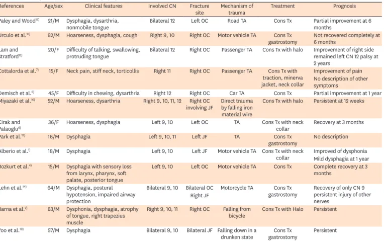
TABLE 1 is a summary of literature reviews1,2,4-8,14-19) of similar cases reported after the 1990s. An asymmetric injury like this case was not reported in literature reviews. The prognosis of most cases was poor such as inserted percutaneous gastrostomy in some reports.14,17-19) Complete recovery has only been reported by Bozkurt et al.4) Unlike previous reports, our case showed injury of asymmetric lower CNs (left CN X and right CN XII) with early significant recovery.
Asymmetric injury of lower CN is related to anatomical location of lower CNs in jugular foramen. When jugular foramen is separated by the jugular spine, the anteromedial portion is pars nervosa including CN IX and inferior petrosal sinus. The posterolateral portion is pars vascularis including CN X, XI and jugular bulb. According to Freitas et al.,10) CN IX emerges from the jugular foramen via the anteromedial portion in front of the vagal nerve. It is curved medially near the posteroinferior aspect of the styloglossus muscle and the anterior wall of the internal carotid artery. CN X emerges from the jugular foramen via the anteromedial TABLE 1. Summary of reports of lower CN injury by skull base fracture
References Age/sex Clinical features Involved CN Fracture
site Mechanism of
trauma Treatment Prognosis
Paley and Wood15) 21/M Dysphagia, dysarthria,
nonmobile tongue Bilateral 12 Left OC Road TA Cons Tx Partial improvement at 6
months Urculo et al.18) 62/M Hoarseness, dysphagia, cough Right 9, 10 Right OC Motor vehicle TA Cons Tx
gastrostomy Not recovered completely at 6 months
Lam and
Stratford13) 20/F Difficulty of talking, swallowing,
protruding tongue Bilateral 12 Right OC Passenger TA Cons Tx with halo Improvement of right side remained left CN 12 palsy at 2 years
Cottalorda et al.7) 15/F Neck pain, stiff neck, torticollis Right 11 Right OC Passenger TA Cons Tx with traction, minerva jacket, neck collar
Improvement of pain No description of other symptoms
Demisch et al.8) 45/F Difficulty in chewing, dysarthria Right 12 Right OC Car TA Cons Tx Partial improvement at 1 year Miyazaki et al.16) 52/M Hoarseness, dysarthria Right 9, 10, 11, 12 Right OC
involving JF Direct trauma by falling iron material wire
Cons Tx with halo Persistent at 12 weeks
Cirak and
Palaoglu6) 36/F Hoarseness, dysphagia Left 9, 10 Left OC TA Cons Tx with neck
collar Recovery at 3 months
Park et al.17) 16/M Dysphagia Left 9, 10, 11 Left JF TA Cons Tx
gastrostomy No description Alberio et al.1) 18/M Dysphagia Left 9, 10 Left JF Motor vehicle TA Cons Tx with neck
collar Improved of dysphonia Mild dysphagia at 1 year Bozkurt et al.4) 15/M Dysphagia with sensory loss
from larynx, pharynx, soft palate, posterior tongue
Left 9, 10 Left OC Motor vehicle TA Cons Tx Complete recovery at 3 months
Lehn et al.14) 64/M Dysphagia, postural hypotension, impaired airway protection
Bilateral 9, 10 Bilateral OC Motorcycle TA Cons Tx
gastrostomy Recovery of only CN 9 persistent injury of other nerves
Right JF Barna et al.2) 63/M Dysphonia, dysphagia, atrophy
of tongue, right trapezius muscle
Right 9, 10, 11 Right OC Falling from
bicycle Cons Tx with Halo Persistent Yoo et al.19) 57/M Dysphagia Bilateral 9, 10 Bilateral JF Falling down in a
drunken state Cons Tx
gastrostomy Persistent M: male, F: female, CN: cranial nerve, OC: occipital condyle, JF: jugular foramen, Cons Tx: conservative treatment, TA: traffic accident.
portion between CN IX and CN XI. CN XI emerges from the jugular foramen via the
anteromedial portion posterior to the vagal nerve. However, in some cases, the nerve passes around the posterior portion. According to Bozkurt et al.,4) cranial IX and X nerves are more perpendicular than cranial XI nerve. The CN XI consists of cranial and spinal parts. Cranial XI has more rootlets than cranial IX and X nerves.
EDx study is a critical diagnostic tool to detect injured nerves. It was also applied in this case as in some prior reports. EMG for tongue, thyroarytenoid, and cricothyroid muscles can assess hypoglossal, recurrent laryngeal, and superior laryngeal nerves. Recurrent laryngeal and superior laryngeal nerves are branches of the CN X. However, EDx study for the CN IX is limited. Stylopharyngeus muscle innervated by that nerve cannot be isolated for electrode placement because it is small in size. In addition, it overlaps with other muscles.12) Because CN IX is anatomically close to CN X, the nerve may be injured. However, we substituted its examination for evaluation of deviation of uvula and sensory on the posterior aspect of the tongue. The uvula did not deviate. In addition, sensory and taste on the posterior aspect of the tongue were intact. VFSS showed severe dysfunction of laryngeal elevation and UES opening controlled by thyrohyoid and cricopharyngeus muscles innervated by CN X. These dysfunctions made an aspiration during and after swallowing.
In this case, compression fracture and subdural hemorrhage with cord compression on the cervical spine were accompanied. We need to evaluate and diagnose carefully because myelopathy or laryngeal nerve injury by cervical spinal problems can make dysphagia or dysphonia. However, any neurologic sign suggesting myelopathy was not shown. EDx study for myelopathy was normal in this case. According to MacEwen et al.,11) laryngeal nerve injury by traction of the spine is possible. Although it means that strong force to the cervical spine may damage the laryngeal nerve, this is extremely rare. It cannot explain simultaneous CN XII injury of the present case.
The prognosis of traumatic lower CN palsy is unfavorable according to previous reports.
Thus, the best treatment is prevention such as wearing a protective device, playing careful activities, and so on. Once accident occurs, treatment should cover various aspects, including symptoms (such as dysphagia, dysarthria, dysphonia, uncontrolled secretion of saliva, weakness of SCM and trapezius muscles), stability and union of fractures, patient's mood, and so on. Treatment in this case was conservative like most of prior cases. It contained a therapy for dysphagia involving oral motor facilitation, compensation maneuver and exercise to strengthen a laryngeal elevation, open UES such as head position, Mendelsohn maneuver, and Shaker's exercise and speech therapy for phonation and articulation. According to prior cases, the main problem is dysphagia that results in tube feeding, compensation technique such as head tilting and rotation for unilateral weakness of swallowing muscles is very useful. It resulted in safe swallowing without aspiration in the present case. VFSS showed improvement of UES opening and the amount of food materials to the esophagus by compensation of head position. Thus, he was allowed to eat oral diet consisting of semi-solid food with compensation techniques rather than considering percutaneous gastrostomy.
CONCLUSION
We report the first case of concomitant injury of left CN X and right CN XII caused by fracture of skull base confirmed by EDx study, laryngoscopy, and swallowing study. Although the prognosis of injury of lower CNs is poor according to previous reports, we believe that the
prognosis can be better if the damage is identified accurately with early rehabilitation for the patient as shown in this case. Therefore, careful and accurate examination about lower CN is important to diagnose and plan a treatment strategy for patients with skull base fracture.
ACKNOWLEDGEMENTS
This work was supported by the Soonchunhyang University Research Fund.
This research was supported by the Bio & Medical Technology Development Program of the National Research Foundation funded by the Korean government (NRF- 2019M3E5D1A02069061, NRF-2020R1F1A1066362).
REFERENCES
1. Alberio N, Cultrera F, Antonelli V, Servadei F. Isolated glossopharyngeal and vagus nerves palsy due to fracture involving the left jugular foramen. Acta Neurochir (Wien) 147:791-794, 2005
PUBMED | CROSSREF
2. Barna M, Štulík J, Kryl J, Vyskočil T, Nesnídal P. Collet-Sicard syndrome due to occipital condyle fracture.
Case report. Acta Chir Orthop Traumatol Cech 82:440-442, 2015 PUBMED
3. Baugnon KL, Hudgins PA. Skull base fractures and their complications. Neuroimaging Clin N Am 24:439-465, vii-viii, 2014
PUBMED | CROSSREF
4. Bozkurt G, Hazer B, Yaman ME, Akbay A, Akalan N. Isolated paralysis of glossopharyngeal and vagus nerve associated with type II occipital condyle fracture: case report. Childs Nerv Syst 26:719-722, 2010 PUBMED | CROSSREF
5. Capuano C, Costagliola C, Shamsaldin M, Maleci A, Di Lorenzo N. Occipital condyle fractures: a hidden nosologic entity. An experience with 10 cases. Acta Neurochir (Wien) 146:779-784, 2004
PUBMED | CROSSREF
6. Cirak B, Akpinar G, Palaoglu S. Traumatic occipital condyle fractures. Neurosurg Rev 23:161-164, 2000 PUBMED | CROSSREF
7. Cottalorda J, Allard D, Dutour N. Fracture of the occipital condyle. J Pediatr Orthop B 5:61-63, 1996 PUBMED | CROSSREF
8. Demisch S, Lindner A, Beck R, Zierz S. The forgotten condyle: delayed hypoglossal nerve palsy caused by fracture of the occipital condyle. Clin Neurol Neurosurg 100:44-45, 1998
PUBMED | CROSSREF
9. Feiz-Erfan I, Horn EM, Theodore N, Zabramski JM, Klopfenstein JD, Lekovic GP, et al. Incidence and pattern of direct blunt neurovascular injury associated with trauma to the skull base. J Neurosurg 107:364-369, 2007
PUBMED | CROSSREF
10. Freitas CA, Santos LR, Santos AN, Amaral Neto AB, Brandão LG. Anatomical study of jugular foramen in the neck. Braz J Otorhinolaryngol 86:44-48, 2020
PUBMED | CROSSREF
11. MacEwen GD, Bunnell WP, Sriram K. Acute neurological complications in the treatment of scoliosis. A report of the Scoliosis Research Society. J Bone Joint Surg Am 57:404-408, 1975
PUBMED
12. Palmer JB, Tanaka E, Siebens AA. Electromyography of the pharyngeal musculature: technical considerations. Arch Phys Med Rehabil 70:283-287, 1989
PUBMED
13. Lam CH, Stratford J. Bilateral hypoglossal nerve injury with occipital condylar fracture. Can J Neurol Sci 23:145-148, 1996
PUBMED | CROSSREF
14. Lehn AC, Lettieri J, Grimley R. A case of bilateral lower cranial nerve palsies after base of skull trauma with complex management issues: case report and review of the literature. Neurologist 18:152-154, 2012 PUBMED | CROSSREF
15. Paley MD, Wood GA. Traumatic bilateral hypoglossal nerve palsy. Br J Oral Maxillofac Surg 33:239-241, 1995 PUBMED | CROSSREF
16. Miyazaki C, Katsume M, Yamazaki T, Aoki K, Kuroki T, Takasu N. Unusual occipital condyle fracture with multiple nerve palsies and Wallenberg syndrome. Clin Neurol Neurosurg 102:255-258, 2000
PUBMED | CROSSREF
17. Park JH, Park IS, Ha JS, Sim JH, Sul SY. Dysphagia associated with jugular foramen syndrome due to traumatic brain injury. J Korean Acad Rehabil Med 25:163-167, 2001
18. Urculo E, Arrazola M Jr, Arrazola M Jr, Riu I, Moyua A. Delayed glossopharyngeal and vagus nerve paralysis following occipital condyle fracture. Case report. J Neurosurg 84:522-525, 1996 PUBMED | CROSSREF
19. Yoo SD, Kim DH, Lee SA, Joo HI, Yeo JA, Chung SJ. Bilateral cranial IX and X nerve palsies after mild traumatic brain injury. Ann Rehabil Med 40:168-171, 2016
PUBMED | CROSSREF