ABSTRACT
Background and Purpose: Neural stem cells (NSCs) have the ability to regenerate, proliferate, and differentiate, enabling them to play important roles in the recovery of the damaged nervous system. However, in neurodegenerative diseases such as Alzheimer's disease (AD), the NSCs are damaged as well. Glia-like cells from human mesenchymal stem cells (ghMSCs) are functionally enhanced adult stem cells. In the present study, we investigated whether ghMSCs could protect NSCs from amyloid beta (Aβ)-mediated toxicity.
Methods: Rat NSCs were obtained from E13–14 fetal rat cortices. NSCs were seeded in pre-coated plates, and the next day, cells were simultaneously treated with 20 μM Aβ and 0.4 μm pore insert well-seeded ghMSCs. After 48 hours of co-treatment, cell viability and proliferation were evaluated. After 2 hours of co-treatment, western blotting was performed to measure inflammasome-related factors, such as NOD-like receptor family pyrin domain containing 3, caspase-1, and interleukin-1β.
Results: The results showed that ghMSCs increased viability and proliferation and reduced the toxicity of NSCs injured by Aβ by reducing the NRLP3 inflammasome activation of NSCs induced by Aβ.
Conclusions: In this study, we confirmed that ghMSCs could protect NSCs in an in vitro model of AD through the regulation of inflammatory response.
Keywords: Alzheimer's Disease; Amyloid; Neural Stem Cells; Mesenchymal Stem Cells;
Inflammasomes
INTRODUCTION
Alzheimer's disease (AD), the most common neurodegenerative disease which is rapidly becoming the most serious social problem worldwide, does not have a cure yet.1 In AD, neurofibrillary tangles and amyloid plaques are the most representative lesions. These lesions lead to neuronal loss, memory loss, and cognitive impairment of the brain.2 In
Original Article
Received: Oct 22, 2020 Revised: Nov 17, 2020 Accepted: Nov 18, 2020 Correspondence to Seong-Ho Koh
Department of Neurology, Hanyang University College of Medicine, 222 Wangsimni-ro, Seongdong-gu, Seoul 04763, Korea.
E-mail: ksh213@hanyang.ac.kr Mi-Sook Chang
Laboratory of Stem Cell & Neurobiology, Department of Oral Anatomy, Dental Research Institute and School of Dentistry, Seoul National University, 101 Daehak-ro, Jongno-gu, Seoul 03080, Korea.
E-mail: mschang@snu.ac.kr
* Mina Hwang and Se hyeon Song contributed equally to this work.
© 2021 Korean Dementia Association This is an Open Access article distributed under the terms of the Creative Commons Attribution Non-Commercial License (https://
creativecommons.org/licenses/by-nc/4.0/) which permits unrestricted non-commercial use, distribution, and reproduction in any medium, provided the original work is properly cited.
ORCID iDs Mina Hwang
https://orcid.org/0000-0002-5866-8906 Se hyeon Song
https://orcid.org/0000-0002-2344-0506 Mi-Sook Chang
https://orcid.org/0000-0002-4379-204X Seong-Ho Koh
https://orcid.org/0000-0001-5419-5761
Mina Hwang ,1,* Se hyeon Song ,2,* Mi-Sook Chang ,2,3 Seong-Ho Koh 1,4
1Department of Neurology, Hanyang University College of Medicine, Seoul, Korea
2 Laboratory of Stem Cell & Neurobiology, Department of Oral Anatomy, Dental Research Institute and School of Dentistry, Seoul National University, Seoul, Korea
3Neuroscience Research Institute, Seoul National University, Seoul, Korea
4 Department of Translational Medicine, Hanyang University Graduate School of Biomedical Science &
Engineering, Seoul, Korea
Glia-Like Cells from Human
Mesenchymal Stem Cells Protect
Neural Stem Cells in an In Vitro Model
of Alzheimer's Disease by Reducing
NLRP-3 Inflammasome
Funding
This research was supported by the Basic Science Research Program of the National Research Foundation of Korea, funded by the Ministry of Science and ICT (2018R1A2A2A15023219, 2019R1A2C1087192);
by a grant from the Korea Health Technology R&D Project through the Korea Health Industry Development Institute (KHIDI) funded by the Ministry of Health & Welfare, Republic of Korea (grant No. HI20C0253); by the Medical Research Center (2017R1A5A2015395); and by the Seoul National University Research Grant (860-20180077).
Conflict of Interest
The authors have no financial conflicts of interest.
Author Contributions
Conceptualization: Hwang M, Song SH, Chang MS, Koh SH; Data curation: Hwang M, Song SH; Funding acquisition: Chang MS, Koh SH; Investigation: Hwang M, Song SH; Methodology: Hwang M, Song SH;
Supervision: Chang MS, Koh SH; Writing - original draft: Hwang M, Song SH; Writing - review & editing: Chang MS, Koh SH.
particular, amyloid beta1-42 (Aβ1-42) oligomers produced from amyloid precursor protein are the most toxic to neurons.3,4 Thus, numerous studies targeting Aβ1–42 have been conducted for the treatment of AD.5
Recently, much emphasis has been laid on the role of neuroinflammation in the pathogenesis of AD. The deposition of Aβ drives the activation of microglia in AD.6 Microglial activation increases interleukin-1β (IL-1β) secretion in response to Aβ, a phenomenon that requires caspase-1. Caspase-1 activity is associated with inflammasomes. The NOD-like receptor family pyrin domain containing 3 (NLRP3) inflammasome induces pyroptotic cell death by regulating several neuroinflammatory responses in AD.7 Heneka et al.7 demonstrated that Aβ-induced invigoration of the NLRP3 inflammasome reinforces AD progression in an in vivo model of AD;
therefore, inhibiting the activity of the NLRP3 inflammasome is important in AD.7
Neural stem cells (NSCs) localize in the subgranular zone of the hippocampal dentate gyrus, hypothalamus, and subventricular zone in the brain.8 NSCs are involved in the course of regeneration of the damaged brain. NSCs can replace lost or damaged neurons through their multipotent nature and ability to self-renew. As such, it has been suggested as a therapeutic strategy for neurological diseases such as AD.9 However, Aβ oligomers also damage NSCs.
Therefore, it is crucial to identify ways of protecting NSCs from Aβ-induced toxicity.
Early-passage human mesenchymal stem cells (hMSCs) exhibit paracrine activity and secrete growth factors and cytokines in the damaged nervous system.10 Early-passage cells have much more protective effects in the damaged brain than late-passage cells; however, obtaining sufficient amounts of early-passage hMSCs for clinical trials can be hard.11 Our previous study confirmed that late-passage hMSCs could be induced into glia-like cells from hMSCs (ghMSCs) with enhanced paracrine activities.11 Since ghMSCs are functionally-enhanced adult stem cells, there is no fear of side effects that could induce either an immune response or tumor formation associated with the use of these cells.11
In the present study, we investigated whether ghMSCs could protect NSCs exposed to an in vitro model of AD, and how ghMSCs affect the NLRP3 inflammasome that is involved in NSC damage in an in vitro model of AD.
METHODS
Culture of NSCs
All procedures involving animals were performed in accordance with the Hanyang University guidelines for the care and use of laboratory animals and were approved by the Institutional Animal Care and Use Committee of Hanyang University (No. 2019-0162A).
For the primary culture of embryonic NSCs, we followed previously published methods.4,12-14 NSCs were obtained from E13–14 fetal rat cortices. Embryos were transferred to a 100-mm petri dish. The embryos were washed 3–4 times with Hank's balanced salt solution (2.5 mM HEPES, 5.6 mM glucose, 0.4 mM KH2PO4, 0.3 mM Na2HPO4, 5.4 mM KCl, 137 mM NaCl; Gibco, BRL, Grand Island, NY, USA). NSCs were isolated from the ventral midbrain, lateral ganglionic eminence, and cerebral cortex of fetal rats. The cells were then seeded in culture dishes. Culture dishes were pre-coated with poly-L-ornithine/fibronectin in Ca2+/Mg2+-free phosphate buffered saline (PBS; Gibco). Cells were cultured in the N2 medium (Dulbecco's modified Eagle's
medium [DMEM]/F12, 20 nM NaHCO3, 8.6 mM D(+) glucose, 2 nM L-glutamine, 0.2 mM ascorbic acid, 20 nM progesterone, 100 μM putrescine, 30 nM selenite, 100 mg/L transferrin, 25 mg/L insulin; Sigma-Aldrich, St. Louis, MO, USA). The culture medium was supplemented with 10 ng/mL basic fibroblast growth factor (bFGF; Gibco) and changed every 2–3 days. Cultures were maintained at 37°C under a humidified 5%-CO2 atmosphere.
Culture of hMSCs
Adult hMSCs were obtained from normal human bone marrow (BM; Cambrex Bioscience, Walkersville, MD, USA). The cells were cultured in low-glucose DMEM supplemented with 10% fetal bovine serum (Gibco). hMSCs (passages 6–12) were cultured at 37°C under a humidified 5%-CO2 atmosphere.
Glia-like cell induction of hMSCs (ghMSCs)
For the induction of ghMSCs, we followed our previously reported protocols.15 First, hMSCs were incubated with 1 mM β-mercaptoethanol (Sigma-Aldrich). After 24 hours of incubation, the cells were added to 0.28 μg/mL all-trans-retinoic acid (Sigma-Aldrich). After 3 days, the cells were treated with a cocktail containing HRG-β1 (200 ng/mL; R&D Systems, Minneapolis, MN, USA), 10 μM forskolin (Sigma-Aldrich), 5 ng/mL PDGF-AA, and 10 ng/mL bFGF (both from PeproTech, Rocky Hill, NJ, USA) for 8 days.
Co-culture with ghMSCs
To assess the effects of ghMSCs on Aβ-injured NSCs, NSCs treated with Aβ oligomers were co-cultured with ghMSCs. NSCs were first seeded in pre-coated 24-well plates and, the next day, simultaneously treated with 20 μM Aβ and 0.4 μm pore insert well-seeded ghMSCs.
The treated cells were co-cultured for 48 hours to evaluate cell viability, proliferation, and cytotoxicity. They were also co-cultured for 2 hours to measure the expression level of inflammasome factors such as NLRP3, caspase-1, and IL-1β by western blotting. NSCs were seeded at 1×10⁵ cells/cm2 for 48 hours co-culture and at 4×10⁵ cells/cm2 for 2 hours co-culture.
ghMSCs were seeded at 2.7×10⁴ cells/cm2 for 48 hours co-culture and at 2.7×10⁴ cells/cm2 for 2 hours co-culture.
Aβ
25–35oligomerization
The Aβ25–35 peptide (Sigma-Aldrich) was re-suspended in dimethyl sulfoxide (Panreac, Barcelona, Spain) at a 5 mM concentration and added to DMEM/F-12 (Gibco) at a final 1 mM concentration. The peptide was incubated for 24 hours at 4°C for oligomerization.
Cell counting kit-8 (CCK-8) and lactate dehydrogenase (LDH) assays
CCK-8 (Dojindo, Kumamoto, Japan) was used to measure the viability of NSCs by combining 1-methoxy-5-methylphenazinium methyl sulfate and water soluble tetrazolium salt-8 [2-(2-methoxy-4-nitrophenyl)-3-(4-nitrophenyl)-5-(2,4-disulfophenyl)-2H-tetrazolium, monosodium salt]. NSCs were co-cultured with Aβ and ghMSCs for 48 hours, and then the cells were replaced with a new culture medium containing CCK-8 reagent according to the manufacturer's instructions. After 2 hours of incubation, the viability of NSCs was assessed using a Synergy H1 Hybrid enzyme-linked immunosorbent assay (ELISA) reader (BioTek, Winooski, VT, USA) at 450 nm and 650 nm.
An LDH cytotoxicity detection kit (Takara, Kusatsu, Japan) was used to evaluate the
cytotoxicity of NSCs releasing LDH. NSCs co-cultured with Aβ and ghMSCs were centrifuged at 200 × g for 10 m, and the supernatant was transferred to a new plate. The supernatant was
added to the mixture of dye solution and a catalyst for 30 minutes without light, according to the manufacturer's instructions. After that, the amount of generated formazan was evaluated using an ELISA reader at 492 nm and 690 nm.
Trypan blue staining
NSCs co-cultured with Aβ and ghMSCs were washed twice using Dulbecco's PBS and transferred to a new culture medium. The cells were then stained with trypan blue solution (Gibco) for 2 minutes. Live and dead cells were counted with a hemocytometer.8
Bromodeoxyuridine (BrdU) cell proliferation assay
A cell-proliferation ELISA BrdU (colorimetric) kit (Roche, Mannheim, Germany) was used to quantify the proliferation of NSCs. According to the manufacturer's instructions in the kit, NSCs co-cultured with Aβ and ghMSCs were labeled with 10 μM BrdU for 18 hours and fixed with a fixation solution for 30 minutes. Then, the fixed cells were added to 300 μL of anti-BrdU-POD working solution. After 2 hours of incubation without light, the samples were washed with a washing solution. Finally, the cells reacted with 300 μL of the substrate. After 10 minutes at room temperature without light, the samples were evaluated using an ELISA reader at 370 nm and 492 nm.
Western blotting
To investigate the expression levels of inflammasome factors such as NLRP3, caspase-1, and IL-1β, in NSCs co-cultured with Aβ and ghMSCs, western blotting was performed. Briefly, cells were collected by scraper and centrifuged at 6,000 × g for 2 minutes at 4°C. Pellets were washed twice and added to lysis buffer (0.5% protease inhibitor cocktail 1×, 1 mM sodium orthovanadate, 1 mM phenylmethylsulfonyl fluoride, RIPA II cell lysis buffer 1× with Triton, without ethylenediaminetetraacetic acid, and 1 mM sodium fluoride [Sigma-Aldrich]). After 30 minutes of incubation on ice, the cells were sonicated several times using a sonicator (Sonoplus; Bandelin Electronics, Berlin, Germany). After another 30 minutes of incubation on ice, the lysates were centrifuged at 21,100 × g for 15 minutes at 4°C, and the protein concentrations of lysates were calculated using a bicinchoninic acid protein assay kit (Sigma- Aldrich). Lysate samples containing equal amounts of protein Proteins were resolved by 4%–
12% sodium dodecyl sulfate-polyacrylamide gel electrophoresis (Invitrogen, Carlsbad, CA, USA). The proteins were then transferred to polyvinylidene fluoride membranes (Millipore, Bedford, MA, USA). The membranes were blocked with 5% skimmed milk. After blocking, the membranes were incubated with a specific primary antibody. The antibodies used in these experiments were as follows: NLRP3 (2 μg/mL; Novus Biologicals, Littleton, CO, USA), caspase-1 (2.5 μg/mL, Novus Biologicals), IL-1β (0.4 μg/mL; Abcam, Burlingame, CA, USA), and β-actin (1:4,000; Cell Signaling Technology, Beverly, MA, USA). The membranes were washed thrice with Tris-buffered saline containing 0.1% Tween-20, followed by incubation with horseradish peroxidase-conjugated anti-rabbit antibody (1:2,000; Jackson ImmunoResearch Laboratories Inc., West Grove, PA, USA). The membranes were stained and visualized using a West-Q Chemiluminescent Substrate Kit (GenDEPOT, Katy, TX, USA). Visualized images were analyzed using ImageQuant LAS 4000 (GE Healthcare, Little Chalfont, UK).
Statistical analysis
All data are expressed as the mean±standard deviation of 3 independent experiments. Statistical comparisons between different treatment groups were performed using Tukey's test after a 1-way analysis of variance. The p-values less than 0.05 were considered statistically significant.
RESULTS
ghMSCs increase the viability and proliferation while reducing the cytotoxicity of NSCs injured by Aβ
To confirm that ghMSCs protect NSCs from Aβ, NSCs seeded at 1×10⁵ cells/cm2 and treated with 20 μM Aβ were co-cultured with ghMSCs seeded at 2.7×10⁴ cells/cm2, and the viability was measured using a CCK-8 assay and trypan blue staining. The results showed that Aβ decreased the viability of NSCs, but co-treatment with ghMSCs increased the viability of NSCs injured by Aβ (p<0.001, Fig. 1A and B). To assess the effect of ghMSCs on the toxicity of Aβ to NSCs, released LDH was evaluated using a cytotoxicity detection assay. The LDH cytotoxicity detection assay demonstrated that Aβ increased the cytotoxicity of NSCs.
However, the Aβ-induced cytotoxicity of NSCs was significantly reduced by co-culturing with ghMSCs (p<0.001, Fig. 1C). Next, we performed a BrdU assay to determine how Aβ and ghMSCs affect NSC proliferation. The results demonstrated that the proliferation of NSCs was reduced in the group treated with Aβ but increased in the group co-treated with Aβ and ghMSCs (p<0.01, Fig. 1D).
ghMSCs reduce NLRP3 inflammasome activation of NSCs induced by Aβ
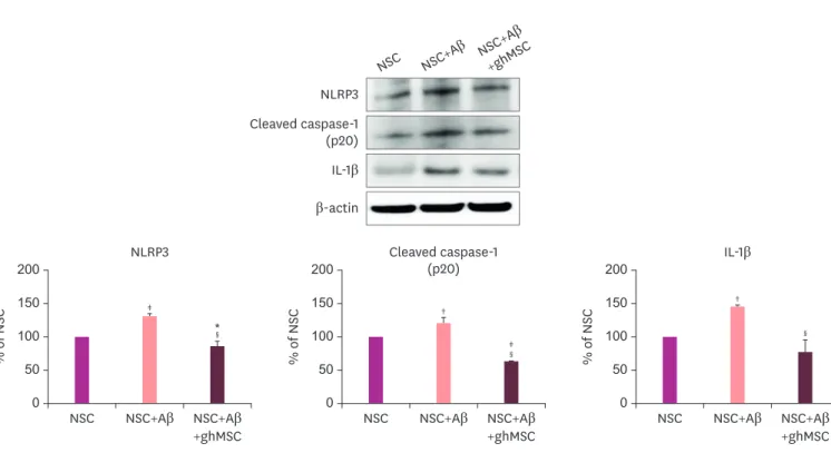
To determine whether increased inflammatory responses in NSCs seeded at 4×10⁵ cells/cm2 and treated with Aβ was affected by co-culture with ghMSCs seeded at 2.7×10⁴ cells/cm2, the expression levels of NLRP3, cleaved caspase-1, and pro-inflammatory cytokine IL-1β were evaluated by western blotting. As shown in Fig. 2, Aβ induced activation of NLRP3, caspase-1† †
‡
*
∥
∥
0 100 200
NSC NSC+Aβ NSC+Aβ +ghMSC CCK-8 assay
A B
Cell viability (% of NSC
)
0 100 200
50 150
50 150
NSC NSC+Aβ NSC+Aβ +ghMSC Trypan blue staining
Cell viability (% of NSC
)
‡
‡ †
∥
§
0 100 200
NSC NSC+Aβ NSC+Aβ +ghMSC LDH assay
C D
Cytotoxicity (% of NSC)
0 100 200
50 150
50 150
NSC NSC+Aβ NSC+Aβ +ghMSC BrdU assay
Cell proliferation (% of NSC)
Fig. 1. Neuroprotective effects of ghMSCs on NSCs injured by Aβ. Viability of NSCs co-treated with Aβ and ghMSCs by CCK-8 assay (A) and trypan blue staining (B). Toxicity and proliferation of NSCs co-cultured with Aβ and ghMSCs by LDH assay (C) and BrdU assay (D). Data are presented as the mean (% of NSC)±standard deviation of 3 independent experiments. Treatment groups were compared with the NSC control group using 1-way analysis of variance followed by Tukey's test.
CCK-8: cell counting kit-8, NSC: neural stem cell, Aβ: amyloid beta, ghMSC: glia-like cell from human mesenchymal stem cells, LDH: lactate dehydrogenase, BrdU: bromodeoxyuridine.
*p<0.05, †p<0.01, ‡p<0.001 (vs. NSC control group); §p<0.01, llp<0.001 (vs. the group that was treated with Aβ alone).
cleavage, and secretion of IL-1β after 2 hours of treatment with NSCs (p<0.01). However, the increased inflammatory responses were significantly reduced by co-culture with ghMSCs.
Expression levels of inflammasome factors such as NLRP3, cleaved caspase-1, and IL-1β were significantly reduced (p<0.001).
DISCUSSION
We investigated whether ghMSCs could protect NSCs from Aβ-mediated cytotoxicity. We had previously reported the effects of Aβ25–35 on NSCs and the mechanism of generation of Aβ25–35
oligomers in NSCs.2 In this study, we demonstrated that Aβ25-35 oligomers decreased both the viability (Fig. 1A) and proliferation (Fig. 1D) of NSCs, while increasing their cytotoxicity (Fig. 1C).
Interestingly, co-treatment of Aβ with ghMSCs resulted in increased viability (Fig. 1A and B), reduced cytotoxicity (Fig. 1C), and increased proliferation of NSCs (Fig. 1D).
We had previously developed an efficient method of inducing late-passage BM-derived hMSCs (passage 5) into ghMSCs with enhanced paracrine activity. We also demonstrated that these ghMSCs significantly protected hypoxia-damaged primary cultured cortical neurons.
In addition, ghMSC transplantation significantly protected the brain from cerebral infarction and dramatically improved neurobehavioral function in an animal model of ischemic stroke.11 Aβ is known to activate the NLRP3 inflammasome with lysosomal damage. The NLRP3 inflammasome can induce the activation of caspase-1 and the release of mature IL-1β. The
NSC NSC+Aβ NSC+Aβ +ghMSC NLRP3
Cleaved caspase-1 (p20) IL-1β β-actin
0 100 200
NSC NSC+Aβ
‡
*
†
†
§
‡
§
§
NSC+Aβ +ghMSC NLRP3
% of NSC
0 100 200
NSC NSC+Aβ NSC+Aβ +ghMSC Cleaved caspase-1
(p20)
% of NSC
0 100 200
50 150
50 150
50 150
NSC NSC+Aβ NSC+Aβ +ghMSC IL-1β
% of NSC
Fig. 2. Reduced expression levels of inflammasome factors by ghMSCs in NSCs damaged by Aβ.
Comparison of the expression levels of NLRP3, cleaved caspase-1, and IL-1β in NSCs simultaneously treated with Aβ and ghMSCs by western blotting. Data are presented as the mean (% of NSC)±standard deviation of 3 independent experiments. Treatment groups were compared with the NSC control group using the 1-way analysis of variance followed by Tukey's test.
NLRP3: NOD-like receptor family pyrin domain containing 3, IL-1β: interleukin-1β, NSC: neural stem cell, Aβ: amyloid beta, ghMSC: glia-like cell from human mesenchymal stem cells.
*p<0.05, †p<0.01, ‡p<0.001 (vs. NSC control group); §p<0.001 (vs. the group that was treated with Aβ alone).
activation of the NLRP3 inflammasome by Aβ is considered an important factor in the inflammatory response in AD.16 We also found that inflammasomes were activated after treating the NSCs with Aβ25–35 oligomers (Fig. 2). Interestingly, the expression levels of NLRP3, cleaved caspase-1, and pro-inflammatory cytokine IL-1β were significantly reduced after co-treatment of Aβ with ghMSCs on NSCs (Fig. 2). Our results suggest that ghMSCs protected NSCs against Aβ-induced cytotoxicity through the regulation of the NRLP3 inflammasome. It has been reported that hMSCs inhibit NLRP3 inflammasome activation and reactive oxygen species production in macrophages by secreting STC-1.17
There are now over 950 registered hMSC clinical trials listed with the Food and Drug Administration due to strong safety profiles and paracrine activities for anti-inflammation in damaged tissue.18 There is also a phase 1 clinical trial of human umbilical cord blood MSCs and one clinical trial (NCT0372413) on AD using BM-MSCs for AD.19
A recent study demonstrated that BM-hMSCs improved cognitive function in AD model mice by ameliorating astrocytic inflammation and synaptogenesis.20 This effect was due to the exosomal transfer of miR-146a from hMSCs to astrocytes. Several studies also showed that hMSC transplantation reduced amyloid load and increased amyloid clearance by enhancing autolysosome formation in animal models of AD.21,22
We had previously demonstrated that insulin-like growth factor binding protein-4 (IGFBP-4) secreted from ghMSCs has significant neuroprotective effects in both in vitro and in vivo models of ischemic stroke.11 We also confirmed that protective signaling of IGFBP-4 is associated with the PI3K/Akt pathway. Our results strongly suggest that the paracrine activity of ghMSCs is clearly involved in the underlying mechanisms for enhanced neuronal survival.
However, further studies are needed to investigate whether IGFBP-4 or the other factor(s) are involved in neuroprotection against Aβ-induced cytotoxicity in both in vitro and in vivo models of AD. It is also necessary to delineate how the neuroprotective effects occurred in these models of AD.
In conclusion, our study demonstrates that ghMSCs induced from late-passage BM-hMSCs can efficiently protect NSCs against Aβ-induced cytotoxicity. Thus, ghMSCs could be used as an effective cell source for clinical trials in AD.
REFERENCES
1. Paspala SA, Murthy TV, Mahaboob VS, Habeeb MA. Pluripotent stem cells - a review of the current status in neural regeneration. Neurol India 2011;59:558-565.
PUBMED | CROSSREF
2. Lee J, Park HH, Koh SH, Choi H. Neural stem cell death mechanisms induced by amyloid beta. Dement Neurocogn Disord 2017;16:121-127.
PUBMED | CROSSREF
3. Haass C, Selkoe DJ. Soluble protein oligomers in neurodegeneration: lessons from the Alzheimer's amyloid beta-peptide. Nat Rev Mol Cell Biol 2007;8:101-112.
PUBMED | CROSSREF
4. Park HH, Lee KY, Kim S, Lee JW, Choi NY, Lee EH, et al. Novel vaccine peptide GV1001 effectively blocks β-amyloid toxicity by mimicking the extra-telomeric functions of human telomerase reverse transcriptase. Neurobiol Aging 2014;35:1255-1274.
PUBMED | CROSSREF
5. Salomone S, Caraci F, Leggio GM, Fedotova J, Drago F. New pharmacological strategies for treatment of Alzheimer's disease: focus on disease modifying drugs. Br J Clin Pharmacol 2012;73:504-517.
PUBMED | CROSSREF
6. Prinz M, Priller J, Sisodia SS, Ransohoff RM. Heterogeneity of CNS myeloid cells and their roles in neurodegeneration. Nat Neurosci 2011;14:1227-1235.
PUBMED | CROSSREF
7. Heneka MT, Kummer MP, Stutz A, Delekate A, Schwartz S, Vieira-Saecker A, et al. NLRP3 is activated in Alzheimer's disease and contributes to pathology in APP/PS1 mice. Nature 2013;493:674-678.
PUBMED | CROSSREF
8. Hwang M, Park HH, Choi H, Lee KY, Lee YJ, Koh SH. Effects of aspirin and clopidogrel on neural stem cells. Cell Biol Toxicol 2018;34:219-232.
PUBMED | CROSSREF
9. Alenzi FQ, Bahkali AH. Stem cells: biology and clinical potential. Afr J Biotechnol 2011;10:19929-19940.
10. Hofer HR, Tuan RS. Secreted trophic factors of mesenchymal stem cells support neurovascular and musculoskeletal therapies. Stem Cell Res Ther 2016;7:131.
PUBMED | CROSSREF
11. Son JW, Park J, Kim YE, Ha J, Park DW, Chang MS, et al. Glia-like cells from late-passage human MSCs protect against ischemic stroke through IGFBP-4. Mol Neurobiol 2019;56:7617-7630.
PUBMED | CROSSREF
12. Chojnacki A, Weiss S. Production of neurons, astrocytes and oligodendrocytes from mammalian CNS stem cells. Nat Protoc 2008;3:935-940.
PUBMED | CROSSREF
13. Currle DS, Hu JS, Kolski-Andreaco A, Monuki ES. Culture of mouse neural stem cell precursors. J Vis Exp 2007:152.
PUBMED | CROSSREF
14. Studer L, Tabar V, McKay RD. Transplantation of expanded mesencephalic precursors leads to recovery in parkinsonian rats. Nat Neurosci 1998;1:290-295.
PUBMED | CROSSREF
15. Park HW, Lim MJ, Jung H, Lee SP, Paik KS, Chang MS. Human mesenchymal stem cell-derived Schwann cell-like cells exhibit neurotrophic effects, via distinct growth factor production, in a model of spinal cord injury. Glia 2010;58:1118-1132.
PUBMED | CROSSREF
16. Halle A, Hornung V, Petzold GC, Stewart CR, Monks BG, Reinheckel T, et al. The NALP3 inflammasome is involved in the innate immune response to amyloid-beta. Nat Immunol 2008;9:857-865.
PUBMED | CROSSREF
17. Oh JY, Ko JH, Lee HJ, Yu JM, Choi H, Kim MK, et al. Mesenchymal stem/stromal cells inhibit the NLRP3 inflammasome by decreasing mitochondrial reactive oxygen species. Stem Cells 2014;32:1553-1563.
PUBMED | CROSSREF
18. Pittenger MF, Discher DE, Péault BM, Phinney DG, Hare JM, Caplan AI. Mesenchymal stem cell perspective: cell biology to clinical progress. NPJ Regen Med 2019;4:22.
PUBMED | CROSSREF
19. Kim HJ, Seo SW, Chang JW, Lee JI, Kim CH, Chin J, et al. Stereotactic brain injection of human umbilical cord blood mesenchymal stem cells in patients with Alzheimer's disease dementia: a phase 1 clinical trial.
Alzheimers Dement (N Y) 2015;1:95-102.
PUBMED | CROSSREF
20. Nakano M, Kubota K, Kobayashi E, Chikenji TS, Saito Y, Konari N, et al. Bone marrow-derived
mesenchymal stem cells improve cognitive impairment in an Alzheimer's disease model by increasing the expression of microRNA-146a in hippocampus. Sci Rep 2020;10:10772.
PUBMED | CROSSREF
21. Lee JK, Jin HK, Endo S, Schuchman EH, Carter JE, Bae JS. Intracerebral transplantation of bone marrow- derived mesenchymal stem cells reduces amyloid-beta deposition and rescues memory deficits in Alzheimer's disease mice by modulation of immune responses. Stem Cells 2010;28:329-343.
PUBMED
22. Shin JY, Park HJ, Kim HN, Oh SH, Bae JS, Ha HJ, et al. Mesenchymal stem cells enhance autophagy and increase β-amyloid clearance in Alzheimer disease models. Autophagy 2014;10:32-44.
PUBMED | CROSSREF