pISSN: 2234-8646 eISSN: 2234-8840
http://dx.doi.org/10.5223/pghn.2015.19.2.143
Pediatr Gastroenterol Hepatol Nutr 2016 June 19(2):143-146
PGHN
Case Report
PEDIATRIC GASTROENTEROLOGY, HEPATOLOGY & NUTRITION
Mesenteric Panniculitis in a Thirteen-Year-Old Korean Boy Treated with Prednisolone: A Case Report
Sun Hwan Bae, Se Jin Park, Wan Seop Kim*, Min Woo Lee
†, and Ji Soo Kim
‡Departments of Pediatrics, *Pathology, †Radiology, and ‡General Surgery, Konkuk University School of Medicine, Seoul, Korea
Pediatric mesenteric panniculitis is an extremely rare disease of unknown etiology characterized by chronic in- flammation, fat necrosis, and fibrosis in the mesenteric adipose tissue. A previously healthy 13-year-old boy was admitted because of right upper abdominal pain. An abdominal computed tomography scan revealed increased at- tenuation and enhancement in the left upper abdominal omental fat and anterior peritoneal wall thickening. A laparo- scopic biopsy showed mesenteric panniculitis with chronic inflammation, adiponecrosis, and septal fibrosis.
Serological tests for autoimmune diseases, nested polymerase chain reaction for Mycobacterium tuberculosis, and special immunohistochemical stains for malignancy were all negative. Symptomatic improvement and improved abnormal findings were achieved after an 8-month treatment with prednisolone according to a follow-up abdominal computed tomography scan. Here, we report a case of pediatric mesenteric panniculitis treated with prednisolone.
Key Words: Mesenteric panniculitis, Prednisolone, Child
Received:July 9, 2015, Revised:October 5, 2015, Accepted:October 27, 2015
Corresponding author: Sun Hwan Bae, Department of Pediatrics, Konkuk University Medical Center, 120-1 Neungdong-ro, Gwangjin-gu, Seoul 05030, Korea. Tel: +82-2-2030-7554, Fax: +82-2-2030-7748, E-mail: baedori@hanafos.com
Copyright ⓒ 2016 by The Korean Society of Pediatric Gastroenterology, Hepatology and Nutrition
This is an openaccess article distributed under the terms of the Creative Commons Attribution NonCommercial License (http://creativecommons.org/licenses/by-nc/4.0/) which permits unrestricted noncommercial use, distribution, and reproduction in any medium, provided the original work is properly cited.
INTRODUCTION
Mesenteric panniculitis is an extremely rare dis- ease of unknown etiology in children [1-6], which is characterized by chronic inflammation, fat necrosis, and fibrosis in the mesenteric adipose tissue.
Although the pathophysiology remains unclear, ma- lignancy, trauma, tuberculosis, drugs, and ante- cedent surgery have been postulated as potential contributing factors in adults [7-9]. No consensus exists on the indications for treatment or an estab-
lished regimen for managing mesenteric pan- niculitis, especially in child. In most of previous pe- diatric cases, the patients were observed without specific treatment or treated surgically [1-4]. Here, we report a case of pediatric mesenteric panniculitis in a 13-year-old boy successfully treated with prednisolone.
CASE REPORT
A 13-year-old boy was admitted because of right
144 Vol. 19, No. 2, June 2016 Pediatr Gastroenterol Hepatol Nutr
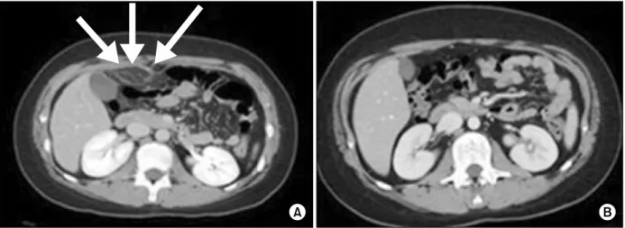
Fig. 1. (A) Initial abdominal computed tomography (CT) scan showed increased attenuation and enhancement of omental fat as well as anterior peritoneal wall thickening and enhancement (arrows). (B) Follow-up abdominal CT scan at 8 months showed marked improvement in intra-abdominal fat attenuation.
upper abdominal pain accompanied by vomiting and diarrhea that developed 3 days before admission. No history of a specific disease, abdominal trauma, or surgery was revealed. Family history was unre- markable. His height was 156.5 cm (50-75 p), weight was 60.3 kg (90-97 p), and BMI was 24.8 kg/m2. Vital signs were normal except for low-grade fever (37.6oC), and tenderness over the right upper abdomen was noted without a palpable mass on a physical exa- mination.
A blood test revealed the following: white blood cells (WBCs), 15,100/μL (neutrophils, 72.9%; lym- phocytes, 18.2%); platelets, 454,000/μL; erythrocyte sedimentation rate, 29 mm/h; and C-reactive protein (CRP), 5.45 mg/dL. Liver function tests, amylase, li- pase, ammonia, prothrombin time, partial thrombo- plastin time, antinuclear antibody, and anti- neutrophil cytoplasmic antibody were all negative. A chest radiograph showed no abnormalities. An ab- dominal computed tomography (CT) scan revealed increased attenuation and enhancement of the omental fat between the gall bladder and epigastric midline, as well as anterior peritoneal wall thicken- ing and enhancement, suggesting omental fat in- flammation or infarction (Fig. 1A). Laparoscopic bi- opsy was performed on hospital day (HD) 5, and the pathology showed mesenteric panniculitis with
chronic inflammation, adiponecrosis, and moderate septal fibrosis (Fig. 2). Nested polymerase chain re- action for Mycobacterium tuberculosis and special immunohistochemical staining for β-catenin, calre- tinin, Ki-67, and P53 for malignancy were all ne- gative.
Prednisolone was started (30 mg/d) on HD 6, and the abdominal pain subsided within 4 days. The blood tests showed improvement: WBCs, 7,320/μL (neutrophils, 55.6%; lymphocytes, 31.7%) and CRP, 1.65 mg/dL. He was discharged on prednisolone (30 mg/d) on HD 12, and the prednisolone was tapered to 10 mg/d over 1 month and continued at that dose for 7 months. An abdominal CT scan performed 8 months after discharge showed improvement from the previous abnormal findings without any sig- nificant sequelae (Fig. 1B). Follow-up blood testing was normal. The patient remained in good health af- ter the 2 year clinical follow-up.
DISCUSSION
Mesenteric panniculitis is a rare disease and usu- ally occurs in adults more than 50 years. Children are much rarely affected [1-6], and as far as we know, this is the first mesenteric panniculitis in Korean child. In the present case, the exact cause was not
www.pghn.org 145
Sun Hwan Bae, et al:Mesenteric Panniculitis in a Thirteen-Year-Old Korean Boy Treated with Prednisolone
Fig. 2. Mesenteric biopsy showed nodular panniculitis, septal fibrosis with hyalinization, chronic inflammation, and fat necrosis (A: H&E, ×200; B: Masson’s trichrome, ×100).
found, even though we performed various tests, in- cluding serological testing for autoimmunity and pathological immunohistochemical staining.
An abdominal CT scan has been proposed to be the most effective diagnostic tool [10]. Two CT scan find- ings, such as a ‘fat ring sign’, reflecting preservation of fat around the mesenteric vessels and ‘a tumoral pseudocapsule’, representing a band of soft tissue that limits the uninvolved mesentery from the in- flamed fat, are considered specific for mesenteric panniculitis [11]. Nevertheless, omental fat in- flammation and infarction cannot be distinguished clearly by a CT scan, and a surgical biopsy and patho- logical analysis are usually necessary to make the di- agnosis, as in the present case.
Three prominent pathological findings establish mesenteric panniculitis in terms of progression:
chronic inflammation, fatty necrosis, and fibrosis.
The clinical course of mesenteric panniculitis is gen- erally favorable, and spontaneous remission has been reported [9,12]. Nevertheless it is associated with significant morbidity, and mortality under the diagnosis of retractile mesenteritis [13,14]. Retractile mesenteritis which is the fibrotic end-stage of mes- enteric panniculitis can result in mesenteric re- traction, adhesion, and distortion of the intestinal loops [3,4,8,15].
No consensus exists about the indications for treatment or an established regimen for managing mesenteric panniculitis. Treatment has been empiric and should be individualized. Immuno-suppressants, such as corticosteroids alone or with azathioprine usually more than 6 months may improve the dis- ease course in adult [16,17]. In most of previous pe- diatric cases, the patients were observed without specific treatment or treated surgically [1-4]. One patient was misdiagnosed as appendicitis initially and diagnosed after surgery [6]. In one special case report, 6-year-old girl with small bowel obstruction was treated successfully with corticosteroid and me- thotrexate for more than 1 year [5]. In the present case, even though the diagnosis was not retractile mesenteritis, moderate fibrosis in the pathology and a high CRP level led us to start treatment, and 8 months of steroid treatment led to an excellent re- sponse in terms of blood and imaging tests. CRP lev- els may serve as a surrogate marker for the ther- apeutic response [18], and decrease in CRP was not- ed 4 days after starting prednisolone in the present case. Complete surgical resection is impossible in most cases, and is recommended only in cases of complications, such as intestinal obstruction or in- testinal ischemia [4,9,19].
The natural history of mesenteric panniculitis re-
146 Vol. 19, No. 2, June 2016 Pediatr Gastroenterol Hepatol Nutr
mains unclear because of its rarity and lack of ad- equate follow-up. However, benign, stable, or slowly progressive courses have been noted in most adult patients [20].
In this case, pediatric mesenteric panniculitis treated with prednisolone successfully. Mesenteric panniculitis is an extremely rare disease in children;
however, it can be considered in children with acute or chronic abdominal pain with an abdominal mass.
REFERENCES
1. Cakmak O, Tanyel FC, Cağlar M, Göğüş S. Mesenteric panniculitis mimicking acute abdomen in a 4-year-old child. Z Kinderchir 1986;41:313-4.
2. Hakgüder G, Akgür FM, Olguner M, Ozer E, Aktug T.
A case of mesenteric panniculitis in a 4-year-old child.
Pediatr Int 2000;42:577-8.
3. Davis CF, Guzzetta PC, Patterson K. Primary (retrac- tile) mesenteritis in a child. J Pediatr Surg 1992;
27:1544-5.
4. Spark RB, Yakovac WC, Wagget J. Retractile scleros- ing mesenteritis. Case report. Clin Pediatr (Phila) 1971;10:119-22.
5. Viswanathan V, Murray KJ. Idiopathic sclerosing mes- enteritis in paediatrics: Report of a successfully treated case and a review of literature. Pediatr Rheumatol Online J 2010;8:5.
6. Rumman N, Rumman G, Sharabati B, Zagha R, Disi N.
Mesenteric panniculitis in a child misdiagnosed as ap- pendicular mass: a case report and review of literature.
Springerplus 2014;3:73.
7. Ferrari TC, Couto CM, Vilaça TS, Xavier MA, Faria LC.
An unusual presentation of mesenteric panniculitis.
Clinics (Sao Paulo) 2008;63:843-4.
8. Ogden WW 2nd, Bradburn DM, Rives JD. Mesenteric panniculitis: review of 27 cases. Ann Surg 1965;161:
864-75.
9. Durst AL, Freund H, Rosenmann E, Birnbaum D.
Mesenteric panniculitis: review of the leterature and presentation of cases. Surgery 1977;81:203-11.
10. Horton KM, Lawler LP, Fishman EK. CT findings in sclerosing mesenteritis (panniculitis): spectrum of disease. Radiographics 2003;23:1561-7.
11. Wat SY, Harish S, Winterbottom A, Choudhary AK, Freeman AH. The CT appearances of sclerosing mesen- teritis and associated diseases. Clin Radiol 2006;
61:652-8.
12. Emory TS, Monihan JM, Carr NJ, Sobin LH. Sclerosing mesenteritis, mesenteric panniculitis and mesenteric lipodystrophy: a single entity? Am J Surg Pathol 1997;21:392-8.
13. Soergel KH, Hensley GT. Fatal mesenteric pan- niculitis. Gastroenterology 1966;51:529-36.
14. Akram S, Pardi DS, Schaffner JA, Smyrk TC. Scleros- ing mesenteritis: clinical features, treatment, and out- come in ninety-two patients. Clin Gastroenterol Hepa- tol 2007;5:589-96.
15. Tedeschi CG, Botta GC. Retractile mesenteritis. N Engl J Med 1962;266:1035-40.
16. Tytgat GN, Roozendaal K, Winter W, Esseveld MR.
Successful treatment of a patient with retractile mesen- teritis with prednisone and azathioprine. Gastroente- rology 1980;79:352-6.
17. Issa I, Baydoun H. Mesenteric panniculitis: various presentations and treatment regimens. World J Gastroenterol 2009;15:3827-30.
18. Ginsburg PM, Ehrenpreis ED. A pilot study of thalido- mide for patients with symptomatic mesenteric pan- niculitis. Aliment Pharmacol Ther 2002;16:2115-22.
19. Parra-Davila E, McKenney MG, Sleeman D, Hartmann R, Rao RK, McKenney K, et al. Mesenteric panniculitis:
case report and literature review. Am Surg 1998;
64:768-71.
20. Coulier B. Mesenteric panniculitis. Part 2: prevalence and natural course: MDCT prospective study. JBR- BTR 2011;94:241-6.