Simple Pulmonary Eosinophilia (Loeffler’ s Sy n d ro m e):
Chest Radiographic and CT Findings
1Kyung-Jae Jung, M.D., Kyung Soo Lee, M.D., Tae Sung Kim, M.D., Man Pyo Chung, M.D.2, Dong-Chull Choi, M.D.2, O Jung Kwon, M.D.2
Purpose :The purpose of our study is to describe the chest radiographic and CT find- ings of simple pulmonary eosinophilia.
Materials and Methods :Twenty-six patients with simple pulmonary eosinophilia un- d e r went chest radiography and CT scanning; the results were analyzed retrospective- ly by two chest radiologists, focusing on the patterns and distribution of the parenchy- mal abnormalities.
Results : The chest radiographs were normal in eight patients (31 %), while among the remaining 18 patients, they showed subtle opacity (n=9), nodules (n=8), consolida- tion (n=2), and mass (n=1). Fo l l ow-up chest radiographs (n=18) demonstrated com- plete (n=16) or partial (n=1) resolution of parenchymal lesions or migratory lesions (n=1). On CT, nodule(s) (n=19) were most commonly seen, followed by ground-glass opacity (n=16), consolidation (n=3), and mass (n=1). A peripheral halo surrounding a nodule or an area of consolidation was seen in 18 patients. The nodules(s) (n=19) were subpleural (n=13) or random (n=6). Areas of ground-glass opacity (n=16) we r e subpleural (n=13), random (n=2), or central (n=1). All lesions were patchy rather than diffuse. Fo l l ow-up CT in nine patients showed complete (n=7) or partial (n=2) resolution of parenchymal lesions.
Conclusion :Chest radiographs of patients with simple pulmonary eosinophilia often r eveal no abnormality. The most common finding is subtle opacity or nodule(s), while CT reveals transient nodule(s) with a surrounding halo or transient areas of ground- glass opacity.
Index words :Lung, abnormalities Lung, CT
Lung, diseases Lung, nodule
Pneumonia, eosinophilic
1Departments of Radiology, Samsung Medical Center, Sungkyunkwan University School of Medicine
2Department of Medicine (Division of Pulmonology), Samsung Medical Center, Sungkyunkwan University School of Medicine Received July 5, 1999 ; Accepted November 5, 1999
Address reprint requests to : Kyung Soo Lee, M.D., Department of Radiology, Samsung Medical Center
#50, Ilwon-Dong, Kangnam-Ku, Seoul 135-710, South Korea.
Tel. 82-2-3410-2511, 2518 Fax. 82-2-3410-2559 E-mail: kslee@ smc.samsung.co.kr
Simple pulmonary eosinophilia, also known as Loef- f l e r’s syndrome, is characterized by the presence on chest radiographs of transient or migratory pulmonary abnormalities, and by peripheral eosinophilia, minimal or no pulmonary symptoms, and rapid spontaneous res- olution (1-5). Pathologically, eosinophils and histiocytes accumulate in alveolar spaces and on alveolar walls (1- 4, 6).
Chest radiographic findings of the disease have been described in several reports; most commonly, unilateral or bilateral nonsegmental areas of consolidation are seen, and these are usually transient and migratory (1, 3, 5). The CT findings of the disease have not, however, been well described. The purpose of this study is to de- scribe both the radiographic and CT findings of simple pulmonary eosinophilia.
Materials and Methods
Between August 1994 and October 1998, 49 consecu- tive patients with simple pulmonary eosinophilia were diagnosed at our institution. Inclusion criteria (1, 2) for the diagnosis of simple pulmonary eosinophilia were peripheral eosinophilia with peripheral blood eosinophil counts greater than 500 cells/microliter (rang- ing from a 6.7 to 72 percent of white blood corpuscle count [540 to 28,706 cells/microliter]) and positive find- ings of parenchymal abnormalities on chest radiographs (n=43) or CT scans (n=26). Both chest radiographic and CT studies of the thorax were available for 26 of these 49 patients . There were 19 men and seven women whose ages ranged from 18 to 74 years (mean, 46 years).
CT scans were obtained within one to 13 (median, 7 ) days of chest radiographs. After a detailed and rigorous review of the patients’medical records (including the t- wo whose imaging studies were obtained in outside in- stitutions), the possibility that the pulmonary lesions were caused by the use of certain drugs or by parasitic infestations was discounted. All drugs used for chemotherapy were evaluated for potential causes of eosinophilia, but none were related to peripheral blood eosinophilia. No patient had a history of asthma. Cases satisfying the diagnostic criteria of acute eosinophilic p- neumonia (n=6), chronic eosinophilic pneumonia (n=3) and idiopathic hypereosinophilic syndrome (n=4) were also excluded (1, 2).
Only seven patients (27 %) complained of mild chest pain (n=3), dyspnea (n=3), or cough (n=2), while the remaining 19 (73 %) were symptom-free. No patient un-
derwent treatment for the alleviation of symptoms relat- ed to simple pulmonary eosinophilia.
The indications of CT scans in these patients were as follows: evaluation of patchy areas of consolidation or subtle opacity on chest radiographs (n=10); evaluation of the nature of solitary pulmonary nodule or mass seen on chest radiographs (n=7); staging work-up or follow- up evaluation of underlying malignancy (n=7) or be- nign lymph node disease (n=2). The identified underly- ing malignancies (n=7) included lung cancer in four pa- tients and primary lymphoma (mucosa-associated lym- phoid tissue origin) of the stomach, breast cancer, and squamous cell carcinoma of the buccal mucosa, each in one patient. Benign lymph node diseases included K i m u r a’s disease (angiolymphoid hyperplasia with eosinophilia) and sarcoidosis, each in one. In all pa- tients, CT scans were obtained before the peripheral eosinophil count was known. Peripheral eosinophilia was present a week before and after the time of initial CT, and as the causes of eosinophilia were not clear, pa- tients with underlying diseases were diagnosed as hav- ing simple pulmonary eosinophilia.
Chest radiographs were obtained with an FCR 9000 or 9501 computed radiographic system (Fuji, Tokyo, Ja- pan), using the following imaging parameters: 120 kVp, 0.6- or 1.2-mm nominal focus, 183-cm film-focus dis- tance, 12:1 oscillating grid, and phototimed exposure.
The default mode of image processing, including dy- namic range compression, gradation enhancement, and edge enhancement, was used. All CT scans were per- formed with a HiSpeed Advantage Scanner (General Electric Medical Systems, Milwaukee, WI). Both helical (7-mm collimation, pitch of 1) and high-resolution CT s- cans(1-mm collimation, 10-mm intervals) were obtained in 18 cases, only high-resolution CT scans in eight pa- tients, and only conventional CT scans from outside hospitals in two. Imaging data were reconstructed using a bone algorithm. Scans were imaged using the lung (window width, 1500 HU; window level, -700 HU) and the mediastinal (window width, 400 HU; window level;
20 HU) window.
Follow-up chest radiographs (range, seven days to six months; median, 18 days) were available in all patients (n=18) in whom initial chest radiographs revealed ab- normalities. In nine cases, follow-up CT scans (range, nine days to six months; mean, 20 days) were obtained after initial CT scans.
The radiologic findings were reviewed retrospectively by two chest radiologists, and final decisions were
reached by consensus. The radiographs and CT scans were reviewed separately at four-week intervals, and analyzed for patterns and distribution of parenchymal abnormalities. The former were categorized as nodules, subtle opacity, air-space consolidation, reticular density,
or mass, as seen on chest radiographs and as nodules, ground-glass opacity, consolidation, irregular linear opacity, or mass, as seen on CT scans. Subtle opacity on chest radiographs was defined as increased density without obscuration of underlying vascular structures,
A B
C D
E
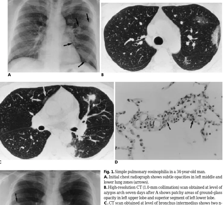
Fig. 1. Simple pulmonary eosinophilia in a 34-year-old man.
A . Initial chest radiograph shows subtle opacities in left middle and lower lung zones (arrows).
B . High-resolution CT (1.0-mm collimation) scan obtained at level of azygos arch seven days after A shows patchy areas of ground-glass opacity in left upper lobe and superior segment of left lower lobe.
C .CT scan obtained at level of bronchus intermedius shows two n- odules with peripheral ground-glass opacity (halo) in lingular seg- ment of left upper lobe and superior segment of left lower lobe.
Subtle area of ground-glass opacity is also seen in anterior segment of right upper lobe (arrows).
D. Photomicrography (H & E, × 400) of pathologic specimen with transbronchial lung biopsy in left lower lobe shows mild interstitial infiltration of eosinophils (arrows) and histiocytes.
E .Follow-up radiograph obtained 20 days after A shows migrating opacity in right middle lung zone (arrows). Note improvement in opacities in left middle lung zone. Opacity still remains in left lower lung zone (curved arrow).
whereas consolidation was defined as markedly in- creased density with obscuration of these structures. If a nodule, mass, or consolidation showed peripheral ground-glass opacity (halo) on CT scans (especially on high-resolution CT), this was recorded. The distribution of each pattern, as seen on radiographs, was divided in- to three zones on the chest radiographs; upper, middle, and lower. The upper lung zone was defined as an area superior to the aortic arch, the lower zone as inferior to the inferior pulmonary vein, and the middle zone as an area between the upper and lower zones. On CT, lesion location was divided into six lobes, the lingular segment being considered a separate lobe. Distribution was clas- sified as predominantly central, subpleural, or random;
and as patchy or diffuse. Lesions were regarded as cen- tral when located within one-third of the lung from the mediastinum, subpleural when they were located with- in the outer third of the lung from the chest wall, and random whey they did not fit either of these categories.
Lesions were considered diffuse when scattered widely and uniformly in both lungs and patchy when inconsis- tent and not uniform.
Two patients underwent both transbronchial lung biopsy and BAL.
R e s u l t s
Transbronchial lung biopsy obtained in two patients demonstrated mild interstitial infiltration of eosinophils and histiocytes (Fig. 1). An increased number of eosinophils (32 % and 7 % of total cell counts, respec- tively) were present in bronchoalveolar lavage fluid.
Chest radiographs were normal in eight patients ( 31 %). The commonest radiographic finding among the remaining 18 was subtle opacity (n=9, 50 %), followed by nodules (n=8, 44 %), airspace consolidations (n=2, 11 %) and mass (n =1, 6 %) (Table 1). Follow-up chest radiographs in 18 patients showed complete resolution in 16 patients (including seven with underlying malig- nancy) within 7 days to 6 months (median, 20 days). In one patient, a migratory lesion was seen on a radiograph obtained 20 days after the initial radiograph (Fig. 1); an- other patient showed partial resolution on a radiograph obtained 14 days after the initial radiograph.
The commonest pattern of parenchymal abnormality observed on CT scans was nodules (n=19; 73 %) (Figs. 1 -3), followed by areas of ground-glass opacity (n=16;
62%) (Figs. 1, 3, and 4), consolidation (n=3; 12 %) (Fig.
3), and mass (n=1; 4 %) (Fig. 5). Peripheral halo of ground-glass opacity was seen in 48 (76 %) of 63 nodules in 18 patients and in all three areas of consolidation (Figs. 1-3). The distribution of nodules (n=19) was sub- pleural (n=13) or random (n=6), that of ground-glass opacity (n=16) was subpleural (n=13), random (n=2)
A
Fig. 2. Simple pulmonary eosinophilia in a 53-year-old man.
A . Initial CT (10-mm collimation) scan obtained at ventricular level shows several nodules with peripheral ground-glass opacity (halo) in both lower lobes and right middle lobe, which are predominantly subpleural in distribution.
B . Follow-up CT (10-mm collimation) scan obtained at same level and 20 days after A shows that nodules disappeared.
Table 1. Radiographic Findings of Simple Pulmonary Eosino- philia in 18 Patients
L a t e r a l i t y Zonal distribution U n i - B i - U p p e r M i d d l e L o w e r
Subtle opacity (n=9; 50%) 4 5 3 5 5
Nodules (n=8; 44%) 5 3 3 7 2
Consolidation (n=2; 11%) 1 1 1 0 2
Mass (n=1; 6%) 1 0 1 0 0
N o t e-Uni:unilateral, Bi: bilateral
B
or central (n=1), and that of areas of consolidation was subpleural (n=2) or random (n=1). For mass, distribu- tion was subpleural. The distribution of all lesions was patchy, and none showed diffuse abnormalities. The predominant lobar distribution was not demonstrated (Table 2).
Follow-up CT scans (n=9) showed complete resolu-
tion of the parenchymal lesions in seven patients within 1 to 6 months (median, 75 days). One patient showed partial resolution of nodules within 20 days, and the other showed almost complete resolution of the pre-ex- isting mass lesion and small new nodules on the CT s- cans obtained 9 days later (Fig. 5).
A
Fig. 3. Simple pulmonary eosinophilia in a 25-year-old woman with breast cancer.
A . High-resolution CT (1.0-mm collimation) scan obtained at level of main bronchi shows nonsegmental area of consolidation with peripheral ground-glass opacity in right upper lobe.
B . CT scan obtained at level of inferior vena cava shows small nodule with halo in lingular segment of left upper lobe. Follow-up chest radiograph (not shown here) 20 days after A and B showed complete disappearance of consolidation and nodule.
Table 2. CT Findings of Simple Pulmonary Eosinophilia in 26 Patients
L a t e r a l i t y Zonal distribution Transverse plane U n i f o r m i t y U n i - B i - U p p e r M i d d l e L o w e r C e n t r a l S u b p l . R a n d o m D i f f u s e P a t c h y Nodules
(n=19; 73%) 9 1 2 1 1 1 2 1 5 0 1 3 6 0 1 9
Ground-glass
opacity (n=16; 62%) 8 08 1 2 07 07 1 1 3 2 0 1 6
Consolidation
(n=3; 12%) 3 00 02 00 01 0 02 1 0 03
M a s s
(n=1; 4%) 1 00 01 00 00 0 01 0 0 01
Note -Uni: unilateral, Bi: bilateral, Subpl: subpleural
A B
Fig. 4. Simple pulmonary eosinophilia in a 33-year-old man.
A . High-resolution CT (1.0-mm colli- mation) scan obtained at level of aortic arch shows area of ground-glass opaci- ty in left upper lobe.
B . CT scan obtained at level of inferior pulmonary vein and on same day with A shows areas of ground-glass opacity in left lower lobe.
B
D i s c u s s i o n
Although numerous classifications of eosinophilic lung diseases have been proposed, there is currently no optimal way of classifying these disorders. They may be classified as entities of unknown causes (simple pul- monary eosinophilia, acute eosinophilic pneumonia, chronic eosinophilic pneumonia, and idiopathic hypere- osinophilic syndrome), known causes (allergic bron- chopulmonary aspergillosis, bronchocentric granulo- matosis, and eosinophilic lung disease with parasitic in- fection and drug reaction); or eosinophilic vasculitis (al- lergic angiitis and granulomatosis or Churg-Strauss syn- drome) (1). Diagnostic criteria for each of these entities have recently been suggested (1, 2).
The eosinophil is a bone marrow-derived polymor- phonuclear leukocyte containing granules within which are a variety of proteins. When released, these proteins are potentially cytotoxic, resulting in pathologic process- es (5, 7 - 9). The term eosinophilia denotes an absolute eosinophil count above 500 cells/microliter (8). In sim- ple pulmonary eosinophilia, the degree of eosinophilia varies, ranging between 1,000 and 50,000 cells/micro- liter (10), but does not appear to correlate with the amount, duration, or intensity of pulmonary infiltration (11, 12). Symptoms are usually lacking and physical findings are often absent (11). In our study, 19 of 26 pa- tients had no symptoms, and in the remaining seven, symptoms were mild. The number of peripheral eosinophils ranged from 540 to 28,706 cells/microliter.
The pathologic changes seen in patients with simple pulmonary eosinophila have been described as areas of pneumonic consolidation consisting of alveolar exudate filled with many eosinophils, mononuclear cells, and occasional foreign body giant cells. Eosinophils and oth- er round cells can also be found in the interstitium, but associated vasculitis is not seen (1 - 4, 6). Because most lesions are transient and mild, lung biopsy is not per- formed.
The radiographic finding commonly reported in pa- tients with simple pulmonary eosinophilia is nonseg- mental consolidation varying in size and either unilater- al or bilateral. Abnormalities are usually transient and migratory (1-3, 11, 12), are often peripheral in nature, and may appear to be subpleural (2, 3). Neither cavita- tion in consolidation nor findings of pleural effusion, lymph node enlargement, or cardiomegaly have been reported (1). Hennell et al (12) described the radiograph- ic findings of five patients with transient eosinophilic lung infiltration as confluent and patchy in appearance which may be radiating from the hilum, and often sym- metric in two lungs. In fact, a patient with probable hy- pereosinophilic syndrome was included in his study and three patients were asthmatics. In our study, the predominant radiographic findings were ground-glass opacity and nodules. Areas of consolidation were infre- quent.
The diagnostic criteria of simple pulmonary eosino- philia include abnormality on chest radiographs, with peripheral eosinophilia. Such abnormality has been a finding in almost all patients with simple pulmonary
A B
Fig. 5. Simple pulmonary eosinophilia mimicking lung cancer in a 56-year-old woman.
A . Mediastinal window setting of CT (10-mm collimation) scan shows about 3-cm sized mass with a small area of air attenuation simulating cavitating mass in left upper lobe. Initially lesion was diagnosed as peripheral lung cancer.
B . Follow-up high-resolution CT (1.0-mm collimation) scan obtained 9 days after A, owing to disappearance of mass on fluoroscopy during percutaneous needle aspiration biopsy, shows almost complete disappearance of mass. Small new nodules in right middle and lower lobes were seen (not shown here).
eosinophilia (5). Our study showed normal chest radi- ographs in eight (31 %) of 26 patients. In simple pul- monary eosinophilia, pulmonary infiltration might not be visible on chest radiographs. In other words, nega- tive chest radiographic findings in patients with periph- eral eosinophilia do not always exclude the possibility of pulmonary infiltration. Under such circumstances, pul- monary infiltration is revealed by CT.
CT findings of simple pulmonary eosinophilia have rarely been described. We described recently the CT findings of the disease in few patients. The lesions ap- peared as single or multiple pulmonary nodules with or without a peripheral halo (1). In the present study, the predominant pattern of parenchymal abnormality seen on CT scans was nodules, distributed mainly in sub- pleural regions. About 76 % of the nodules were accom- panied by a peripheral halo of ground-glass opacity.
Patchy areas of ground-glass opacity were the second most frequent finding.
Patients with simple pulmonary eosinophilia have an excellent prognosis and because pulmonary infiltrates and blood eosinophilia resolve spontaneously within one month, do not need treatment(1, 2, 6). The condi- tion can, however, also be associated with parasitic or other infestation, drug reaction, environmental expo- sure, or malignancy. Infections may be parasistic (ame- biasis, ascariasis, trichomosis, filariasis, paragonimiasis, strongyloidiasis), fungal (histoplasmosis, coccidioidomy- cosis, brucellosis), or protozoan (Entamoeba histolytica, Entamoeba coli), while the drugs involved in adverse re- actions include acetylsalicylic acid, para-aminosalicylic acid, penicillin, mephensen carbonate, nitrofurantoin and potentially almost all classes of pharmaceuticals.
Examples of environmental exposure are smoke inhala- tion, poison ivy desensitization, contact with nickel and pollen inhalation. About one half of the patients in whom simple pulmonary eosinophilia was initially diag- nosed will eventually be shown to be affected by infes- tation, drug reaction, or environmental exposure (1, 2, 5). The etiology is quite obscure; the evidence to date suggests that an allergy is involved (3, 4, 6, 10, 12). In light of the above, the possibility of parasitic infestation, drug reaction, environmental exposure, and underlying malignancy should be carefully investigated (2).
It is also well known that malignant diseases (bron- chogenic carcinoma, gastrointestinal tract tumors, H o d g k i n’s and non-Hodgkin’s lymphoma), chronic granulomatous disorders (tuberculosis, sarcoidosis), and collagen vascular disease (rheumatoid arthritis, pol-
yarteritis nodosa, lupus erythematosus) can be associat- ed with peripheral eosinophilia (3-7, 10, 13). In the p- resence of these conditions, the cause of eosinophilia is not clear, however. In our study, simple pulmonary eosinophilia was associated with an underlying malig- nant disease in nine patients. In two patients, the dis- ease was associated with a Kimura’s disease (angiolym- phoid hyperplasia with eosinophilia) and a sarcoidosis, one in each patient. In these patients, chest radiograph revealed no abnormalities. Parenchymal abnormalities were seen only on CT scans, which were obtained in or- der to evaluate tumor staging or to search for pul- monary or mediastinal complications caused by under- lying diseases. There are some diagnostic problems in- volved in detecting nodule(s) with or without a halo sign of simple pulmonary eosinophilia in patients with known underlying malignancies. The nodules should not be confused with metastatic pulmonary nodule(s), and to this end, an early differential leukocyte count and careful recording of any parasitic or fungal infesta- tion and drug intake are required.
A peripheral halo surrounding the nodule or consoli- dation can be seen under a variety of conditions: hemor- rhagic nodules of infectious (aspergillosis, mucormyco- sis, candidiasis, coccidioidomycosis) and noninfectious origin (Wegener’s granulomatosis, metastatic angiosar- coma, and Kaposi sarcoma); primary (bronchioloalveo- lar carcinoma or primary lymphoma) or secondary lung malignancy, and inflammatory disease such as organiz- ing pneumonia (14, 15). We believe that the halo repre- sents pathologically loose interstitial infiltration of eosinophils and histiocytes around dense eosinophilic consolidation. Diagnosis is usually possible on the basis of a complete history, a simple differential count of white blood cells in peripheral blood, and a follow-up radiologic study.
Our study was limited by several drawbacks. This suffers from the limitations of a retrospective study.
Although we included patients (especially those with underlying malignancy) with increased peripheral blood eosinophil count and parenchymal abnormalities at CT, specific causes were not identified in these pa- tients. Possible causes may have been unidentified para- sitic or other infections, drug intake, and environmental exposure. Pathologic evaluation was performed in only two patients and not all patients underwent a follow-up radiologic examination. Radiologic-pathologic correla- tion would give detailed information, especially regard- ing the pathologic basis of the peripheral halo of ground-
glass opacity seen on CT. We included only 26/49 pa- tients with simple pulmonary eosinophilia, for whom both chest radiographs and CT scans were obtained, and this may have led to a selection bias.
In summary, simple pulmonary eosinophilia is char- acterized clinically by no or minimal symptoms and spontaneous resolution within a month. The condition most commonly involves subtle opacity of nodules on chest radiographs. Pulmonary lesions are more readily detected by CT scans than by chest radiographs. The predominant CT findings of simple pulmonary eosinophilia are subpleural nodules of patchy distribu- tion with surrounding ground-glass opacity (halo sign) or patchy areas of ground-glass opacity.
R e f e r e n c e s
1 . Kim Y, Lee KS, Choi D-C, Primack SL, Im JG. The spectrum of eosinophilic lung disease: radiologic findings. J Comput Assist T o m o g r 1 9 9 7 ; 2 1 : 9 2 0 - 9 3 0
2 . Allen JN, Davis WB. Eosinophilic lung disease. Am J Respir Crit Care Med 1 9 9 4 ; 1 5 0 : 1 4 2 3 - 1 4 3 8
3 . Citro LA, Gordon ME, Miller WT. Eosinophilic lung disease (or
how to slice P.I.E.). AJR 1 9 7 3 ; 1 1 7 : 7 8 7 - 7 9 7
4 . Bedrossian CWM, Greenberg SD, Williams LJ. Ultrastructure of the lung in Loeffler’s pneumonia. Am J Med 1 9 7 5 ; 5 8 : 4 3 8 - 4 4 3 5 . Bain GA, Flower CDR. Pulmonary eosinophilia. Eur J Radiol
1 9 9 6 ; 2 3 : 3 - 8
6 . Ford RM. Transient pulmonary eosinophilia and asthma. Am Rev Respir Dis 1 9 6 6 ; 9 3 : 7 9 7 - 8 0 3
7 . Chapman B, Capewell S, Gibson R, Greening AP, Crompton GK.
Pulmonary eosinophilia with and without allergic bronchopul- monary aspergillosis. Thorax 1 9 8 9 ; 4 4 : 9 1 9 - 9 2 4
8 . Schats M, Wasserman S, Patterson R. The eosinophil and the lung.
Arch Intern Med 1 9 8 2 ; 1 4 2 : 1 5 1 5 - 1 5 1 9
9 . Weller PF. The immunobiology of eosinophils. N Engl J Med 1 9 9 1 ; 3 2 4 : 1 1 1 0 - 1 1 1 8
1 0 . Marcy TW. Eosinophilia in patients presenting with pulmonary infiltrates and fever. Semin Respir Inf 1 9 8 8 ; 3 : 2 4 7 - 2 5 7
1 1 . Reeder WH, Goodrich BE. Pulmonary infiltration with eosinophil- ia (PIE syndrome). Ann Intern Med 1 9 5 2 ; 3 6 : 1 2 1 7 - 1 2 4 0
1 2 . Hennell H, Sussman ML. The roentgen features of eosinophilic in- filtrations in the lungs. Radiology 1 9 4 5 ; 4 4 : 3 2 8 - 3 3 4
1 3 . Schats M, Wasserman S, Patterson R. Eosinophils and immuno- logic lung disease. Med Clin North Am 1 9 8 1 ; 6 5 : 1 0 5 5 - 1 0 7 1 1 4 . Primack SL, Hartman TE, Lee KS, Muller NL. Pulmonary nodules
and the CT halo sign. Radiology 1 9 9 4 ; 1 9 0 : 5 1 3 - 5 1 5
1 5 . Kim Y, Lee KS, Jung K-J, Han J, Kim JS, Suh JS. Halo sign of pul- monary nodule on high-resolution CT: findings in spectrum of pulmonary diseases with pathologic correlation. J Comput Assist T o m o g r (in press)
대한방사선의학회지 2 0 00;42:83- 9 0
단순 폐 호산구증 (레플러 증후군):
흉부단순촬영 및 CT 소견11성균관대학교 의과대학 삼성서울병원 방사선과
2성균관대학교 의과대학 삼성서울병원 호흡기내과
정경재・이경수・김태성・정만표2・최동철2・권오정2
목적 : 단순 폐 호산구증의 흉부단순촬영 및 CT 소견을 기술하고자 하였다.
대상 및 방법 : 단순 폐 호산구증으로 진단된 환자 26 명을 대상으로 흉부단순촬영과 CT 를 시행하였다. 흉부단순
촬영 및 CT 소견을 폐실질소견의 양상과 분포를 중심으로 두명의 흉부방사선과 전문의가 후향적으로 분석하였다.
결과 : 8명 ( 3 1 % )의 환자는 흉부단순촬영상 정상이었다. 나머지 1 8명의 환자들에서 흉부단순촬영 소견은 미약한 음영증가 (n=9), 결절 (n=8), 경결 (n=2), 그리고 종괴 (n=1) 등이었다. 추적 흉부단순촬영 (n=18) 상 폐실질 소견의 완전 (n=16) 혹은 부분 (n=1) 호전이나 이동하는 양상 ( n = 1 )이 관찰되었다. 결절 ( n = 1 9 )이 CT 상 가 장 흔한 양상이었으며 간유리음영 (n=16), 경결 (n=3), 그리고 종괴 ( n = 1 )의 순서로 관찰되었다. 결절이나 경 결 주위 달무리음영이 18 명의 환자에서 관찰되었다. 결절 ( n = 1 9 )의 분포는 늑막하 ( n = 1 6 )나 무작위 (n=6) 양 상이 흔하였다. 간유리음영 ( n = 1 6 )의 분포 역시 늑막하 (n=13), 무작위 (n=2), 중심성 (n=1) 순이었다. 모든 병변은 미만성보다는 군집성으로 분포하였다. 9명에서 시행한 추적 C T상 폐병변들은 완전히 (n=7) 혹은 부분 적으로 (n=2) 호전되었다.
결론 : 단순폐호산구증은 단순촬영상 종종 정상으로 나타나며, 단순촬영에서는 미약한 음영증가나 결절이, CT 상은 달무리를 동반한 일과성 결절이나 간유리음영이 가장 흔한 소견이다.