233
Received June 8, 2007
Accepted for publication June 1, 2008
Reprint request to: Mi Woo Lee, M.D., Department of Dermatology, Asan Medical Center, University of Ulsan College of Medicine, 388-1, Pungnap 2-dong, Songpa-gu, Seoul 138-736, Korea. Tel: 82-2-3010-3460, Fax: 82-2- 486-7831, E-mail: miumiu@amc.seoul.kr
Papular Mucinosis Associated with Systemic Lupus Erythematosus
Woo Jin Lee, M.D., Gyeong Hun Park, M.D., Sung Eun Chang, M.D., Mi Woo Lee, M.D., Jee Ho Choi, M.D., Kee Chan Moon, M.D., Jai Kyoung Koh, M.D.
Department of Dermatology, Asan Medical Center, University of Ulsan College of Medicine, Seoul, Korea
Papulonodular mucinosis (PNM) is a rare variant of lupus erythematosus (LE) eruptions, and PNM is characterized histologically by diffuse dermal mucin without any typical epidermal inflammatory changes. We herein describe a case of papular mucinosis that was characterized by several erythematous papules on the lower back of a 32-year-old man with systemic LE. It is interesting that he didn't display any other skin manifestations of LE such as malar rash, discoid rash and photosensitivity during the previous 2 years. He achieved remission of his PNM without recurrence after 5 months treatment with topical steroids, in addition to receiving systemic antimalarials and steroids.
(Ann Dermatol (Seoul) 20(4) 233∼236, 2008)
Key Words: Papulonodular mucinosis, Systemic lupus erythematosus
INTRODUCTION
Papulonodular mucinosis (PNM) is a variant of lupus erythematosus (LE), and PNM is charac- terized by dermal mucin deposition without any of the other histological features of LE. Although this malady was first recognized as early as 19541, PNM associated with LE has been reported for fewer than 50 patients. There has been only one reported case of PNM associated with systemic LE in the Korean dermatologic literature2. A recent review reported that about 75% of the PNM cases were associated with systemic LE, 20% were associated with discoid LE and 5% were associated with subacute cutaneous LE3. PNM is primarily associated with other cu- taneous eruptions in patients with established LE and the PNM fluctuates with the LE disease ac-
tivity4. At times, however, PNM can be the only cutaneous manifestation of LE. We herein describe a patient who experienced papular mucinosis as the only cutaneous lesion of his systemic LE.
CASE REPORT
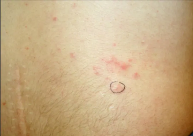
A 32-year-old man presented with a 1-week history of several erythematous papules on the lower back. They had started as tiny skin-colored papules, and these progressively enlarged to form confluent papules. During this time, several new lesions developed. There were no subjective symptoms, and the patient denied any history of injection or trauma. The physical examination showed multiple, relatively well-demarcated, variable sized erythematous papules scattered over his lower back (Fig. 1).
Two years earlier, he had begun to develop arthralgia in his knees and shoulders. He had also noticed joint stiffness in the morning, Raynaud's phenomenon and oral ulcers. He had showed no skin lesions, including malar rash, discoid rash or photosensitivity during the previous 2 years. The significant laboratory values included leukocyto- penia (2,200/mm3), an antinuclear antibody titer of
Annals of Dermatology
234 WJ Lee, et al. Vol. 20, No. 4, December 2008
Fig. 1. Multiple, relatively well-demarcated, flesh or erythematous colored papules on the lower back.
1:1280 (a speckled pattern), hypocomplementemia (C3: 53.1 mg/dl [normal range: 88∼201 mg/dl], C4: 10.0 mg/dl [normal range: 16∼47 mg/dl]) and
positivity for antibodies against Ro/SSA, La/SSB and Scl-70. However, the anti-ds DNA antibodies, the hemoglobin level, the erythrocyte sedimentation rate, the kidney and liver function tests and urinalysis were within normal limits. He had been diagnosed with systemic LE and then treated with an antimalarial agent (hydroxylchloroquine 100 mg/day) and prednisolone (7.5∼10 mg/day) for 2 years, which resulted in substantial improvement.
A 4 mm punch biopsy of skin from a papule on the patient's lower back showed deposition of bluish materials around the sweat glands and between the collagen bundles in the dermis (Fig. 2A, B). The mucinous material was stained with alcian blue at pH 2.5 (Fig. 2C). A scant perivascular lymphocytic infiltrate was observed. The number of fibroblasts in the dermis was not significantly increased. Any histopathologic findings of cutaneous LE were not detected, such as epidermal involvement and va- cuolar degeneration at the dermoepidermal junction.
Fig. 2. A skin biopsy specimen showing (A) intact epidermis, deposition of basophilic mucinous materials and scant infiltration of mononuclear cells in the dermis (H&E, ×40). (B) Deposition of mucinous materials between the collagen bundles and around the sweat glands (H&E, ×100). (C) Alcian blue staining (pH 2.5) showing the deposits of mucin in the dermis (×40).
Papular Mucinosis Associated with Systemic Lupus Erythematosus 235
Based on these findings, the skin lesions were diagnosed as papular mucinosis in a patient with systemic LE. He was treated with topical steroids in addition to receiving systemic antimalarials and prednisolone. After 2 weeks treatment with the medications, he achieved remission of his PNM without recurrence for 5 months.
DISCUSSION
PNM is a distinctive, although unusual cutaneous manifestation of LE and PNM is due to a diffuse deposit of mucin in the dermis. It is clinically characterized by asymptomatic flesh-colored papules and nodules, which occur in the presence or ab- sence of the typical cutaneous lesions of LE. PNM has a predilection for the trunk and upper extremities, but the face and other areas of the body such as the neck and thighs may also be affected5. This condition must be distinguished from other cutaneous mucinoses, such as myxedema, reticular erythematous mucinosis, scleredema, alopecia mu- cinosa and cutaneous focal mucinosis. PNM is histopathologically characterized by mucin deposi- tion between the collagen bundles in the papillary and mid-reticular dermis and there is also mild superficial perivascular lymphocytic infiltrates, whereas the epidermis is normal and it is without basal vaculopathy. The classic epidermal and dermal inflammatory changes characteristic of LE are not typically seen in PNM.
To the best of our knowledge, of the 33 reported cases of PNM in the English medical literature, 28 were accompanied by other typical cutaneous eruptions of LE, such as discoid lesions, macular erythema, annular atropic lesions, papulosquamous eruptions, vasculopathic lesions or alopecia5-9. It is interesting that our patient had no skin lesions for 2 years after being diagnosed with systemic LE, and papular mucinosis was the only cutaneous mani- festation of his systemic LE. PNM may be a pre- senting sign of LE3 or it may be the first cutaneous manifestation of LE. Yet for our patient, PNM did not precede the onset of LE, but the PNM occurred after the clinical manifestations of systemic LE.
Several factors may contribute to the patho- genesis of this condition. UV light may aggravate skin lesions. In addition, androgens or other sex- related factors may have a role in the pathogenesis
of PNM because the male-to-female ratio of patients with PNM is much greater than that of patients with systemic LE4. Furthermore, serum factors, such as cytokines and circulating antibodies, have been found to upregulate the synthesis of mucin from fibroblasts10.
Many therapeutic modalities have been proposed to treat this condition, including glucocorticoids, antimalarial agents, retinoids, cyclophosphamide and methotrexate; plasmapheresis and surgical op- tions such as dermabrasion, laser and excision have also been tried, but these are usually unsatisfactory3. However, the skin lesions on our patient responded to medication comprised of topical steroids, systemic antimalarials and prednisolone.
In conclusion, we describe here a patient with papular mucinosis associated with systemic LE, and the patient had no LE-specific skin lesions after being diagnosed with systemic LE.
REFERENCES
1. Gold SC. An unusual papular eruption associated with lupus erythematosus. Br J Dermatol 1954;
66:429-433.
2. Park J, Park CO, Lee KH. Papulonodular mucinosis associated with systemic lupus erythematosus: cu- taneous lupus mucinosis. Korean J Dermatol 2004;
42:1073-1075.
3. Sonntag M, Lehmann P, Megahed M, Ruzicka T, Kuhn A. Papulonodular mucinosis associated with subacute cutaneous lupus erythematosus. Derma- tology 2003;206:326-329.
4. Lowe L, Rapini RP, Golitz LE, Johnson TM.
Papulonodular dermal mucinosis in lupus erythe- matosus. J Am Acad Dermatol 1992;27:312-315.
5. Choi EH, Hann SK, Chung KY, Park YK. Pa- pulonodular cutaneous mucinosis associated with systemic lupus erythematosus. Int J Dermatol 1992;31:649-652.
6. Kanda N, Tsuchida T, Watanabe T, Tamaki K.
Cutaneous lupus mucinosis: a review of our cases and the possible pathogenesis. J Cutan Pathol 1997;24:553-558.
7. Williams WL, Ramos-Caro FA. Acute periorbital mucinosis in discoid lupus erythematosus. J Am Acad Dermatol 1999;41:871-873.
8. Kobayashi T, Shimizu H, Shimizu S, Harada T, Nishikawa T. Plaquelike cutaneous lupus muci-
Annals of Dermatology
236 WJ Lee, et al. Vol. 20, No. 4, December 2008
nosis. Arch Dermatol 1993;129:383-384.
9. Kano Y, Sagawa Y, Yagita A, Nagashima M.
Nodular cutaneous lupus mucinosis: report of a case and review of previously reported cases. Cutis 1996;57:441-444.
10. Pandya AG, Sontheimer RD, Cockerell CJ, Taka-
shima A, Piepkorn M. Papulonodular mucinosis associated with systemic lupus erythematosus: pos- sible mechanisms of increased glycosaminoglycan accumulation. J Am Acad Dermatol 1995;32:199- 205.