ISSN 2234-3806 • eISSN 2234-3814
https://doi.org/10.3343/alm.2019.39.4.345
The Platelet-to-Lymphocyte Ratio as an Inflammatory Marker in Rheumatic Diseases
Armen Yuri Gasparyan , M.D., Ph.D.1, Lilit Ayvazyan , Ph.D.2, Ulzhan Mukanova , M.D., Ph.D.3, Marlen Yessirkepov , M.D., Ph.D.4, and George D. Kitas , M.D., Ph.D., F.R.C.P.1,5
1Departments of Rheumatology and Research and Development, Dudley Group NHS Foundation Trust (Teaching Trust of the University of Birmingham, UK), Russells Hall Hospital, Dudley, West Midlands, UK; 2Department of Medical Chemistry, Yerevan State Medical University, Yerevan, Armenia;
3Department of Surgical Disciplines, South Kazakhstan Medical Academy, Shymkent, Kazakhstan; 4Department of Biology and Biochemistry, South Kazakhstan Medical Academy, Shymkent, Kazakhstan; 5Arthritis Research UK Epidemiology Unit, University of Manchester, Manchester, UK
The platelet-to-lymphocyte ratio (PLR) has emerged as an informative marker revealing shifts in platelet and lymphocyte counts due to acute inflammatory and prothrombotic states.
PLR has been extensively examined in neoplastic diseases accompanied by immune sup- pression and thrombosis, which can be predicted by combined blood cell counts and their ratios. Several large observational studies have demonstrated the value of shifts in PLR in evaluating the severity of systemic inflammation and predicting infections and other comorbidities, in inflammatory rheumatic diseases. The value of PLR as an inflammatory marker increases when its fluctuations are interpreted along with other complementary hematologic indices, particularly the neutrophil-to-lymphocyte ratio (NLR), which provides additional information about the disease activity, presence of neutrophilic inflammation, infectious complications, and severe organ damage in systemic lupus erythematosus. PLR and NLR have high predictive value in rheumatic diseases with predominantly neutro- philic inflammation (e.g., Behçet disease and familial Mediterranean fever). High PLR, along with elevated platelet count, is potentially useful in diagnosing some systemic vas- culitides, particularly giant-cell arteritis. A few longitudinal studies on rheumatic diseases have demonstrated a decrease in PLR in response to anti-inflammatory therapies. The main limitations of PLR studies are preanalytical faults, inadequate standardization of lab- oratory measurements, and inappropriate subject selection. Nonetheless, accumulating evidence suggests that PLR can provide valuable information to clinicians who encounter multisystem manifestations of rheumatic diseases, which are reflected in shifts in platelet, lymphocyte, neutrophil, or monocyte counts. Interpretation of PLR combined with com- plementary hematologic indices is advisable to more accurately diagnose inflammatory rheumatic diseases and predict related comorbidities.
Key Words: Platelet count, Lymphocyte count, Neutrophil count, Inflammation, Rheumatic diseases, Markers, Platelet-to-lymphocyte ratio
Received: November 14, 2018 Revision received: December 12, 2018 Accepted: February 7, 2019
Corresponding author:
Armen Yuri Gasparyan, M.D., Ph.D.
https://orcid.org/0000-0001-8749-6018 Departments of Rheumatology and Research and Development, Dudley Group NHS Foundation Trust (Teaching Trust of the University of Birmingham, UK) Russells Hall Hospital, Pensnett Road, Dudley DY1 2HQ, West Midlands, UK
Tel: +44-(0)-1384-456111 Fax: +44-(0)-1384-244272 E-mail: [email protected]
© Korean Society for Laboratory Medicine This is an Open Access article distributed under the terms of the Creative Commons Attribution Non-Commercial License (http://creativecom- mons.org/licenses/by-nc/4.0) which permits unrestricted non-commercial use, distribution, and reproduction in any medium, provided the original work is properly cited.
INTRODUCTION
Blood cell interactions are essential in the pathophysiology of in- flammation, immune responses, hemostasis, and oncogenesis.
These interactions are multifaceted, and it is often difficult to
distinguish primary triggering signals and the specific roles of each cell type in the development and progression of disease states.
Platelets are rich in proinflammatory agents and are capable of releasing highly active microparticles, and they are intimately
2017-03-16 https://crossmark-cdn.crossref.org/widget/v2.0/logos/CROSSMARK_Color_square.svg
involved in the development and perpetuation of various inflam- matory rheumatic diseases, which often manifest with arthritis and may be complicated by cardiovascular, metabolic, infec- tious, and lymphoproliferative, among many other, comorbidities [1-3]. Fluctuations in platelet counts in rheumatic diseases re- flect nonspecific inflammatory thrombopoiesis, with abundant release of reactive cells from the bone marrow to the bloodstream, their migration to and excessive consumption at inflammatory sites, and their destruction via binding to anti-platelet antibod- ies. Platelet counts correlate with platelet volume and reactivity and may indicate autoimmune disease activity, response to anti- inflammatory therapies, and presence of various comorbidities [4-6].
Observational studies have demonstrated that in rheumatoid arthritis (RA), platelet counts gradually increase with radiological disease progression, whereas leukocyte and erythrocyte count variations are unremarkable [7, 8]. RA patients with increased platelet counts (>400×109/L) appear to show more substantial improvements in disease activity score in 28 joints (DAS28) and acute-phase reactants in response to biological agents, such as tocilizumab, than do RA patients with “normal” platelet counts (<400×109/L) [9].
In systemic lupus erythematosus (SLE), thrombocytopenia is more prevalent (15–27%) than leuco- (14%) or erythrocytope- nia (5%) [5, 10]. Platelet counts negatively correlate with lupus activity, whereas thrombocytopenia, which is often mild (70 –150×109/L), is associated with arthritis, neurological disturban- ces, and lupus nephritis. Moderate and severe thrombocytope- nia in SLE may manifest with major hemorrhage (25% and 76%, respectively), resulting in death due to blood loss or infection [11]. Although there is no direct evidence of an association of autoimmune markers with hematologic parameters, the antici- pated low platelet count in SLE is likely due to disease-specific disturbances, resulting in excessive production of anti-platelet antibodies and intensified platelet destruction in the spleen (hy- persplenism) [10].
A high platelet count is of diagnostic value in elderly patients with temporal artery inflammation suggestive of giant-cell arteri- tis (GCA) [12]. Accordingly, a recent study suggested that a high platelet count predicts diagnosis of GCA even in the absence of characteristic temporal artery involvement [13].
Interestingly, in children with systemic vasculitides, particularly Kawasaki disease (KD), platelet counts may predict disease out- comes at various stages. An increase in platelet count accom- panied by an increase in lymphocyte count, a decrease in neu- trophil count, and amelioration of immunoglobulin G, M, and A
levels suggests a good response to intravenous immunoglobulin and aspirin therapy at the early, in-hospital stages of convales- cence in KD [14]. However, persistently high platelet counts may indicate a complicated course and the development of coronary aneurysms in KD [15]. A high platelet count is also associated with renal involvement in Henoch-Schönlein purpura, another common systemic vasculitis in childhood [16].
In-vitro and in-vivo studies have elucidated the origin of acti- vated circulating platelets, which display numerous membrane receptors and release multiple biologically active substances from their granules that can regulate cellular interactions and contribute to immune, inflammatory, and thrombotic diseases [17-19]. Circulating platelets can interact with erythrocytes, neu- trophils, and lymphocytes in the vessel lumen at sites of vascu- lar damage [20, 21]. The interaction of platelets with T-lympho- cytes, mediated by P-selectin, reduces lymphocyte proliferation, resulting in a decrease in proinflammatory cytokines, such as interferon-alpha (IFN-α), tumor necrosis factor-alpha (TNF-α), and interleukin (IL)-17, and an increase in anti-inflammatory cytokines, such as IL-10 [22]. Drug-induced modulation of such intercellular interactions and subsequent cytokinergic reactions, which depend on platelet and lymphocyte counts, dampens im- mune inflammation in the synovial space of patients with RA, and can be monitored by employing combined parameters of hemograms, such as the platelet-to-lymphocyte ratio (PLR).
Platelets of healthy subjects modulate the activities of neutro- phils and monocytes by responding to stimulation of their plate- let-bound Toll-like receptors (TLR) in a dose-dependent man- ner. Low doses of TLR agonists, such as lipopolysaccharide and fibroblast-stimulating lipopeptide-1, reduce CD66b expression on neutrophils and related granulocyte elastase secretion in plate- let-neutrophil coculture [23]. Platelets activated by fibroblast- stimulating lipopeptide-1 in coculture increase IL-6 and IL-10 and reduce TNF-α production.
Activated platelets release microparticles that can interact with neutrophils via the expression of platelet-type lipoxygenase and activation of the eicosanoid pathway [24]. Synovial neutrophils of patients with RA internalize platelet microparticles and thereby intensify synovial inflammation. Currently available anti-cytokin- ergic drugs, such as anti-TNFα, restrict the ability of platelets to bind to and activate leukocytes in RA [25], which may decrease the risk of thrombotic events [26].
Several in-vitro studies have revealed that platelets regulate chemotactic and cytotoxic activities of neutrophils via CD62P (P-selectin) expression on their surface and subsequent plate- let-neutrophil conjugate formation [27, 28]. Notably, P-selectin
expression cascades intercellular interactions in response to anti-citrullinated protein antibodies (ACPA) and may induce in- flammatory polyarthritis [29]. Recombinant human vimentin has been shown to bind to P-selectin with subsequent inhibition of neutrophils and disruption of their interactions with endothe- lial cells and platelets under shear stress [30]. However, prelimi- nary evidence suggests that the P-selectin pathway of platelet- neutrophil interactions does not play a role in SLE and lupus-re- lated organ damage; instead, vascular cell adhesion molecule 1 (VCAM-1) may mediate the adhesion of neutrophils to lympho- cytes with subsequent exacerbation of lupus nephritis [31].
Based on the available evidence of the roles of platelet inter- actions with other blood cells, we attempted to explore the value of PLR as a potential marker of inflammation in rheumatic dis- eases. Given the importance of neoplastic, prothrombotic, and metabolic states in inflammatory rheumatic diseases, we also analyzed PLR in these potentially comorbid states.
ANALYSIS OF PUBLICATION ACTIVITY
The global interest in the value of PLR in laboratory diagnostics of a wide variety of diseases is reflected in a snapshot Scopus- based bibliographic analysis of relevant publication activity. As of October 30, 2018, 1,186 articles were tagged with the term
“platelet lymphocyte ratio” in the title, abstract, or keywords, with the publication date ranging from 1972 to 2018. The analysis revealed some of the closely related keywords, such as “neutro- phil lymphocyte ratio” (tagged in 778 articles [66%]), “cancer prognosis” (426, 36%), “overall survival” (403, 34%), “inflam- mation” (321, 27%), and “cancer staging” (306, 26%). A rela- tively small number of articles were related to inflammatory rheu- matic diseases. Thirteen articles were tagged with the terms “plate- let lymphocyte ratio” and “rheumatoid arthritis,” six with “sys- temic lupus erythematosus,” two with “systemic sclerosis,” two with “spondyloarthritis,” two with “psoriatic arthritis,” 10 with
“Behçet disease,” three with “familial Mediterranean fever,” four with “vasculitis,” one with “giant cell arteritis,” and two with “Taka- yasu arteritis”. The exponential annual increase in the number of articles tagged with “platelet lymphocyte ratio” started in 2008 (two articles) and peaked in 2017 (331 articles) (Fig. 1).
The top five journals covering PLR were Oncotarget (50 arti- cles), Medicine (United States; 35), Angiology (29), Annals of Surgical Oncology (21), and Oncotargets and Therapy (20). Of the indexed rheumatology journals, only Archives of Rheumatol- ogy (5), Clinical Rheumatology (4), Egyptian Rheumatologist (2), International Journal of Rheumatic Diseases (2), and Modern
Rheumatology (2) published tagged articles. Turkey was the leading country in this field, with 382 articles, closely followed by China (373), the United States (76), Japan (69), and Korea (65). A substantial number of the articles were systematic re- views with meta-analyses (59), predominantly cross-linked to the terms “overall survival” (48), “cancer prognosis” (43), “can- cer survival” (25), “cancer staging” (17), and “colorectal can- cer” (13).
Ten articles in the field were cited 141–450 times, with over- views of the values of PLR, neutrophil-to-lymphocyte ratio (NLR), and other inflammatory markers for predicting survival in colorec- tal and other types of cancer topping the list [32]. Finally, 45 ar- ticles were tagged with the keyword “platelet lymphocyte ratio”
and cited at least 45 times (h-index=45).
PLR IN NEOPLASTIC, PROTHROMBOTIC, AND METABOLIC DISEASES
Over the past decade, PLR has emerged as a universal labora- tory marker for predicting various neoplastic, prothrombotic, and metabolic diseases [33-37]. PLR fluctuations can be interpreted in the context of the underlying multifaceted immune-inflamma- tory reactions. Shifts in this parameter correlate positively with other markers of systemic inflammation, particularly with NLR.
PLR better predicts clinical outcomes in patients with systemic inflammation than either platelet or lymphocyte count. Basically, the magnitude of stress-induced hypercortisolemia with subse- quent release of platelets into the bloodstream and transient lymphopenia influence the degree of elevation of PLR across Fig. 1. Number of Scopus-indexed articles tagged with the term
“platelet lymphocyte ratio” in 2008–2018 (as of October 30, 2018).
331 284 275 194 58 18 12 4 3 2
2008 2009 2010 2011 2012 2013 2014 2015 2016 2017 2018 Years of publication activity
Number of indexed documents
numerous proinflammatory and prothrombotic disease states [38]. Such a nonspecific mechanism of PLR elevation can be counteracted by intensified platelet destruction or consumption at the sites of immune inflammation and thrombosis, necessi- tating cross-checks of all blood cell counts and other inflamma- tory and immune markers [39].
In colorectal cancer, PLR often increases along with NLR and platelet volume, and decreases after tumor resection [40]. High PLR is associated with poor cancer-specific survival, as deter- mined by ROC curve analysis [41]. The significance of PLR as an inflammatory marker of cancer has been extensively exam- ined; one exemplary study on patients with colorectal cancer (N =200) suggested both PLR and NLR as predictors of the outcome of surgical intervention and PLR as an independent predictor of overall survival (hazard ratio [HR], 1.971, 95% con- fidence interval [CI], 1.102–3.335) [42].
Importantly, based on the largest analysis of medical records of patients with colorectal cancer (N=1,868), high PLR and NLR have prognostic value at only advanced stages [43]. Nonethe- less, the predictive value of high PLR in terms of overall survival is greater in cancer patients with comorbidities, particularly those with metabolic syndrome, which doubles the risk of cancer-re- lated mortality [44].
In a cohort of 1,646 patients with stable coronary artery dis- ease who underwent coronary angiography, PLR correlated with the Gensini score (r=0.37, P <0.001), which reflects the sever- ity of coronary atherosclerosis, and C-reactive protein (CRP) (r=0.31, P <0.001) [45]. It was also independently associated with severe coronary atherosclerosis in multivariate logistic re- gression (odds ratio [OR], 1.043, 95% CI, 1.036–1.049) [45].
In another study involving 1,146 patients from cardiology outpa- tient clinics (539 patients with and 607 without metabolic syn- drome), platelet count, CRP, and PLR were all significantly ele- vated in the metabolic syndrome group [46]. The highest PLR values were noted for patients with several components of meta- bolic syndrome, and PLR, but not platelet or lymphocyte count, independently predicted the presence of metabolic syndrome in multivariate logistic regression (OR, 1.121, 95% CI, 1.113–1.135) [46].
A recent analysis of eight pooled cohorts with a total of 6,627 patients with acute coronary syndrome demonstrated that high PLR (>150) doubles the risk of in-hospital all-cause and cardio- vascular mortality (pooled relative risk, 2.15, 95% CI, 1.73–2.67 and 1.95, 95% CI, 1.30–2.91, respectively) [47].
Some evidence suggests that high PLR is associated with ve- nous thrombosis. In particular, large cohort studies have revealed
links between high PLR and cerebral vein thromboses [48, 49].
The association was strong in cohorts of cancer patients in whom high PLR predicted venous thromboembolism (OR, 2.757, 95%
CI, 1.655–3.862) [50, 51].
Finally, in a cohort of postmenopausal women (N=211), PLR, but not platelet count or NLR, was significantly elevated in women with low bone mineral density (BMD) than in women with nor- mal BMD (P <0.05); PLR was also negatively correlated with femoral BMD (r=-0.21, P <0.002) [52]. A potential mechanism of the association between PLR and bone loss may be related to persistent systemic inflammation with impaired calcium trans- port and vitamin D metabolism, leading to osteoporosis and bone fractures [52]. Clinical implications of such a mechanism in postmenopausal women with and without chronic inflamma- tory diseases remain to be elucidated in specifically designed prospective studies.
PLR IN INFLAMMATORY RHEUMATIC DISEASES
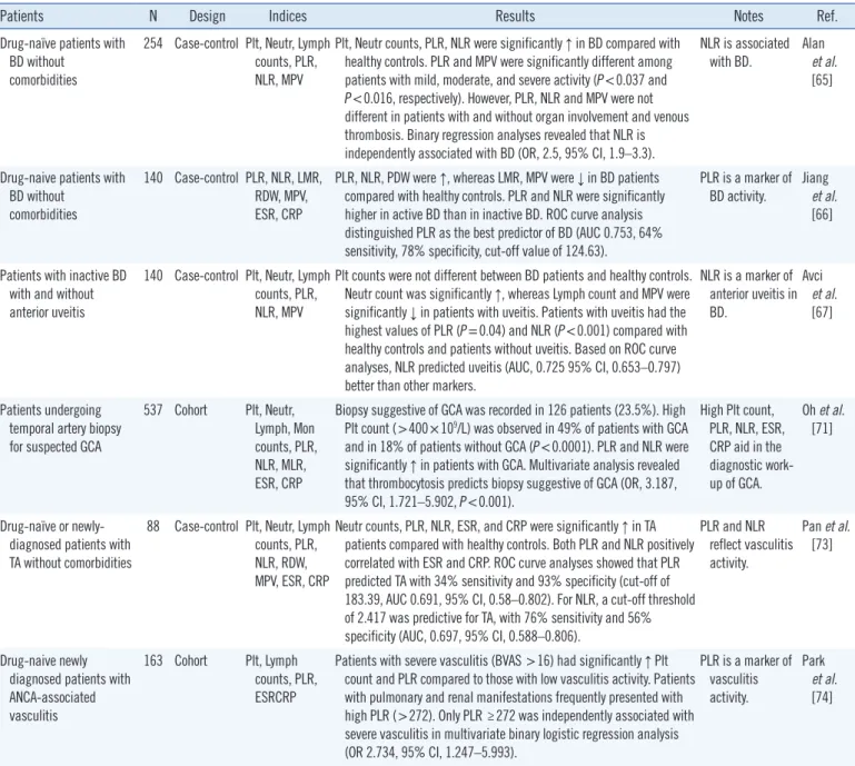
We comprehensively analyzed research reports on PLR across inflammatory rheumatic diseases, employing Scopus database searches and following recommendations on comprehensive and systematic searches of evidence-based data [53]. Large adult-cohort studies on newly-diagnosed and drug-naïve rheu- matic patients reported in English-language journals were pro- cessed without time limits. The synthesized qualitative data are summarized in Table 1.
Shifts in PLR were examined in four large retrospective co- horts of patients with RA [54-57]. In all four studies, laboratory analyses were carried out in single centers. Patients with cardio- vascular, endocrine, hematologic, neoplastic, gastrointestinal, autoimmune comorbidities, as well as those on corticosterioids were reportedly excluded to concentrate on potentially specific associations between PLR and rheumatoid activity. Only one study pointed to an exclusive association of PLR with RA [54], whereas the remaining studies [55-57] considered PLR in com- bination with NLR as potentially valuable in the accurate evalua- tion of inflammatory activity. Combined analysis of shifts in PLR and NLR suggested an increase in platelet and neutrophil counts and a decrease in lymphocyte count during the active stage of RA. Shifts in separate blood cell counts in RA were moderate and within normal ranges in all these studies. PLR and NLR val- ues in patients with RA differed from those in non-RA controls and correlated positively with laboratory (erythrocyte sedimenta- tion rate [ESR], CRP) and clinical (DAS28) markers of rheuma-
Table 1. Studies on PLR and other laboratory indices in adults with inflammatory rheumatic diseases
Patients N Design Indices Results Notes Ref.
Newly diagnosed, drug- naïve RA patients without comorbidities
104 Case-control Plt, Neutr, Lymph counts, PLR, NLR, CRP, RF
Plt and Neutr counts, PLR, and NLR were significantly ↑, and Lymph count was significantly ↓ in patients with RA compared with healthy controls. In an adjusted stepwise logistic regression analysis PLR was associated with RA (OR, 1.026, 95% CI, 1.007–1.045). ROC curve analyses revealed predictive value of PLR: AUC, 0.847, sensitivity, 82.5%, sensitivity, 74.8%, 95% CI, 0.794–0.901.
PLR, but not NLR, is associated with RA.
Peng et al.
[54]
Patients with active RA
without comorbidities 128 Case-control Plt, Neutr, Lymph counts, PLR, NLR, CRP, ESR
Plt, Neutr counts, PLR, and NLR were significantly ↑ in RA. Lymph count was significantly ↓. PLR and NLR were positively correlated with CRP and DAS28.
Both PLR and NLR reflect activity of RA.
Fu et al.
[55]
Patients with RA with low (<2.6) and high DAS28 (>2.6) without comorbidities and corticosteroid-naive
104 Case-control Plt, Neutr, Lymph counts, PLR, NLR, CRP, ESR
Neutr count, PLR, and NLR were significantly ↑ in RA. PLR and NLR were
positively correlated with DAS28. Both PLR and NLR
are markers of RA activity.
Uslu et al.
[56]
Patients with active and
non-active RA 125 Case-control Plt, Neutr, Lymph counts, PLR, NLR, CRP, ESR
Plt, Lymph, Neutr counts were not different between RA and healthy control groups. PLR and NLR were significantly ↑ in RA. PLR and NLR were positively correlated with ESR. ROC curve analyses revealed better predictive value of the combined NLR-PLR curves for distinguishing patients with active RA from controls: AUC for NLR, 0.813, 95% CI, 0.739–0.888, for PLR, 0.762, 95% CI 0.673–0.85, and for NLR-PLR, 0.88, 95% CI, 0.819–0.94.
Combined analyses of PLR and NLR are valuable for detecting activity of RA.
Zhang et al.
[57]
Drug-naïve patients without comorbidities with mild (SLEDAI − 2K<9) and severe SLE (SLEDAI − 2K >9)
116 Case-control Plt, Neutr, Lymph counts, PLR, NLR, CRP, ESR
Plt, Neutr, Lymph counts were significantly ↓, whereas PLR and NLR were significantly ↑ in patients with SLE compared with healthy controls. Both PLR and NLR were positively correlated with SLEDAI-2K (r=0.298, P <0.001 and r=0.312, P <0.001, respectively). NLR, but not PLR, correlated with C3 (r=-0.218, P <0.05) and C4 (r=-0.211, P <0.05). Patients with nephritis had a significantly higher NLR (P =0.027). The ROC curve analyses revealed that PLR had 42%
sensitivity and 84% specificity to predict high lupus activity.
Both PLR and NLR reflect activity of SLE. High NLR reflects nephritis.
Wu et al.
[58]
Drug-naive patients with SLE without
comorbidities
154 Case-control Plt, Neutr, Lymph counts, PLR, NLR, MPV, CRP, ESR
Neutr count was ↑, whereas Lymph and Plt counts were ↓ in patients with SLE compared with healthy controls. PLR, NLR, and MPV were significantly ↑ in SLE. Both PLR and NLR positively correlated with SLEDAI and were significantly higher in patients with lupus nephritis.
ROC curve analysis revealed that NLR had the best sensitivity (71%) and specificity (64%), with a cut-off value of 2.664 (AUC, 0.715, 95%
CI, 0.616–0.787) for predicting lupus nephritis.
Both PLR and NLR reflect activity of SLE. NLR is more valuable than PLR for predicting nephritis.
Qin et al.
[59]
Patients with SLE with flares and concurrent infections
120 Cohort (flares vs infections)
Plt, Neutr, Mon, Lymph counts, PLR, NLR, MLR, CRP, ESR
Plt counts were not different between SLE groups. Neutr count, NLR, and PLR were significantly ↑ in patients with infections. ROC curve analyses showed that NLR (cut-off value of 5.7) had 75% sensitivity and 90% specificity for predicting infections.
NLR is a tool for distinguishing infections from flares in SLE.
Kim et al.
[60]
Patients with AS without
comorbidities 148 Case-control Plt, Neutr, Mon, Lymph, Erythr counts, PLR, NLR, MLR, RDW, CRP, ESR
Plt, Neutr, Mon counts, PLR, NLR, MLR were ↑, whereas Lymph count was ↓ in AS. Only MLR and RDW were significantly different between patients with AS and nonradiographic axial spondyloarthritis. ROC curve analyses revealed the highest AUC of 0.768 for MLR in AS (cut- off value of 0.22, 95% CI 0.696–0.839, 71% sensitivity, 68%
specificity).
MLR is a marker of severe axial AS. Huang
et al.
[61]
Drug-naïve patients with Ps and PsA without comorbidities
136 Case-control Plt, Neutr, Lymph, Mon, Eos, Bas counts, PLR, NLR, ESR, CRP
Plt, Neutr, Mon counts, and NLR were significantly ↑ in PsA compared with Ps and healthy controls. Multivariate binary logistic regression analysis showed that NLR is the strongest predictor of PsA: OR, 3.351, 95% CI, 1.785–6.292 and 1.012, 1.003–1.021 (NLR vs. PLR). Based on ROC curve analysis, the optimal cut-off value of NLR for predicting arthritis was 2.274 (79% sensitivity and 71% specificity).
NLR is the strongest predictor of PsA.
Kim et al.
[64]
(Continued to the next page)
toid activity. ROC curve analyses suggested that combined PLR- NLR curves more accurately distinguish patients with active RA from healthy controls. The same trend was observed when non- active RA patients were compared with healthy controls [57].
In adult SLE cohorts, blood cell count ratios appeared more informative than blood cell counts per se because of a tendency
toward pancytopenia and thrombocytopenia [58-60]. In a co- hort consisting of 116 patients, SLE disease activity index (SLE- DAI) positively and significantly correlated with NLR (r=0.312) and PLR (r =0.298); further, NLR, but not PLR, significantly correlated with C3 (r =-0.218) and C4 complement fractions (r=-0.211) [58]. In the same study, patients with nephritis had
Patients N Design Indices Results Notes Ref.
Drug-naïve patients with BD without
comorbidities
254 Case-control Plt, Neutr, Lymph counts, PLR, NLR, MPV
Plt, Neutr counts, PLR, NLR were significantly ↑ in BD compared with healthy controls. PLR and MPV were significantly different among patients with mild, moderate, and severe activity (P <0.037 and P <0.016, respectively). However, PLR, NLR and MPV were not different in patients with and without organ involvement and venous thrombosis. Binary regression analyses revealed that NLR is independently associated with BD (OR, 2.5, 95% CI, 1.9–3.3).
NLR is associated with BD.
Alan et al.
[65]
Drug-naive patients with BD without
comorbidities
140 Case-control PLR, NLR, LMR, RDW, MPV, ESR, CRP
PLR, NLR, PDW were ↑, whereas LMR, MPV were ↓ in BD patients compared with healthy controls. PLR and NLR were significantly higher in active BD than in inactive BD. ROC curve analysis distinguished PLR as the best predictor of BD (AUC 0.753, 64%
sensitivity, 78% specificity, cut-off value of 124.63).
PLR is a marker of BD activity.
Jiang et al.
[66]
Patients with inactive BD with and without anterior uveitis
140 Case-control Plt, Neutr, Lymph counts, PLR, NLR, MPV
Plt counts were not different between BD patients and healthy controls.
Neutr count was significantly ↑, whereas Lymph count and MPV were significantly ↓ in patients with uveitis. Patients with uveitis had the highest values of PLR (P =0.04) and NLR (P <0.001) compared with healthy controls and patients without uveitis. Based on ROC curve analyses, NLR predicted uveitis (AUC, 0.725 95% CI, 0.653–0.797) better than other markers.
NLR is a marker of anterior uveitis in BD.
Avci et al.
[67]
Patients undergoing temporal artery biopsy for suspected GCA
537 Cohort Plt, Neutr, Lymph, Mon counts, PLR, NLR, MLR, ESR, CRP
Biopsy suggestive of GCA was recorded in 126 patients (23.5%). High Plt count (>400×109/L) was observed in 49% of patients with GCA and in 18% of patients without GCA (P <0.0001). PLR and NLR were significantly ↑ in patients with GCA. Multivariate analysis revealed that thrombocytosis predicts biopsy suggestive of GCA (OR, 3.187, 95% CI, 1.721–5.902, P <0.001).
High Plt count, PLR, NLR, ESR, CRP aid in the diagnostic work- up of GCA.
Oh et al.
[71]
Drug-naïve or newly- diagnosed patients with TA without comorbidities
88 Case-control Plt, Neutr, Lymph counts, PLR, NLR, RDW, MPV, ESR, CRP
Neutr counts, PLR, NLR, ESR, and CRP were significantly ↑ in TA patients compared with healthy controls. Both PLR and NLR positively correlated with ESR and CRP. ROC curve analyses showed that PLR predicted TA with 34% sensitivity and 93% specificity (cut-off of 183.39, AUC 0.691, 95% CI, 0.58–0.802). For NLR, a cut-off threshold of 2.417 was predictive for TA, with 76% sensitivity and 56%
specificity (AUC, 0.697, 95% CI, 0.588–0.806).
PLR and NLR reflect vasculitis activity.
Pan et al.
[73]
Drug-naive newly diagnosed patients with ANCA-associated vasculitis
163 Cohort Plt, Lymph counts, PLR, ESRCRP
Patients with severe vasculitis (BVAS >16) had significantly ↑ Plt count and PLR compared to those with low vasculitis activity. Patients with pulmonary and renal manifestations frequently presented with high PLR (>272). Only PLR ≥272 was independently associated with severe vasculitis in multivariate binary logistic regression analysis (OR 2.734, 95% CI, 1.247–5.993).
PLR is a marker of vasculitis activity.
Park et al.
[74]
Abbreviations: PLR, platelet-to-lymphocyte ratio; RA, rheumatoid arthritis; Plt; platelet; Neutr, neutrophil; Lymph, lymphocyte; NLR, neutrophil-to-lymphocyte ratio; ↑, increased; ↓, decreased; CRP, C-reactive protein; RF, rheumatoid factor; OR, odds ratio; AUC, area under the curve; CI, confidence interval; ESR, erythrocyte sedimentation rate; DAS28, disease activity score in 28 joints; SLE, systemic lupus erythematosus; SLEDAI-2K, SLE disease activity index 2000;
Mon, monocyte; MLR, monocyte-to-lymphocyte ratio; AS, ankylosing spondylitis; Erythr, erythrocyte; RDW, red cell distribution width; Ps, psoriasis; PsA, pso- riatic arthritis; Eos, eosinophil; Bas, basophil; BD, Behçet disease; MPV, mean platelet volume; LMR, lymphocyte-to-monocyte ratio; PDW, platelet distribu- tion width; GCA, giant-cell arteritis; TA, Takayasu arteritis; ANCA, anti-neutrophil cytoplasmic antibodies; BVAS, Birmingham vasculitis activity score.
Table 1. Continued
higher NLR than those without nephritis (P =0.027). In another study involving 154 patients, both PLR and NLR levels were sig- nificantly higher in patients with nephritis than in those without nephritis (P =0.03 and P <0.01, respectively) [59]. However, ROC curve analysis revealed that high NLR was predictive of lu- pus nephritis (cut-off value of 2.664, 71% sensitivity, 64% spec- ificity, area under the curve [AUC], 0.715, 95% CI, 0.616–0.787) [59]. Finally, in a cohort of 120 lupus patients with and without concurrent infections, NLR, with a cut-off value of 5.7 and an AUC of 0.872, had high sensitivity (75%) and specificity (90%) for predicting infections, whereas individual blood cell counts and PLR were less informative [60]. Overall, high levels of both PLR and NLR were found to reflect lupus activity, whereas only high NLR was predictive of lupus nephritis and concurrent in- fections [58-60].
A recent study extensively examined blood cell counts and ra- tios in 148 patients with ankylosing spondylitis (AS) [61]. Alth- ough various cell ratios significantly differed between patients and healthy controls and positively correlated with inflammatory markers (P <0.05), only the monocyte-to-lymphocyte ratio (MLR) differed significantly (P <0.05) between patients with and with- out radiographic sacroiliitis, a feature of severe disease. In addi- tion, ROC curve analyses indicated that MLR yielded a higher AUC (0.768) than PLR and NLR. Thus, MLR, but not PLR or NLR, was suggested as a marker of severe axial spondyloarthri- tis. Indirect evidence from two other cohort studies in patients with AS with sensorineural hearing loss and cardiovascular dis- eases corroborated that neither PLR nor NLR reflects inflamma- tory activity and severity in this disease [62, 63].
An analysis of hemograms of patients with psoriasis (N=111) and psoriatic arthritis (N =25) also demonstrated increases in neutrophil, monocyte, and platelet counts in psoriatic disease, and NLR, not PLR, was the strongest predictor of the presence of spondyloarthritis (OR, 3.351, 95% CI, 1.785–6.292) [64].
A series of specifically designed adult-cohort studies have ex- amined the values of blood cell ratios in systemic vasculitides, focusing on their associations with clinical manifestations. Three studies have examined shifts in Behçet disease (BD) [65-67], which is an autoinflammatory disease with neutrophilic inflam- mation, predominantly venous thromboses, and other manifes- tations resembling those of spondyloarthritides and systemic vasculitides [68, 69]. PLR was not associated with joint, eye, central nervous system, large vessel, or gastrointestinal involve- ment in BD [65]. Neither PLR nor NLR was associated with ve- nous thromboses. However, based on ROC curve analyses, NLR was a more informative predictor of anterior uveitis than PLR
(AUC, 0.725, 95% CI 0.653–0.797, P <0.001, and AUC, 0.6, 95% CI, 0.523–0.676, P =0.012, respectively) [67]. These re- sults are partly in line with the results of another study that high- lighted the value of NLR as a marker of BD-related inflammatory activity, but not thrombosis [70].
Interestingly, a high platelet count and PLR may aid in con- firming diagnosis in patients with large-vessel vasculitides, par- ticularly in those with GCA. Analysis of a large number of hemo- grams and temporal artery biopsies of 537 patients with sus- pected GCA between 1992 and 2015 revealed that positive bi- opsies were twice more likely in patients with thrombocytosis, and that a high platelet count and PLR were more valuable for predicting positive results on a temporal artery biopsy than were NLR and other inflammatory markers [71]. Reportedly, 49% of patients with GCA, but only 18% of patients without GCA, had an elevated platelet count (OR, 4.36, 95% CI, 2.61–7.30) [71].
Likewise, an elevated PLR was found in 44% of patients with GCA but only in 19.4% of those without GCA (OR 3.34, 95% CI, 2.00–5.58) [71]. These results are in line with a population-based study that pointed out thrombocytosis as an informative clue in the differential diagnosis of GCA [72].
In patients with Takayasu arteritis (TA), both PLR and NLR are predictive of vasculitis, albeit with low sensitivity and speci- ficity [73]. One study that examined solely PLR in anti-neutro- phil cytoplasmic antibody (ANCA)-associated vasculitis revealed that pulmonary and renal manifestations were associated with high PLR values (above the cut-off of 272) and that values above the cut-off independently predicted severe vasculitis at diagno- sis (OR 2.7) [74].
Additional information on diagnostic and predictive values of blood cell ratios can be derived from reports from pediatric vas- culitis cohorts. In patients with KD, small- and medium-size ar- teritis with platelet count fluctuations is a hallmark of disease activity and coronary artery dilation [75, 76]. The combination of PLR and NLR was predictive of a severe course, intravenous immunoglobulin resistance, and coronary artery aneurysm de- velopment in KD [77-80]. A retrospective analysis of hemograms of 217 patients with KD from the period 2004–2014 pointed out an association of both low (≤300×109/L) and high (≥550×109/L) platelet counts with a severe course, resulting in treatment re- sistance and coronary artery aneurysms, respectively [80].
Finally, two studies analyzed PLR, NLR, and other inflamma- tory markers in cohorts of pediatric patients with familial Medi- terranean fever (FMF) [81, 82], an autoinflammatory disease with characteristic periodic attacks of polyserositis due to neu- trophilic inflammation and diverse rheumatic manifestations and
vascular comorbidities [83]. Despite some discrepancies, both studies examined hemograms of children with FMF regularly treated with colchicine (0.5–2 mg/daily) and did not recommend PLR as a predictor of inflammation at attacks and during attack-free periods [81, 82]. NLR performed better as an inflammatory marker of attacks in these studies.
LIMITATIONS OF STUDIES ON PLR
The reliability of the specificity of shifts in blood cell ratios, and particularly, PLR in laboratory studies is highly dependent on several confounding factors (Box 1). These factors are some- times overlooked and not reported. The most important one is perhaps the enrollment of patients with high-grade inflammatory diseases with history of drug therapies affecting the maturation of blood cells in the bone marrow and their release into the blood- stream. In this regard, information on the use of corticosteroids and manifestations of hypercortisolemia, associated with decre- ased lymphocyte and increased neutrophil counts, should be derived from the patients’ medical records to correct for these acute effects [84]. There can be also diurnal fluctuations in blood cell counts. Patients with immune-mediated inflammatory dis- eases chronically treated with oral methylprednisolone experi- ence transient acute lymphopenia within eight hours of cortico- steroid administration, followed by morning lymphocytosis [85].
Lymphocyte and platelet counts can vary in response to short- and long-term therapies with methotrexate, a frontline drug for the treatment of RA, other rheumatic diseases, and malignan- cies. A single high dose of methotrexate (50 mg/m2 intramuscu- lar injection) rarely causes thrombocytopenia [86]. However, chronic exposure to regular therapeutic doses of the drug re- sults in fluctuations in lymphocyte counts that may trigger lym- phoproliferative diseases in some cases [87]. Recovery of the
absolute lymphocyte count in methotrexate-induced immuno- deficiency with lymphoproliferative diseases takes from two to 76 weeks after cessation of the therapy [88, 89].
The cross-sectional, single-center design of and single mea- surements of laboratory parameters in the absolute majority of the studies on PLR limit their clinical significance. One can ask in what way and to what extent the observed associations are helpful in routine practice. Are there validated cut-offs one could rely on to suspect a severe course or a complication of an in- flammatory disease? Cut-offs derived from a single study are not generalizable.
Ideally, PLR values should be reported along with values for NLR and other inflammatory markers, which may facilitate draw- ing the whole picture of inflammatory diseases and their infec- tious, thrombotic, or neoplastic complications. Associations be- tween PLR and the course of an inflammatory disease can be more accurately explored in prospective studies with repeated measurements of laboratory parameters and information on mor- bidity and mortality in follow-up reports. There currently are only two small trial reports on PLR in inflammatory rheumatic dis- eases treated with anti-inflammatory drugs. A recent study on 38 patients with RA reported a significant decrease in PLR along with a drop in DAS28 and markers of systemic inflammation in response to six months of therapy with rituximab [90]. Likewise, 12-month-apart repeated measurements of NLR, PLR, and CRP in patients with psoriatic arthritis (N=50) revealed decreases in all these inflammatory markers in the course of therapies with infliximab, adalimumab, and ustekinumab [91]. However, re- peated measurements of platelet and other blood counts in a period longer than three to six months require controlling for seasonal variations and other confounding factors, such as pro- gressive cardiovascular, metabolic, autoimmune, hematologic, and neoplastic comorbidities. Both of the above studies [90, 91]
Box 1. Main limitations of studies on the platelet-to-lymphocyte ratio
• Confounding effects of corticosteroids and other anti-rheumatic drugs that affect blood cell counts are sometimes overlooked.
• Results of retrospective studies tell nothing about causal relationships of shifts in PLR.
• Single measurements of laboratory parameters do not reflect their dynamics.
• Long duration of subjects’ enrollment (more than three to six months) requires correction for seasonal variability of hemogram indexes.
• Specificity of the shifts in PLR is often overlooked in patients with long-standing diseases who may suffer from occult progressive vascular, metabolic, autoimmune, and neoplastic comorbidities.
• Molecular markers of activation of platelets and other blood cells are not measured to reveal associations with cellular markers of inflammation.
• Preanalytical faults with inadequate anticoagulation of blood samples and use of ethylenediaminetetraacetic acid may result in in-vitro platelet agglutination and pseudothrombocytopenia, which is particularly an issue in patients with malignancies.
• There are no race-, age-, and sex-specific recommendations for the use of hemograms to date.
had limitations related to the lack of controls and corrections for confounders.
Although a single-center design limits the generalizability of results, multicenter studies may have their own drawbacks, in- cluding variable standardization of blood processing techniques among laboratories [92].
To some extent, the issue of nonspecific shifts in hemograms relates to the so-called pseudothrombocytopenia and is primar- ily due to the anticoagulation agent EDTA and in-vitro-mediated platelet agglutination [93]. Although the incidence of pseudo- thrombocytopenia is generally low (0.07–2%), and sometimes relates to technical faults, its likelihood increases in patients with cancer [94].
Finally, numerous observational studies have analyzed ROC curves and reported predictive cut-off thresholds of PLR and other blood cell ratios. The use of any of such cut-off values is limited, given the linear origin of inflammatory markers and the necessity to correct for race-, age-, and sex-specific confound- ers [95-97].
CONCLUDING REMARKS
Accumulating evidence points to potentially great diagnostic and prognostic values of complementary components of com- plete blood count, and particularly, blood cell count ratios. Nu- merous observational studies have suggested that PLR is an in- flammatory marker of immune-mediated, metabolic, prothrom- botic, and neoplastic diseases. Bibliographic searches suggested that the absolute majority of articles in this field are related to the value of PLR in cancer. The value of PLR as a predictor of severe course and development of concurrent infections and aneurismal coronary artery disease has been also highlighted in large cohort studies on various inflammatory rheumatic diseases [60, 77-80], although the number of related articles is relatively small. To date, the major rheumatology journals have rarely cov- ered PLR and other hematologic indices, which are readily avail- able clinical data.
Although available studies on PLR are heterogenous in terms of background disease, race, gender, patient age and enrollment, and analytical methods, we can draw some general conclusions.
The majority of these studies, analyzed in this article, focus on the association of PLR with other laboratory markers of inflam- mation. The main finding is that combined evaluation of shifts in PLR and NLR is advisable for predicting the severity of inflam- mation, infectious complications, and other comorbidities. The recording of other blood cell ratios is also desirable as immune
inflammation is multifaceted and may lead to predominant in- volvement of certain blood cells (e.g., monocytes in spondyloar- thritis, and neutrophils in BD and FMF). Indirect evidence from these studies suggests that monitoring of PLR and other blood cell ratios may aid in the diagnosis of comorbidities of rheumatic diseases developing in the course of long-term anti-inflamma- tory and immunosuppressive therapies. In particular, hemograms may point to overt lymphoproliferative and other neoplastic co- morbidities. However, the value of PLR in the early detection of these comorbidities remains uncertain [43], and properly de- signed, large longitudinal studies with serial laboratory measure- ments are warranted. Such longitudinal studies may also shed light on the potential clinical significance of reported cut-off thresh- olds of PLR, NLR, and other complementary markers.
Additional information can be obtained from future analyses of PLR in combination with the closely related mean platelet vol- ume (MPV) and red cell distribution width (RDW). These pa- rameters can be easily calculated from hemograms based on related indices. Some of the observational studies analyzed in this article have indirectly reflected on shifts in MPV and RDW in inflammation and thrombosis, without drawing conclusions on their predictive values [59, 61, 65-67, 73]. The main issue is that MPV and platelet distribution width (PDW) are more vari- able than platelet and other blood cell counts and related ratios [98, 99], limiting their attractiveness for prospective studies. None- theless, the interpretation of combined hematologic indices may uncover patients at increased risk of vascular comorbidities and thrombotic events in rheumatic diseases, particularly in light of the closely related pathobiological shifts in platelets and erythro- cytes in high-grade inflammatory diseases [20]. A few prelimi- nary studies have already examined the abovementioned pa- rameters in combination. In a recent study, an increased MPV- to-lymphocyte ratio reliably distinguished patients with diabetic nephropathy from control subjects [100]. An observational study of pregnant subjects with and without preeclampsia indicated that elevation of PLR, NLR, MPV, and RDW predicted arterial hypertension and high-risk pregnancy [101]. Finally, a pilot study in RA patients (N=57), which was not included in our analysis, demonstrated elevated NLR, MPV, and PDW, but not PLR, in patients with active rheumatoid disease; MPV and PDW nega- tively correlated with ESR [102]. These results are partly in line with previous studies on RA and SLE that reported an associa- tion between high disease activity and low MPV [103, 104]. How- ever, a major limitation of the above pilot study was that the pa- tients were on corticosteroids, methotrexate, and other anti-in- flammatory drugs [102], making it difficult to draw a conclusion.
In summary, PLR is an emerging inflammatory marker that, in combination with NLR and other hematologic indices, can help in the diagnosis and assessment of the activity and severity of several rheumatic diseases, in early detection of various comor- bidities at a subclinical stage, and in monitoring the response to anti-inflammatory therapies. More research is warranted to elu- cidate the role of serial measurements of PLR in the diagnosis and monitoring of proinflammatory and prothrombotic disease states. The design of future studies should take into account confounding effects of numerous clinical, drug-related, and pre- analytical factors that affect the variability of PLR and other indi- ces. Prospective enrollment of subjects in these studies and standardized measurements of all laboratory parameters at a single time point are advisable. Race, age, and sex of the sub- jects, among many other confounders, should be considered when interpreting results.
Authors’ Disclosures of Potential Conflicts of Interest
The authors have no conflicts of interest to declare.
REFERENCES
1. Gasparyan AY, Stavropoulos-Kalinoglou A, Mikhailidis DP, Douglas KM, Kitas GD. Platelet function in rheumatoid arthritis: arthritic and cardio- vascular implications. Rheumatol Int 2011;31:153-64.
2. Olumuyiwa-Akeredolu OO and Pretorius E. Rheumatoid arthritis: nota- ble biomarkers linking to chronic systemic conditions and cancer. Curr Pharm Des 2016;22:918-24.
3. Scherlinger M, Guillotin V, Truchetet ME, Contin-Bordes C, Sisirak V, Duffau P, et al. Systemic lupus erythematosus and systemic sclerosis:
all roads lead to platelets. Autoimmun Rev 2018;17:625-35.
4. Gasparyan AY, Sandoo A, Stavropoulos-Kalinoglou A, Kitas GD. Mean platelet volume in patients with rheumatoid arthritis: the effect of anti- TNF-α therapy. Rheumatol Int 2010;30:1125-9.
5. Abdel Galil SM, Edrees AM, Ajeeb AK, Aldoobi GS, El-Boshy M, Hus- sain W. Prognostic significance of platelet count in SLE patients. Plate- lets 2017;28:203-7.
6. Lood C, Tydén H, Gullstrand B, Nielsen CT, Heegaard NHH, Linge P, et al. Decreased platelet size is associated with platelet activation and anti-phospholipid syndrome in systemic lupus erythematosus. Rheu- matol Oxf Engl 2017;56:408-16.
7. Milovanovic M, Nilsson E, Järemo P. Relationships between platelets and inflammatory markers in rheumatoid arthritis. Clin Chim Acta 2004;343:237-40.
8. Zha Q, He Y, Lu Y, Lu A. Relationship between platelet counts and cartilage erosion in 436 cases of rheumatoid arthritis. Clin Chim Acta 2006;371:194-5.
9. Matsuno H. Remarkable efficacy of tocilizumab for treating rheuma- toid arthritis in patients with high platelet counts. Mod Rheumatol 2015;25:38-42.
10. Üsküdar Cansu D, Üsküdar Teke H, Musmul A, Korkmaz C. Is throm-
bocytosis always an indicator of autosplenectomy in patients with sys- temic lupus erythematosus? Rheumatol Int 2018;38:239-47.
11. Li J, Pan Z, Liu H, Ding F, Shu Q, Li X. Retrospective analysis of the risk of hemorrhage associated with moderate and severe thrombocyto- penia of 173 patients with systemic lupus erythematosus. Medicine (Baltimore) 2018;97.
12. El-Dairi MA, Chang L, Proia AD, Cummings TJ, Stinnett SS, Bhatti MT.
Diagnostic algorithm for patients with suspected giant cell arteritis. J Neuroophthalmol 2015;35:246-53.
13. Bornstein G, Barshack I, Koren-Morag N, Ben-Zvi I, Furie N, Gross- man C. Negative temporal artery biopsy: predictive factors for giant cell arteritis diagnosis and alternate diagnoses of patients without arte- ritis. Clin Rheumatol 2018;37:2819-24.
14. Han JW, Oh JH, Rhim JW, Lee KY. Correlation between elevated plate- let count and immunoglobulin levels in the early convalescent stage of Kawasaki disease. Medicine (Baltimore) 2017;96.
15. Maric LS, Knezovic I, Papic N, Mise B, Roglic S, Markovinovic L, et al.
Risk factors for coronary artery abnormalities in children with Kawasaki disease: a 10-year experience. Rheumatol Int 2015;35:1053-8.
16. Elmas AT and Tabel Y. Platelet counts in children with Henoch-Schon- lein purpura—relationship to renal involvement. J Clin Lab Anal 2016;
30:71-4.
17. Cognasse F, Hamzeh-Cognasse H, Lafarge S, Chavarin P, Cogné M, Richard Y, et al. Human platelets can activate peripheral blood B cells and increase production of immunoglobulins. Exp Hematol 2007;35:
1376-87.
18. Koupenova M, Clancy L, Corkrey HA, Freedman JE. Circulating plate- lets as mediators of immunity, inflammation, and thrombosis. Circ Res 2018;122:337-51.
19. Yeung J, Li W, Holinstat M. Platelet signaling and disease: targeted therapy for thrombosis and other related diseases. Pharmacol Rev 2018;70:526-48.
20. Olumuyiwa-Akeredolu OO and Pretorius E. Platelet and red blood cell interactions and their role in rheumatoid arthritis. Rheumatol Int 2015;
35:1955-64.
21. Łukasik ZM, Makowski M, Makowska JS. From blood coagulation to innate and adaptive immunity: the role of platelets in the physiology and pathology of autoimmune disorders. Rheumatol Int 2018;38:959- 74.
22. Zamora C, Cantó E, Nieto JC, Bardina J, Diaz-Torné C, Moya P, et al.
Binding of platelets to lymphocytes: a potential anti-inflammatory ther- apy in rheumatoid arthritis. J Immunol 2017;198:3099-108.
23. Hally KE, La Flamme AC, Harding SA, Larsen PD. Platelets regulate leucocyte responses to Toll-like receptor stimulation. Clin Transl Im- munol 2018;7:e1036.
24. Duchez AC, Boudreau LH, Naika GS, Bollinger J, Belleannée C, Clout- ier N, et al. Platelet microparticles are internalized in neutrophils via the concerted activity of 12-lipoxygenase and secreted phospholipase A2-IIA. Proc Natl Acad Sci U S A 2015;112:E3564-73.
25. Bunescu A, Seideman P, Lenkei R, Levin K, Egberg N. Enhanced Fc- gamma receptor I. alphaMbeta2 integrin receptor expression by mono- cytes and neutrophils in rheumatoid arthritis: interaction with platelets.
J Rheumatol 2004;31:2347-55.
26. Manfredi AA, Baldini M, Camera M, Baldissera E, Brambilla M, Peretti G, et al. Anti-TNFα agents curb platelet activation in patients with rheu- matoid arthritis. Ann Rheum Dis 2016;75:1511-20.
27. Kornerup KN, Salmon GP, Pitchford SC, Liu WL, Page CP. Circulating platelet-neutrophil complexes are important for subsequent neutrophil activation and migration. J Appl Physiol (1985) 2010;109:758-67.
28. Maugeri N, Rovere-Querini P, Evangelista V, Godino C, Demetrio M, Bal-
dini M, et al. An intense and short-lasting burst of neutrophil activation differentiates early acute myocardial infarction from systemic inflam- matory syndromes. PLoS One 2012;7:e39484.
29. Habets KL, Trouw LA, Levarht EW, Korporaal SJ, Habets PA, de Groot P, et al. Anti-citrullinated protein antibodies contribute to platelet activa- tion in rheumatoid arthritis. Arthritis Res Ther 2015;17:209.
30. Lam FW, Da Q, Guillory B, Cruz MA. Recombinant human vimentin binds to p-selectin and blocks neutrophil capture and rolling on platelets and endothelium. J Immunol 2018;200:1718-26.
31. Cheng Q, Hoi A, Hickey MJ, Morand EF. Lymphocytes from systemic lupus erythematosus patients display increased spreading on VCAM- 1, an effect associated with active renal involvement. Lupus 2012;21:
632-41.
32. Roxburgh CS and McMillan DC. Role of systemic inflammatory response in predicting survival in patients with primary operable cancer. Future Oncol 2010;6:149-63.
33. Gary T, Pichler M, Belaj K, Hafner F, Gerger A, Froehlich H, et al. Plate- let-to-lymphocyte ratio: a novel marker for critical limb ischemia in pe- ripheral arterial occlusive disease patients. PLoS One 2013;8:e67888.
34. Templeton AJ, Ace O, McNamara MG, Al-Mubarak M, Vera-Badillo FE, Hermanns T, et al. Prognostic role of platelet: lymphocyte ratio in solid tumors: a systematic review and meta-analysis. Cancer Epidemiol Biomarkers Prev 2014;23:1204-12.
35. Tan D, Fu Y, Su Q, Wang H. Prognostic role of platelet: lymphocyte ra- tio in colorectal cancer: a systematic review and meta-analysis. Medi- cine (Baltimore) 2016;95:e3837.
36. Li W, Liu Q, Tang Y. Platelet to lymphocyte ratio in the prediction of ad- verse outcomes after acute coronary syndrome: a meta-analysis. Sci Rep 2017;7:40426.
37. Wang Q, Ma J, Jiang Z, Ming L. Prognostic value of neutrophil-to-lym- phocyte ratio and platelet-to-lymphocyte ratio in acute pulmonary em- bolism: a systematic review and meta-analysis. Int Angiol 2018;37:4- 11.
38. Mischler K, Fischer JE, Zgraggen L, Kudielka BM, Preckel D, von Kä- nel R. The effect of repeated acute mental stress on habituation and recovery responses in hemoconcentration and blood cells in healthy men. Life Sci 2005;77:1166-79.
39. Aktar F and Tekin R. Mean platelet volume, neutrophil to lymphocyte ratio and platelet: lymphocyte ratio in determining the diagnosis or outcome in children with snakebite. Arch Argent Pediatr 2017;115:
576-80.
40. Kilincalp S, Çoban Ş, Akinci H, Hamamcı M, Karaahmet F, Coşkun Y, et al. Neutrophil/lymphocyte ratio, platelet/lymphocyte ratio, and mean platelet volume as potential biomarkers for early detection and moni- toring of colorectal adenocarcinoma. Eur J Cancer Prev 2015;24:328- 33.
41. Ozawa T, Ishihara S, Nishikawa T, Tanaka T, Tanaka J, Kiyomatsu T, et al. The preoperative platelet to lymphocyte ratio is a prognostic marker in patients with stage II colorectal cancer. Int J Colorectal Dis 2015;30:
1165-71.
42. Kwon HC, Kim SH, Oh SY, Lee S, Lee JH, Choi HJ, et al. Clinical sig- nificance of preoperative neutrophil-lymphocyte versus platelet-lym- phocyte ratio in patients with operable colorectal cancer. Biomarkers 2012;17:216-22.
43. Kim JH, Lee JY, Kim HK, Lee JW, Jung SG, Jung K, et al. Prognostic significance of the neutrophil-to-lymphocyte ratio and platelet-to-lym- phocyte ratio in patients with stage III and IV colorectal cancer. World J Gastroenterol 2017;23:505-15.
44. You J, Zhang H, Shen Y, Chen C, Liu W, Zheng M, et al. Impact of plate- let to lymphocyte ratio and metabolic syndrome on the prognosis of color-
ectal cancer patients. OncoTargets Ther 2017;10:2199-208.
45. Akboga MK, Canpolat U, Yayla C, Ozcan F, Ozeke O, Topaloglu S, et al. Association of platelet to lymphocyte ratio with inflammation and severity of coronary atherosclerosis in patients with stable coronary ar- tery disease. Angiology 2016;67:89-95.
46. Akboga MK, Canpolat U, Yuksel M, Yayla C, Yilmaz S, Turak O, et al.
Platelet to lymphocyte ratio as a novel indicator of inflammation is cor- related with the severity of metabolic syndrome: a single center large- scale study. Platelets 2016;27:178-83.
47. Li H, Zhou Y, Ma Y, Han S, Zhou L. The prognostic value of the plate- let: lymphocyte ratio in acute coronary syndrome: a systematic review and meta-analysis. Kardiol Pol 2017;75:666-73.
48. Akboga YE, Bektas H, Anlar O. Usefulness of platelet to lymphocyte and neutrophil to lymphocyte ratios in predicting the presence of cere- bral venous sinus thrombosis and in-hospital major adverse cerebral events. J Neurol Sci 2017;380:226-9.
49. Artoni A, Abbattista M, Bucciarelli P, Gianniello F, Scalambrino E, Pap- palardo E, et al. Platelet to lymphocyte ratio and neutrophil to lympho- cyte ratio as risk factors for venous thrombosis. Clin Appl Thromb He- most 2018;24:808-14.
50. Yang W and Liu Y. Platelet-lymphocyte ratio is a predictor of venous thromboembolism in cancer patients. Thromb Res 2015;136:212-5.
51. Tham T, Rahman L, Persaud C, Olson C, Costantino P. Venous throm- boembolism risk in head and neck cancer: significance of the preop- erative platelet-to-lymphocyte ratio. Otolaryngol Head Neck Surg 2018;
159:85-91.
52. Koseoglu SB. Bone loss and platelet-to-lymphocyte ratio. Biomark Med 2017;11:5-10.
53. Gasparyan AY, Ayvazyan L, Blackmore H, Kitas GD. Writing a narrative biomedical review: considerations for authors, peer reviewers, and edi- tors. Rheumatol Int 2011;31:1409-17.
54. Peng YF, Cao L, Zeng YH, Zhang ZX, Chen D, Zhang Q, et al. Platelet to lymphocyte ratio and neutrophil to lymphocyte ratio in patients with rheumatoid arthritis. Open Med (Wars) 2015;10:249-53.
55. Fu H, Qin B, Hu Z, Ma N, Yang M, Wei T, et al. Neutrophil- and plate- let-to-lymphocyte ratios are correlated with disease activity in rheuma- toid arthritis. Clin Lab 2015;61:269-73.
56. Uslu AU, Küçük A, Şahin A, Ugan Y, Yılmaz R, Güngör T, et al. Two new inflammatory markers associated with Disease Activity Score-28 in patients with rheumatoid arthritis: neutrophil-lymphocyte ratio and platelet-lymphocyte ratio. Int J Rheum Dis 2015;18:731-5.
57. Zhang Y, Yin Y, Kuai S, Shan Z, Pei H, Wang J. Combination of neutro- phil to lymphocyte ratio and platelet to lymphocyte ratio as diagnostic biomarker for rheumatoid arthritis. Int J Clin Exp Med 2016;9:22076- 81.
58. Wu Y, Chen Y, Yang X, Chen L, Yang Y. Neutrophil-to-lymphocyte ratio (NLR) and platelet-to-lymphocyte ratio (PLR) were associated with dis- ease activity in patients with systemic lupus erythematosus. Int Immu- nopharmacol 2016;36:94-9.
59. Qin B, Ma N, Tang Q, Wei T, Yang M, Fu H, et al. Neutrophil to lym- phocyte ratio (NLR) and platelet: lymphocyte ratio (PLR) were useful markers in assessment of inflammatory response and disease activity in SLE patients. Mod Rheumatol 2016;26:372-6.
60. Kim HA, Jung JY, Suh CH. Usefulness of neutrophil-to-lymphocyte ra- tio as a biomarker for diagnosing infections in patients with systemic lupus erythematosus. Clin Rheumatol 2017;36:2479-85.
61. Huang Y, Deng W, Zheng S, Feng F, Huang Z, Huang Q, et al. Rela- tionship between monocytes to lymphocytes ratio and axial spondylo- arthritis. Int Immunopharmacol 2018;57:43-6.
62. Bozan N, Alpaycı M, Aslan M, Cankaya H, Kıroglu AF, Turan M, et al.