Salvage of Failed Femoral Neck Fracture Fixation with Conversion Total Hip Arthroplasty Using
the Direct Anterior Approach
Andrew Yun, MD, MBA, Marilena Qutami, PA-C, MMSc, Kory B. Dylan Pasko, BS Department of Orthopedic Surgery, Center for Hip and Knee Replacement,
Providence Saint John’s Health Center, Santa Monica, CA, USA
Purpose: Failed femoral neck fracture (FNF) fixation with in situ pinning presents a surgical challenge. Osteoporotic bone, retained hardware, and a typically elderly population magnify the risks of surgery. Here, outcomes of con- version total hip arthroplasty (THA) using two separate incisions in these high-risk patients were examined.
Materials and Methods: Medical records for 42 patients with a prior history of FNF fixation who underwent conversion THA with hardware removal between 2009 and 2019 were retrospectively reviewed. Surgery was performed by a single surgeon at a single institution. All patients underwent hardware removal followed by direct anterior approach (DAA) THA using two separate incisions. Clinical outcomes, radiographic findings, and perioperative morbidity and mortality are reported.
Results: Clinically, there were no postoperative dislocations, periprosthetic fractures, or infections at follow-up.
After a mean follow-up of 4 years, the mean hip disability and osteoarthritis outcome score, junior (HOOS, Jr) was 91. Radiographically, the mean postoperative cup abduction was 44 degrees and the mean cup anteversion was 21 degrees with an improvement in preoperative leg length discrepancy. Perioperative complications includ- ed one case of immediate foot drop and two readmissions for medical issues. One patient died one month after conversion THA.
Conclusion: Salvage of failed FNF treatment may be managed with conversion THA and DAA with a separate incision for hardware removal. Preservation of posterior soft tissues using a DAA and intraoperative fluoroscopy may mitigate well-known complications related to fracture and dislocation. While favorable clinical outcomes are possible, salvage surgery is still not without substantial surgical and medical risks.
Key Words: Total hip arthroplasty, Femoral neck fracture, Posttraumatic arthritis, Hardware removal, Direct anterior approach
Hip Pelvis 32(4): 199-206, 2020 http://dx.doi.org/10.5371/hp.2020.32.4.199
Submitted:May 22, 2020 1st revision:June 3, 2020 Final acceptance:July 13, 2020
Address reprint request to Kory B. Dylan Pasko, BS
(https://orcid.org/0000-0001-8669-344X)
Department of Orthopedic Surgery, Center for Hip and Knee Replacement, Providence Saint John’s Health Center, 2121 Santa Monica Blvd, Santa Monica, CA 90404, USA
TEL:+1-310-582-7474 FAX:+1-310-582-7481 E-mail:[email protected]
This is an Open Access article distributed under the terms of the Creative Commons Attribution Non-Commercial License (http://creativecommons.
org/licenses/by-nc/4.0) which permits unrestricted non-commercial use, dis- tribution, and reproduction in any medium, provided the original work is properly cited.
and dislocation compared to primary THA2-4).
Weak and osteoporotic bone, retained hardware, and a fragile elderly population compound the risks of surgery after prior closed reduction and pinning2). Standard tech- nical recommendations (e.g., use of longer stems, cement fixation, extensile approaches) have not reliably reduced the rate of worse outcomes. Consequently, conversion THA has been found to be more comparable to a revision THA in terms of blood loss, rate of dislocation, length of stay, and resource utilization2).
An alternative technique was explored to potentially min- imize these risks. In this study, a conversion using a direct anterior approach (DAA) THA with two incisions was con- ducted and long-term outcomes, perioperative course and complications were examined.
MATERIALS AND METHODS
A total of 42 consecutive patients with failed in situ pin- ning of femoral neck fractures (FNF) who required con-
sion. Surgery was performed by a single surgeon at a sin- gle institution with a minimum of 1-year follow-up. Charts were reviewed for indications, medical history, and mode of FNF treatment failure. The primary outcome measures were radiographic findings and the acute perioperative complications of fracture and dislocation. Secondary outcomes measured were patient reported hip disability and osteoarthritis outcome score, junior (HOOS, Jr), at follow-up, hospital course, infection, and death5). The study was approved by the Institutional Review Board (STUDY2020000184).
1. Surgical Technique
Patients were placed on the anterior hip replacement table (Hana; Mizuho, Union City, CA, USA) in the standard fash- ion. The leg was prepped to include the original lateral inci- sion used for pinning. A 3-cm incision was made through the old incision with dissection taken past the iliotibial band and vastus laterals to the level of the screws. Screws
F
Fiigg.. 11.. Failed femoral neck fracture fixation requiring conversion total hip arthroplasty. (AA) Anteroposterior (AP) radiograph with femoral neck nonunion 9 months after in situ pinning of left hip fracture. (BB) AP radiograph with right hip osteonecrosis after pinning. Cortical collapse is visible. (CC) AP radiograph of left hip post traumatic arthritis 6 years after pinning.
A
C
B
were removed by hand and with power tools. Fluoroscopy was used if there was difficulty locating the hardware. The wound was closed in layers and an occlusive dressing was placed over the closed incision.
The table was leveled, and attention was turned to the hip. In the method described by Matta et al.6), an anterior approach was used with a Hana table, fluoroscopy, and digital radiography. After an H-shaped capsulotomy with preservation of all capsular tissue, the head was drilled with a corkscrew to gain control of the proximal femur. With the boot unlocked, the femoral head was dislocated through rotation and traction on the head itself with the corkscrew.
Importantly, minimal to no torsional stress was placed through the femoral shaft. A provisional osteotomy was made based on preoperative and intraoperative templating.
A cementless, titanium, hemispherical, acetabular shell Reflection 3 (Smith & Nephew, London, UK) prior to 2018;
Trident II (Stryker, Kalamazoo, MI, USA) from 2018, was used with a neutral, highly cross-linked, fixed bearing poly- ethylene liner to accommodate the largest femoral head.
On the femoral side, a standard length cementless titanium stem using a triple taper hydroxyapatite-coated design Corail (DePuy Synthes, Warsaw, IN, USA) was used for all patients. Intraoperative imaging was used to guide com- ponent position and sizing as well as hip length and off- set. The capsule was completely repaired by formally reap- proximating the capsular leaves at the end of the procedure.
In this series, patients were allowed to progress with weight bearing as tolerated and without dislocation precautions.
Patients were anticoagulated with aspirin for 3 weeks unless stronger anticoagulation was specifically indicated based on risk stratification, degree of immobility, and/or prior med- ical history.
2. Radiographic Measurement
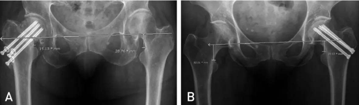
Postoperatively, radiographs were evaluated for cup ori- entation and leg length discrepancy. Leg length discrepan- cy was determined by the vertical distance from a similar point on the lesser trochanters to a horizontal line drawn across the bottom of each radiographic teardrop (Fig. 2).
Anteversion was calculated using the angle of the opening of the ellipse as calculated by the Radlink software (Radlink Inc., El Segundo, CA, USA). At follow-up, implants were evaluated for loosening, migration, and radiolucent lines.
RESULTS
The mean age of patients at the time of index surgery was 71 years (range: 44-89 years). Patients included 28 female and 14 male. There were 24 right hips and 18 left hips. The mean body mass index at the time of index surgery was 23.8 kg/m2(range: 18-36.2 kg/m2). Demographics are summa- rized in Table 1. The mean time from FNF to conversion THA was 11 years (range: 1-42 years). The most common method of fixation was 3 screws (range: 1-4). The indications for F
Fiigg.. 22.. Radiographic measurement of leg length discrepancy. (AA) Anteroposterior (AP) radiograph with 14 mm of shortening in right hip and nonunion. (BB) AP radiograph with 10 mm of shortening in left hip and malunion.
A B
Table 1. Demographic Information
Measurement Value
No. of cases 42
Age (yr) 71±±12.6 (44-89)0
Sex
Male 14 (33.3)
Female 28 (66.7)
Body mass index (kg/m2) 23.8±±4.2 (18-36.2) Laterality
Right 24 (57.1)
Left 18 (42.9)
Values are presented as number only, mean±±standard deviation (range), or number (%).
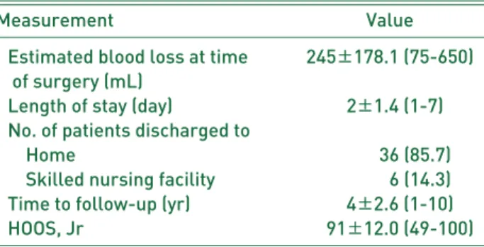
fusion (7%) and no patients required care in the intensive care unit. The mean length of stay was 2 days (range: 1-7 days). As shown in Table 3, 36 patients were discharged to home, and 6 were discharged to a skilled nursing facility.
1. Clinical Outcomes
There were no known fractures or dislocations at a mean follow-up of 4 years (range: 1-10 years). At final follow- up, the average HOOS, Jr was 91 (range: 49-100). Due to the recent transition to patient reported outcome using the HOOS, Jr score, a preoperative score was not available for comparison. There was one reoperation in the immediate postoperative period; this patient was noted to have a post- operative foot drop and was immediately brought back to the operating room. On exploration, a large hematoma was evacuated. A postoperative magnetic resonance imaging revealed that the sciatic nerve was intact. After consultation with neurology, possible causes for the nerve palsy includ- ed compression from the hematoma or excessive traction on the sciatic nerve during femoral preparation. The foot drop did not resolve, and the patient was managed with an ankle foot orthosis. This patients’ HOOS, Jr score was 85
unclear etiology one month after index surgery at a skilled nursing facility.
At final follow-up, there were 5 deaths, one acute, as noted. The time to death after conversion THA for the other 4 patients was 2 to 6 years. The causes of death in the latter group were unrelated to conversion THA.
2. Radiographic Outcomes
For the acetabular implant, the mean abduction angle was 44 degrees (range: 42-50 degrees) and the mean antever- sion was 21 degrees (range: 16-26 degrees). In terms of leg length discrepancy, the mean amount of preoperative shortening was 7 mm (range: 0-19 mm). The mean amount of operative limb lengthening was 5 mm (range: 0-19 mm).
Postoperatively, the mean limb length inequality was 2 mm (range: 0-11 mm). At final follow-up, all hip implants appeared stable without loosening or subsidence (Fig. 3).
Radiographic results are summarized in Table 4.
DISCUSSION
Although the success of in situ pinning for FNFs remains high, there is a subset of patients who require further sur- gical management. Similar to the findings in the series described here, the reported incidence of complications Table 2. Mechanism of Failed Femoral Neck Fracture Fixation
and Mean Time from Fracture to Index Surgery for Each Mode of Failure
Variable Value
Preoperative diagnosis (no. of cases)
Osteonecrosis 20 (47.6)
Post-traumatic arthritis 11 (26.2)
Malunion 07 (16.7)
Nonunion 4 (9.5)
Mean time from fracture to index surgery (yr)
Osteonecrosis 9.5±±9.8
Post-traumatic arthritis 13.2±±160.
Malunion 16.9±±15.1
Nonunion 2.5±±1.7
Values are presented as number (%) or mean±±standard deviation.
Table 3. Clinical Outcomes
Measurement Value
Estimated blood loss at time 245±±178.1 (75-650) of surgery (mL)
Length of stay (day) 02±±1.4 (1-7) No. of patients discharged to
Home 0000036 (85.7)
Skilled nursing facility 0000006 (14.3) Time to follow-up (yr) 004±±2.6 (1-10)
HOOS, Jr 0091±±12.0 (49-100)
Values are presented as mean±±standard deviation (range) or number (%).
HOOS, Jr: hip disability and osteoarthritis disability score, junior.
after FNF treatment are as high as 44% for osteonecrosis, 35% for posttraumatic arthritis, and 21% for nonunion4). A database analysis of 9,962 patients with FNF’s revealed that up to 10% of patients will fail treatment within a year1). Interestingly, the time to failure after FNF correlated with diagnosis in our series. Hence, patients with osteonecro- sis and nonunion experienced early failure at a mean of 9.5 and 2.5 years, respectively; whereas patient with post-trau- matic arthritis and malunion experienced late failure at a mean of 13.2 and 16.9 years.
Patients similar to those described here often complain of intractable pain, weakness, limp, and a shortened leg.
Damage to either the femoral head or neck is too extensive for pin removal alone. Therefore, conversion THA becomes an appropriate salvage option.
Conversion THA after failure of in situ pinning, however, is fraught with a greater risk of surgical complications. Two series reported a fracture rate of 3-5%3,7). To protect against the risk of proximal fracture through the screw holes, these authors recommend a sequence of femoral head dislocation with the hardware in place for torsional stability, reduction of the hip, hardware removal, and then secondary disloca- tion. While certainly reasonable, we have also found that hardware can be safely removed prior to head dislocation as long as the torsional force on the femur is controlled through the femoral head. Additionally, the concern of stress risers from the screw holes has also led some authors to suggest further protective measures. Hernandez et al.4)recommend- ed bone grafting the screw holes. In the series described here, the compaction broaching technique for the triple taper stem may have autografted the residual screw holes.
Chen and Hozack8) has suggested that the screw holes should be bypassed by 3 cm with use of revision-style femoral implants. Interestingly, it was noted here that stan- dard length implants performed as a reasonable alternative without any increased incidence of postoperative peripros- thetic fractures. Careful examination of the radiographs reveals that the pins are typically placed in the thin cortex of metaphyseal bone at or above the level of the lesser trochanter, and standard-length stems easily bypass these F
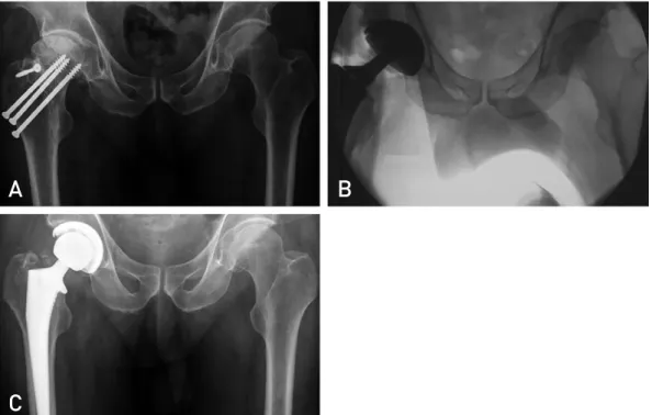
Fiigg.. 33.. Sequence of deformity and correction in conversion total hip arthroplasty. (AA) Anteroposterior (AP) radiograph of failed treatment with posttraumatic arthritis and retained hardware. (BB) Intraoperative imaging to assess implant position and ori- entation. (CC) Follow-up AP radiograph at 4 years with well-fixed components.
A
C
B
Table 4. Pre- and Postoperative Radiographic Results
Measurement Value
Preoperative leg length discrepancy 7±±5.3 (0-19) (mm)
Postoperative leg length discrepancy 2±±2.5 (0-11) (mm)
Acetabular cup abduction (degrees) 44±±2.6 (42-50) Acetabular cup anteversion (degrees) 21±±2.7 (16-26) Values are presented as mean±±standard deviation (range).
F
Fiigg.. 44.. Standard length femoral components easily bypass screw holes. (AA) Close-up anteroposterior (AP) view of left hip shows stem bypassing screw holes noted by area of bone overgrowth. (BB) Close-up AP view of right hip shows stem bypass- ing screw holes marked by retained washers. Because each screwhead had loosened from the bone beyond 1 cm, no further dissection to remove the washers was done.
A B
F
Fiigg.. 55.. Intraoperative imaging aided implant positioning. (AA) Fluoroscopy of right hip conversion. Note cup orientation. (BB) Fluoroscopy of left hip conversion. Note hip offset symmetry. (CC) Fluoroscopy of right hip conversion. Note hip length sym- metry.
A
C
B
proximal screw holes by at least 3 cm (Fig. 4). Unlike dia- physeal holes that remain after dynamic hip screws or short cephalomedullary nails, the more superior metaphyseal holes from in situ pinning may be less susceptible to fracture over- all, as also theorized by Hernandez et al.4)and Mortazavi et al.9).
Postoperative dislocation rates of up to 20% represent another potential complication of conversion after FNF10). There is concern over associated damage to the soft tissue structures after a fall, and further surgery may further reduce the intrinsic stability around the hip. Given that prior pub- lications have reported a dislocation rate of 5-20% with the posterior approach, it has been recommended that hemi- arthroplasty or a dual mobility construct be used to augment stability in high risk patients3,4). In the group described here, however, fixed-bearing constructs with a large head were chosen instead. Our series revealed no postoperative dis- locations and it was noted that the DAA window did not require an extensile dissection and preserved the posterior soft tissues. A formal capsular repair rather than capsulec- tomy also augmented postoperative stability. It is possi- ble that the absence of dislocations may be partly related to avoiding dissection of the posterior capsule associated with an anterior approach. Similarly, Mortazavi et al.9)reduced the dislocation rate to 0% using an anterolateral approach further demonstrating that maintaining the posterior soft tissues may play an important role in decreasing disloca- tion risk in this vulnerable cohort. Additionally, we found the use of intraoperative imaging for cup orientation, implant sizing, and hip length helps minimize the risk of outliers that may contribute to the risk of dislocation (Fig. 5).
The underlying health, cognitive decline, and overall fragili- ty of these patients is also of concern. Hernandez et al.4) noted a mortality rate of 23% at two years after conversion.
Although the average age of patients in our series was 71 years, eight were 80 years or older at time of conversion THA. Many had significant comorbidities typical of this age group. Two patients required acute rehospitalization for med- ical issues, and one died shortly after surgery. Fortunately, the recovery of the remaining patients after conversion THA was otherwise uneventful. Although most of the patients described here recovered without major incident, we agree that conversion THA carries an increased risk of compli- cations and morbidity and that there is a clear concern for greater resource utilization, as warned by Schwarzkopf et al.2).
The major limitation of this study is that it is a small ret- rospective series describing the experience of a single sur-
geon. With a single surgeon there is an intrinsic risk of bias, so the aim of this analysis was to focus on objective points of reference (e.g., complications and radiographic out- comes). With the limitation of only 42 patients, a larger sample population may reveal a complication profile simi- lar to that of other series. Another major limitation is the lack of a control group for comparison. A comparative cohort of patients treated with a posterior approach and intraop- erative imaging could potentially demonstrate similarly favorable findings. Alternatively, future comparative stud- ies with computer navigation or robotic assisted conversion THA may provide equivalent or even improved results compared to intraoperative fluoroscopy. Although a mini- mum 2-year follow-up is more common, we chose a mini- mum 1-year follow-up to include 4 additional patients for a greater pool for analysis, since the primary outcome mea- sures were perioperative complications, fractures, and dis- locations. Finally, as stated previously, a preoperative HOOS, Jr score was not available for comparison.
CONCLUSION
Failed FNF management remains perilous and techni- cally challenging. Patients with osteonecrosis, nonunion, or secondary changes are substantially disabled and with limited options. While reasonable outcomes with conver- sion THA are possible, these salvage procedures are still not without substantial risk. The series described here is intended to highlight a potential alternative approach to con- ventional technical recommendations for these high-risk patients in the hope of minimizing future complications.
Further investigation is warranted to reproduce these results.
CONFLICT OF INTEREST
The authors declare that there is no potential conflict of interest relevant to this article.
REFERENCES
01. Kahlenberg CA, Richardson SS, Schairer WW, Cross MB.
Rates and risk factors of conversion hip arthroplasty after closed reduction percutaneous hip pinning for femoral neck fractures-a population analysis. J Arthroplasty. 2018;33:
771-6.
02. Schwarzkopf R, Manzano G, Woolwine S, Slover J. Salvage treatment of hip fractures after failure of surgical fixation:
a systematic review. Orthop Knowl Online J. 2015;13. doi:
10.5435/OKOJ-13-3-3
03. Morice A, Ducellier F, Bizot P. Total hip arthroplasty after
06. Matta JM, Shahrdar C, Ferguson T. Single-incision anteri- or approach for total hip arthroplasty on an orthopaedic table.
Clin Orthop Relat Res. 2005;441:115-24.
07. Archibeck MJ, Carothers JT, Tripuraneni KR, White RE Jr.
ison of primary arthroplasty with early salvage arthroplasty after failed internal fixation. J Bone Joint Surg Am. 2002;
84:2010-5.