대한소화기학회지 2005;45:52-59
ꠏꠏꠏꠏꠏꠏꠏꠏꠏꠏꠏꠏꠏꠏꠏꠏꠏꠏꠏꠏꠏꠏꠏꠏꠏꠏꠏꠏꠏꠏꠏꠏꠏꠏ Correspondence to: Jae Seon Kim, M.D.
Division of Gastroenterology, Korea University Guro Hospital 97 Guro-dong gil, Guro-gu, Seoul 152-150, Korea
Tel: +82-2-818-6631, Fax: +82-2-866-1643 E-mail: kimjs@kumc.or.kr
ꠏꠏꠏꠏꠏꠏꠏꠏꠏꠏꠏꠏꠏꠏꠏꠏꠏꠏꠏꠏꠏꠏꠏꠏꠏꠏꠏꠏꠏꠏꠏꠏꠏꠏ 접수: 2004년 9월 2일, 승인: 2004년 12월 24일 연락처: 김재선, 152-150, 서울시 구로구 구로동길 97
고려대학교 구로병원 내과
Tel: (02) 818-6631, Fax: (02) 866-1643 E-mail: kimjs@kumc.or.kr
간내담석증 및 간내담관암에서 Epidermal Growth Factor Receptor, ErbB2, Matrix Metalloproteinase-9 발현
강원대학교 의과대학 내과학교실*, 고려대학교 의과대학 내과학교실
김효정*⋅김재선⋅강창돈*⋅이성준*⋅김진용⋅연종은⋅박종재⋅심재정⋅변관수⋅박영태⋅이창홍
Expression of Epidermal Growth Factor Receptor, ErbB2 and Matrix Metalloproteinase-9 in Hepatolithiasis and Cholangiocarcinoma
Hyo Jung Kim, M.D.*, Jae Seon Kim, M.D., Chang Don Kang, M.D.*, Sung Joon Lee, M.D.*, Jin Yong Kim, M.D., Jong Eun Yeon, M.D., Jong-Jae Park, M.D., Jae Jeong Shim, M.D., Kwan Soo Byun, M.D.,
Young-Tae Bak, M.D, and Chang Hong Lee, M.D.
Department of Internal Medicine, Kangwon National University College of Medicine*, Chuncheon;
Department of Internal Medicine, Korea University College of Medicine, Seoul, Korea
Background/Aims: Hepatolithiasis is a common disease in East Asia and presents as a histological feature of proliferative glands containing mucin. 5-10% of hepatolithiasis is known to be associated with cholangiocarcinoma.
Recent studies reported that epidermal growth factor receptor (EGFR) could be activated through heparin binding- EGF cleavage by metalloproteinases. Matrix metalloproteinases (MMPs) which digest the extracellular matrix are required for cancer cell invasion and the expression of MMP-9 is known to be increased in cholangiocarcinoma.
However, there has been few studies on the expressions and roles of EGFR and MMP in hepatolithiasis. This study was performed to clarify and compare the expressions of EGFR, erbB2 and MMP-9 in hepatolithiasis and cholangiocarcinoma. Methods: Surgically resected liver tissues with hepatolithiasis (n=14), cholangiocarcinoma (n=20) and trauma (n=2 as controls) were included. The expressions of EGFR, erbB2 and MMP-9 in tissue samples were examined by immunohistochemistry using respective monoclonal antibodies. Results: In traumatic livers, the expressions of EGFR, erbB2 and MMP-9 were all negative. The expression of EGFR was increased in hepatolithiasis group (79%, 11/14) compared with cholangiocarcinoma group (25%, 5/20) (p<0.05). The expres- sion of erbB2 was detected only in cholangiocarcinoma (25%, 5/20). MMP-9 was increased in both hepatolithiasis (79%, 11/14) and cholangiocarcinoma (95%, 19/20) (p>0.05). Conclusions: EGFR expression appears to be the dominant component in periductular hyperplasia of hepatolithiasis and MMP-9 is upregulated not only in cholangiocarcinoma but also in hepatolithiasis. This study suggests that EGFR and MMP-9 are associated with cholangiocarcinoma and hepatolithiasis. (Korean J Gastroenterol 2005;45:52-59)
ꠏꠏꠏꠏꠏꠏꠏꠏꠏꠏꠏꠏꠏꠏꠏꠏꠏꠏꠏꠏꠏꠏꠏꠏꠏꠏꠏꠏꠏꠏꠏꠏꠏꠏꠏꠏꠏꠏꠏꠏꠏꠏꠏꠏꠏꠏꠏꠏꠏꠏꠏꠏꠏꠏꠏꠏꠏꠏꠏꠏꠏꠏꠏꠏꠏꠏꠏꠏꠏꠏꠏꠏꠏꠏꠏꠏꠏꠏꠏꠏꠏꠏꠏꠏꠏꠏꠏꠏꠏꠏꠏꠏꠏꠏꠏꠏꠏꠏꠏꠏꠏꠏꠏꠏꠏꠏꠏꠏꠏꠏꠏꠏꠏ
Key Words: Stone, Intrahepatic; Recepotor, Epidermal growth factor; Genes, erbB-2; Matrix metalloproteinases;
Cholangiocarcinoma
김효정 외 10인. 간내담석증 및 간내담관암에서의 EGFR, ErbB2, MMP-9 발현 53
서 론
간내담석증은 동양에서 비교적 흔한 질환으로, 우리나라 에서 간내담석증의 빈도는 전체 담석증의 14.1%이다.1 간내 담관의 원발 간내담석은 담낭담석이나 총수담관담석과는 달리 담석이 제거된 후에도 간내 여러 부위에 담관의 협착 및 확장, 담석의 재발을 일으키고, 반복적인 담도염, 간농양, 그리고 간실질의 파괴 등을 초래하며 조절 불가능한 경과를 보이기도 한다.2,3 또한 담관암의 위험인자로서 간내담석증 환자의 약 5-10%에서 담관암이 동반된다.4
간내담관암은 간에 발생하는 원발 간암의 10%를 차지한 다. 하지만 조기발견이 어렵고 국소적으로 상당히 진행하여 담관 주위 조직 및 담관벽을 따른 직접 침윤, 간-십이지장 인대에 미만성 침윤, 주위 림프절 전이, 신경 또는 신경 주 위 침윤 등을 동반하는 경우가 적지 않아, 외과 절제율이 낮 고 예후가 매우 불량하다. 담관암의 원인은 잘 알려져 있지 않지만 만성 염증을 유발하며 장기간 담도 손상을 일으키는 간내담석, 원발 경화 담도염, 간흡충 등이 위험인자이다.5 이 중 간내담석은 주위 담관 상피에 반복적인 궤양 및 치유 과정을 일으켜 담도 상피의 비정형 비후 및 이형성을 유발 하고 간내담관암을 일으킨다고 추정되나,6 이에 관한 정확 한 기전에 대해서는 잘 알려진 바가 없다.
종양에서 epidermal growth factor receptor (EGFR) 발현이 증가하며, EGFR이 EGF-like growth factor들을 유도하여 암 발생에 있어 중요한 역할을 한다. 담관암에서도 EGFR의 발 현이 증가되며,7 EGFR family 중 원발 암유전자(proto- oncogene)로 알려진 erbB2와 erbB3이 과발현되어 담관암의 발암 과정에 EGFR이 관여한다.8
Matrix metalloproteinase (MMP)는 인체 내 정상적인 조직 재생 과정과 종양 전이 등의 병적인 조직 재형성 과정에 중 요한 역할을 하는 단백분해효소로, MMP와 MMP 활성인자 및 억제인자들이 정상적인 간내담도 발생 과정에서 관찰되 며, 담관암에서도 발현이 증가된다.9 또한, MMP가 EGFR의 리간드 중의 하나인 heparin binding EGF를 활성화시켜 MMP 가 EGFR을 활성화시킬 수 있다.10-12 그리고 type IV colla- genase인 MMP-9은 다형핵 백혈구에서 분비되고, 여러 성장 인자와 싸이토카인 등에 의해서 발현이 유도되어, 담관암의 전이 및 예후와 관련이 있다.13 하지만 간내담석증에서의 MMP-9 발현에 대해서는 아직 보고된 바가 없다.
이번 연구는 담관암의 발생 또는 진행에 관여한다고 알려 진 EGFR, erbB2, MMP-9의 발현을 간내담석증에서 알아보 고 이를 간내담관암과 비교하고자 하였다.
대상 및 방법
1. 대상
간내담석증이나 간내담관암으로 진단받고 간절제술을 시 행받은 환자들의 수술 조직을 이용하였다. 간내담석증만 있 었던 군은 14예(남녀 비 6:8, 평균 연령 47.5±8.6세)였다. 간 내담관암이 있었던 군은 20예(남녀 비 9:11, 평균 연령 60.2
±6.3세)였다. 간내담관암군 20예 중 5예에서는 간내담석이 동반되었으며, 15예에서는 간내담석이 동반되지 않았다. 그 리고 정상 대조군으로는 간 손상으로 간절제술을 시행받은 2예를 이용하였다.
2. 방법
1) EGFR, ErbB2, MMP-9에 대한 면역조직화학검사 파라핀으로 처리된 간 조직을 5μm 두께의 절편의 조직 슬라이드로 만들어 xylene으로 파라핀을 제거시키고, 100%, 95%, 70%, 50% 알코올로 단계별로 함수시켰다. 조직 내의 내인성 과산화효소 활성을 제거하기 위하여 0.3% 메탄올로 30분 간 처리하였다. Phosphate-buffered saline (PBS)로 씻어 낸 후, 항체의 비특이 결합을 억제하기 위하여 0.05% Tween- 20과 1%의 정상 염소 혈청을 함유한 PBS를 이용하여 20분 간 반응시켰다. EGFR, erbB2 그리고 MMP-9에 대한 각각의 단클론 항체(Oncogene, Boston, MA, USA)는 모두 1:100의 비율로 사용하였으며 실온에서 1시간 동안 반응시켰다.
PBS로 5분 간 3차례 씻어 낸 후, 실온에서 1시간 동안 bio- tinylated 이차 항체와 반응시켰다. 결합된 항체를 avidin- biotin-peroxidase complex method을 이용하여 관찰하였으며, 헤마톡실린으로 대조 염색 후 광학현미경으로 검경하였다.
조직의 처리는 환자군과 대조군을 동시에 시행하여 비교하 였으며, 음성 대조군은 일차 항체 대신 PBS를 이용하였다.
2) 결과 판정
각 항체에 대한 염색 판정은 2명의 병리의사가 따로 판독 하였으며, 간내담석증의 경우에는 증식된 담도주위선에서, 담관암의 경우에는 종양 부위에서, 광학현미경 200배 시야 에서 적갈색의 과립이 분명히 관찰되는 부분이 전체의 10%
이상으로 염색될 때 양성으로 판정하였다.8
3) 통계 분석
통계 분석은 Fisher's exact test를 이용하였으며, 통계 유의 성은 p값이 0.05보다 작은 경우에 유의한 것으로 판정하였 다.
54 The Korean Journal of Gastroenterology: Vol. 45, No. 1, 2005
결 과
1. 정상 대조군, 간내담석군, 담관암군에서의 EGFR의 발현
정상 대조군에서 EGFR은 발현되지 않으나, 간내담석 환 자군 14예 중 11예(79%)에서 증식된 담도주위선에서
EGFR이 발현되었다(Table 1, Fig. 1). 간내담관암 환자군에 서는 20예 중 5예(25%)에서만 종양 부위에서 발현되어, EGFR 은 간내담석증에서 유의하게 높게 발현되었다(p=0.003, Table 1, Fig. 2). 간내담관암 환자들에서 간내담석 유무에 따른 EGFR 발현은, 담석이 있었던 5예에서는 1예(20%)에서, 그 리고 담석이 동반되지 않았던 15예에서는 4예(27%)로 차이 가 없었다(Table 1). 또한 간내담석증의 EGFR 발현은 담석
Fig. 1. Immunohistochemical stains in a hepatolithiasis case (×100). Proliferated peribiliary glandular and bile duct epithelial cells are stained for EGFR and MMP-9. (A) Negative staining for EGFR. (B) Positive staining for EGFR. (C) Negative staining for MMP-9.
Gallstone is showed in left of picture. (D) Positive staining for MMP-9.
EGFR, epidermal growth factor receptor; MMP-9, matrix metalloproteinase-9.
Table 1. Expression of EGFR in Control, Hepatolithiasis, and Cholangiocarcinoma Groups
Control (n=2)
Hepatolithiasis (n=14)
Cholangiocarcinoma With stones
(n=5)
Without stones (n=15)
Subtotal (n=20) Positive (%) 0 11 (79)* 1 (20)† 4 (27)‡ 5 (25)§ Negative (%) 2 3 (21) 4 (80) 11 (73) 15 (75)
* vs †, p=0.038; * vs ‡, p=0.007; * vs §, p=0.003; †vs ‡, p=0.634.
EGFR, epidermal growth factor receptor.
Kim HJ, et al. Expression of EGFR, ErbB2 and MMP-9 in Hepatolithiasis and Cholangiocarcinoma 55
을 동반한 간내담관암이나 담석을 동반하지 않은 간내담관 암 모두에 비해 높은 발현율을 보였다(각각 p=0.038, p=
0.007).
2. 정상 대조군, 간내담석군, 담관암군에서의 ErbB2 발현
정상 대조군 2예 모두에서 erbB2는 발현되지 않았다. 간내 담석군에서 erbB2 발현은 14예 모두에서 관찰되지 않았으 며, 담관암군에서는 20예 중 5예(25%)에서 발현되었다(Table 2, Fig. 2). 담관암군에서 간내담석 동반 유무에 따른 erbB2 발현은 간내담석이 동반되었던 5예 중 2예(40%), 간내담석 이 동반되지 않았던 15예 중 3예(20%)에서 발현되었다
(Table 2). 담관암군에서 간내담석군에 비해 erbB2의 발현율 이 높았으나(p=0.056), 담관암군에서 간내담석 동반 유무에 따른 erbB2의 발현율, 간내담석군과 간내담석이 동반된 담 관암군에서의 erbB2의 발현율, 간내담석군과 간내담석이 동 반되지 않았던 담관암군에서의 erbB2의 발현율은 통계적으 로 유의한 차이가 없었다(Table 2).
3. 정상 대조군, 간내담석군, 담관암군에서의 MMP-9 발현
정상 대조군 2예 모두에서 MMP-9은 발현되지 않았다. 간 내담석군과 담관암군에서의 MMP-9 발현은 간내담석군의
Table 2. Expression of erbB2 in Control, Hepatolithiasis, and Cholangiocarcinoma Groups
Control (n=2)
Hepatolithiasis (n=14)
Cholangiocarcinoma With stones
(n=5)
Without stones (n=15)
Subtotal (n=20) Positive (%) 0 0 (0)* 2 (40)† 3 (20)‡ 5 (25)§ Negative (%) 2 14 (100) 3 (60) 12 (80) 15 (75)
* vs †, p=0.058; * vs ‡, p=0.125; * vs §, p=0.056; †vs ‡, p=0.366.
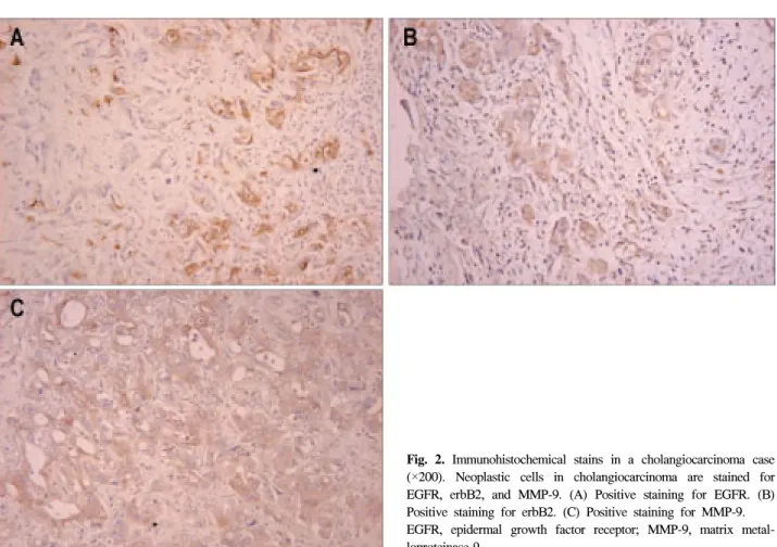
Fig. 2. Immunohistochemical stains in a cholangiocarcinoma case (×200). Neoplastic cells in cholangiocarcinoma are stained for EGFR, erbB2, and MMP-9. (A) Positive staining for EGFR. (B) Positive staining for erbB2. (C) Positive staining for MMP-9.
EGFR, epidermal growth factor receptor; MMP-9, matrix metal- loproteinase-9.
56 대한소화기학회지: 제45권 제1호, 2005
경우 14예 중 11예(79%), 담관암군의 경우 20예 중 19예 (95%)에서 발현되었다(Table 3, Fig. 1, 2). 담관암군에서 간 내담석 동반 유무에 따른 MMP-9 발현은 간내담석이 동반 되었던 5예 중 5예(100%), 간내담석이 동반되지 않았던 15 예 중 14예(93%)에서 발현되었다(Table 3). 간내담석군 중 MMP-9에 발현을 보였던 11예 중 10예에서는 EGFR이 발현 되었으며, 간내담관암 중 MMP-9에 발현을 보였던 19예 중 5예에서 EGFR이 발현되었다(Table 3). 간내담석군과 담관암 군에서의 MMP-9의 발현율, 담관암군에서 간내담석 동반 유 무에 따른 MMP-9의 발현율, 간내담석군과 간내담석이 동반 된 담관암군에서의 MMP-9의 발현율, 간내담석군과 간내담 석이 동반되지 않았던 담관암군에서의 MMP-9의 발현율은 유의한 차이가 없었다(Table 3).
고 찰
간내담석증은 재발 혹은 반복적인 담관염을 일으켜서 주 위 조직의 섬유화 및 협착을 초래하고 일부에서는 담관암으 로 진행하기 때문에 적극적인 진단 및 치료가 요구되는 질 환이다.2-4 담즙의 구성 성분 변화, 세균 감염, 담즙 정체 등 이 간내담석의 형성에 있어 중요한 역할을 하지만,14 담석 발생의 원인 및 발생기전은 명확하지 않다. 일반적으로 간 내담석증은 간내담석이 있는 담도 및 담도 주위에 염증세포 침윤, 담도주위선들의 증식 및 섬유화 등의 특징적인 조직 소견을 보이며,15-17 담도 및 담도주위선들에서의 점액 과분 비가 간내담석 발생에 있어 중요한 역할을 한다.18,19 이번 연구에서도 담도주위선의 증식은 대조군 2예에서는 관찰되 지 않았으나, 간내담석군 14예 전 예에서 담도주위선의 증 식을 관찰할 수 있었다.
간내담관암은 간내담관 상피세포에서 기원하는 원발 간 암으로, 원발 경화 담관염, 간흡충, 간내담석증 등 여러 가 지 위험인자들이 있다.5 이 중 간내담석은 주위 담관 상피의 반복적인 궤양 및 치유 과정을 일으켜, 만성 염증이 주위 담 관 상피의 비정형 비후 및 이형성을 유발시켜 간내담관암이
발생한다고 알려져 있으나6 논란이 많다.
점액의 주요 단백 성분인 apomucin에 대한 여러 유전자들 이 밝혀져20 간내담석증과 담관암에서는 발현이 변한다.21,22 또한 간내담석증의 증식된 담도주위선에서는 증식 핵항원 (proliferating cell nuclear antigen)의 발현이 증가되어 있어 담 관암뿐만 아니라 간내담석증에서도 담관 상피가 증식된 다.23
담관 상피의 성장과 분화에 관여한다고 알려진 물질로는 hepatocyte growth factor (HGF), transforming growth factor- alpha (TGF-α), insulin-like growth factor (IGF), proline, chol- ecystokinin, EGF 등이 있으며,24,25 이들 성장인자에 대한 수 용체가 존재한다. 이 중 EGFR은 자체의 단백 타이로신 키 나제를 가지고 있으면서 세포 성장을 일으키는 세포막 수용 체 집합체로서, 여기에는 통상 EGFR로 알려진 erbB1 외에 도 erbB2 (HER-2), erbB3 (HER-3), erbB4 (HER-4)가 있다.26 이들 EGFR family는 모두 단량체 형태로 세포막에 존재하 나 EGF 등의 여러 리간드들과 반응하여 세포 신호전달을 활성화시키고, 세포 증식 및 이온통로 조절 등 세포 내 여러 가지 변화를 일으킨다.27
종양에서의 EGFR 발현 증가에 대해서는 유방암, 폐암, 췌장암 등 여러 종양에서 활발히 연구되어 왔으며, EGFR이 EGF-like growth factor들을 유도하여 암 발생에 있어 중요한 역할을 한다.27 담낭암 및 담도암에서도 EGFR 발현이 증가 되고,7 EGFR 발현 증가가 담도암 발생에 관여하며,28 특히 erbB2는 원발 암유전자로도 잘 알려져 있어,8,29-31 간내담관 암 발생에 있어 EGFR의 역할 및 임상 특징에 대한 연구가 많은 반면, 간내담석증에서의 EGFR 및 erbB2의 발현 증가 유무 및 역할에 대해서는 현재까지 연구된 바가 거의 없어 이에 대한 규명이 필요한 실정이다.
면역조직화학염색을 이용하여 간내담석증과 간내담관암 에서의 EGFR, erbB2의 발현을 관찰한 이번 연구에서, EGFR 는 간내담석증과 간내담관암 모두에서 발현되지만, 간내담 석증군의 경우 79%로, 담관암군의 25%에 비해 높게 발현되 고, 담석을 동반한 간내담관암이나 담석을 동반하지 않은 Table 3. Expression of MMP-9 in Control, Hepatolithiasis, and Cholangiocarcinoma Groups
Control (n=2)
Hepatolithiasis (n=14)
Cholangiocarcinoma With stones
(n=5)
Without stones (n=15)
Subtotal (n=20) Positive (%) 0 11 (79)* 5 (100)† 14 (95)‡ 19 (95)§
Negative (%) 2 3 (21) 0 (0) 1 (5) 1 (5)
* 10 of 11 cases were EGFR (+); §5 of 19 cases were EGFR (+); * vs †, p=0.376; * vs ‡, p=0.272; * vs §, p=0.179; †vs
‡, p=0.750.
EGFR, epidermal growth factor receptor; MMP-9, matrix metalloproteinase-9.
김효정 외 10인. 간내담석증 및 간내담관암에서의 EGFR, ErbB2, MMP-9 발현 57
간내담관암 모두에 비해 높은 발현율을 보였다. 그러나 담 관암군에서 간내담석 동반 유무에 따른 EGFR의 발현율은 유의한 차이가 없었다. 간내담관암의 EGFR 발현율은 이전 연구들과 큰 차이를 보이지 않았으나,7 간내담석증의 EGFR 발현율은 간내담관암에 비해 매우 높았다. 이는 EGFR이 담 관암에서의 재생 증식, 이형성과는 달리 간내담석증에서는 주로 담관 주위의 염증반응에 의해 발현되어 담관주위선의 증식 및 점액 변화에 관여할 것으로 생각되나, 간내담석증 과 연관된 암 발생 과정에 있어 EGFR 역할에 대해서는 지 속적인 연구가 필요하다.
ErbB2는 간내담관암에서 30-70%까지 다양하게 발현되
며,8,29-31 이번 연구에서 erbB2는 간내담석군에서는 발현되지
않았으나, 간내담관암군에서는 25%에서 발현되었다. 간내 담관암군이 간내담석군에 비해 erbB2의 발현율이 높은 경 향을 보였으나, 간내담관암군 내에서 간내담석 동반 유무에 따른 erbB2의 발현율은 유의한 차이를 보이지 않았다. 비록 erbB2의 발현율은 검사방법에 따라 많은 차이를 보이지만32 간내담석증의 암 발생 과정에 erbB2는 관여하지 않을 것으 로 생각된다.
MMP는 zinc dependent endopeptidase로 세포외 기질을 분 해하는 기능을 가진 효소로, 약 20가지의 아형이 존재하며 정상 조직의 재형성, 염증반응, 그리고 종양의 진행에 관여
한다.9,33-35 호중구, 단핵구, 대식세포, 호산구 및 림프구와 같
은 염증세포에서 주로 분비되고, 여러 성장인자, 싸이토카 인 등에 의해 발현이 유도되는 MMP-9 (gelatinase-B)은 담관 암의 전이 및 예후와 관련이 있다.13,35,36 간내담석증에서의 MMP-9 발현에 대한 연구는 아직 보고된 바가 없으며, 이번 연구에서 MMP-9 발현은 간내담석군에서 79%, 간내담관암 군에서 95%로, 간내담석증과 간내담관암 모두에서 높게 발 현되었다. 그러나 간내담석군과 간내담관암군에서의 MMP-9 의 발현율, 담관암군 내에서 간내담석 동반 유무에 따른 MMP-9의 발현율, 간내담석군과 간내담석이 동반된 담관암 군에서의 MMP-9의 발현율, 간내담석군과 간내담석이 동반 되지 않았던 담관암군에서의 MMP-9의 발현율은 유의한 차 이가 없었다. 그러나 간내담석군의 높은 MMP-9 발현율은 간내담석증에 동반되어 있는 만성 염증으로 인한 발현율 증 가를 배제할 수는 없다. MMP가 EGFR을 활성화시킬 수 있
으며10-12 MMP-9에 양성을 보였던 11예 중 10예에서 EGFR
에도 양성을 보인 이번 연구를 고려해 볼 때, MMP-9도 EGFR과 함께 간내담석증에서 관찰되는 담관 및 담관주위 선 변화와 연관이 있을 것으로 생각된다.
그러나 간내담관암에서는 MMP-9에 양성을 보인 19예 중 5예에서만 EGFR에 양성을 보여 MMP-9이 EGFR을 활성화 시키는지에 대해서는 확실하지 않은 결과를 보였고, 간내담 석증에서 증가된 MMP-9이 담관암 발생에 관여를 하는지에
대해서는 지속적인 연구가 필요하다.
요 약
목적: 간내담석증은 동양에서 비교적 흔한 질환으로, 점 액을 분비하는 담도주위선이 증식된 조직학적 특징을 가지 고 있으며, 약 5-10%에서 담관암이 동반된다. ErbB2를 포함 한 EGFR family는 담관암의 발암 과정에 관여할 수 있으며, MMP도 담관암에서 발현이 증가된다. 하지만 간내담석증에 서의 EGFR, erbB2, MMP-9에 대한 연구는 잘 알려져 있지 않다. 이번 연구는 담관암의 발생 또는 진행에 관여한다고 알려진 EGFR, erbB2, MMP-9의 발현을 간내담석증에서 알 아보고 이를 간내담관암과 비교하고자 하였다. 대상 및 방 법: 간내담석증(14예)이나 간내담관암(20예)으로 진단받고 간절제술을 시행받은 환자들의 수술 조직을 이용하였고, 정 상 대조군으로는 간 손상으로 간절제술을 시행받은 2예를 이용하였다. EGFR, erbB2, MMP-9의 발현은 면역조직화학 검사로 분석하였다. 결과: 정상 대조군에서는 EGFR, erbB2, MMP-9의 발현이 관찰되지 않았고, EGFR은 간내담관암군 (25%, 5/20)보다 간내담석군(79%, 11/14)에서 발현율이 증가 되었다(p<0.05). ErbB2 발현은 간내담관암군(25%, 5/20)에 서만 관찰되었고, MMP-9는 간내담석군(79%, 11/14)과 간내 담관암군(95%, 19/20) 모두에서 발현이 증가되었다(p>0.05).
결론: 간내담관암군에서는 EGFR, erbB2, MMP-9의 발현이 모두 증가되었고, 간내담석군에서는 EGFR와 MMP-9의 발 현이 증가되었다. EGFR와 MMP-9이 간내담석증과 간내담 관암 모두에 관련이 있고, 간내담석증에서는 담도주위선 증 식과 연관이 있을 것으로 생각되며, 간내담석증이 담관암 발생에 관여를 하는지에 대해서는 지속적인 연구가 필요하 다.
ꠏꠏꠏꠏꠏꠏꠏꠏꠏꠏꠏꠏꠏꠏꠏꠏꠏꠏꠏꠏꠏꠏꠏꠏꠏꠏꠏꠏꠏꠏꠏꠏꠏꠏꠏꠏꠏꠏꠏꠏꠏꠏꠏꠏꠏꠏꠏꠏꠏꠏꠏꠏꠏꠏ 색인단어: 간내담석, Epidermal growth factor 수용체, ErbB2,
Matrix metalloproteinases, 담관암
참고문헌
1. Kim MH, Lim BC, Myung SJ, et al. Epidemiologic study on Korean gallstone disease: a nationwide cooperative study.
Dig Dis Sci 1999;44:1674-1683.
2. Kim HY, Choi CS, Choi YK. Clinicopathologic features and prognosis in peripheral cholangiocarcinoma. Korean J Gas- troenterol 1999;33:268-275.
3. Kim MH, Sekijima J, Lee SP. Primary intrahepatic stones.
Am J Gastroenterol 1995;90:540-548.
4. Chen MF, Jan YY, Wang CS, et al. A reappraisal of cholan- giocarcinoma in patient with hepatolithiasis. Cancer 1993;71:
58 The Korean Journal of Gastroenterology: Vol. 45, No. 1, 2005
2461-2465.
5. Okuda K, Nakanuma Y, Miyazaki M. Cholangiocarcinoma:
recent progress. Part1: epidemiology and etiology. J Gastro- enterol Hepatol 2002;17:1049-1055.
6. Shimonishi T, Sasaki M, Nakanuma Y. Precancerous lesions of intrahepatic cholangiocarcinoma. J Hepatobiliary Pancreat Surg 2000;7:542-550.
7. Lee CS, Pirdas A. Epidermal growth factor receptor im- munoreactivity in gallbladder and extrahepatic biliary tract tumors. Pathol Res Pract 1995;191:1087-1091.
8. Ito Y, Takeda T, Sasaki Y, et al. Expression and clinical significance of the erbB family in intrahepatic cholangiocel- lular carcinoma. Pathol Res Pract 2001;197:95-100.
9. Coussens LM, Fingleton B, Matrisian LM. Matrix metal- loproteinase inhibitors and cancer: trials and tribulations.
Science 2002;295:2387-2392.
10. Prenzel N, Zwick E, Daub H, et al. EGF receptor trans- activation by G-protein-coupled receptors requires metallo- proteinase cleavage of proHB-EGF. Nature 1999;402:884- 888.
11. Umata T, Hirata M, Takahashi T, et al. A dual signaling cascade that regulates the ectodomain shedding of heparin- binding epidermal growth factor-like growth factor. J Biol Chem 2001;276:30475-30482.
12. Nath D, Williamson NJ, Jarvis R, Murphy G. Shedding of c-Met is regulated by crosstalk between a G-protein coupled receptor and the EGF receptor and is mediated by a TIMP-3 sensitive metalloproteinase. J Cell Sci 2001;114:1213-1220.
13. Shirabe K, Shimada M, Kajiyama K, et al. Expression of matrix metalloproteinase-9 in surgically resected intrahepatic cholangiocarcinoma. Surgery 1999;126:842-846.
14. Kim MH, Choi HS, Lee SK, et al. Is the composition of primary intrahepatic stones changing in Korea? Korean J Gastroenterol 1996;28:85-91.
15. Nakanuma Y, Yamaguchi K, Ohta G, Terada T. Pathologic features of hepatolithiasis in Japan. Hum Pathol 1988;19:
1181-1186.
16. Nakanuma Y, Sasaki M, Terada T, Harada K. Intrahepatic peribiliary glands of humans. II. Pathological spectrum. J Gastroenterol Hepatol 1994;9:80-86.
17. Nakanuma Y, Katayanagi K, Terada T, Saito K. Intrahepatic peribiliary glands of humans. I. Anatomy, development and presumed functions. J Gastroenterol Hepatol 1994;9:75-79.
18. Sasaki M, Nakanuma Y, Kim YS. Expression of apomucins in the intrahepatic biliary tree in hepatolithiasis differs from that in normal liver and extrahepatic biliary obstruction.
Hepatology 1998;27:54-61.
19. Kim HJ, Jung GM, Kim YH, et al. Relation between intrahe- patic peribiliary glandular proliferations and expression of epidermal growth factor receptor in hepatolithiasis. Korean J Gastroenterol 2002;40:120-125.
20. Sasaki M, Nakanuma Y, Kim YS. Characterization of apo- mucin expression in intrahepatic cholangiocarcinomas and their precursor lesions: an immunohistochemical study. He- patology 1996;24:1074-1078.
21. Vandenhaute B, Buisine MP, Debailleul V, et al. Mucin gene expression in biliary epithelial cells. J Hepatol 1997;
27:1057-1066.
22. Lee KT, Liu TS. Altered mucin gene expression in stone- containing intrahepatic bile ducts and cholangiocarcinomas.
Dig Dis Sci 2001;46:2166-2172.
23. Lee KT, Sheen PC. Proliferating cell nuclear antigen expres- sion in peribiliary glands of stone-containing intrahepatic bile ducts. Dig Dis Sci 1999;44:2251-2256.
24. Roberts SK, Ludwig J, Larusso NF. The pathobiology of bi- liary epithelia. Gastroenterology 1997;112:269-279.
25. Harada K, Terada T, Nakanuma Y. Detection of transform- ing growth factor-alpha protein and messenger RNA in he- patobiliary diseases by immunohistochemical and in situ hybridization techniques. Hum Pathol 1996;27:787-792.
26. Bogdan S, Klambt C. Epidermal growth factor receptor sig- naling. Curr Biol 2001;10:R292-R295.
27. Boulougouris P, Elder J. Epidermal growth factor receptor structure, regulation, mitogenic signalling and effects of acti- vation. Anticancer Res 2001;21:2769-2775.
28. Yoon JH, Higuchi H, Werneburg NW, Kaufmann SH, Gores GJ. Bile acids induce cyclooxygenase-2 expression via the epidermal growth factor receptor in a human cholangiocar- cinoma cell line. Gastroenterology 2002;122:985-993.
29. Endo K, Yoon BI, Pairojkul C, Demetris AJ, Sirica AE.
ERBB-2 overexpression and cyclooxygenase-2 up-regulation in human cholangiocarcinoma and risk conditions. Hepat- ology 2002;36:439-450.
30. Su WC, Shiesh SC, Liu HS, Chen CY, Chow NH, Lin XZ.
Expression of oncogene products HER2/Neu and Ras and fibrosis-related growth factors bFGF, TGF-beta and PDGF in bile from biliary malignancies and inflammatory disor- ders. Dig Dis Sci 2001;46:1387-1392.
31. Terada T, Ashida K, Endo K, et al. c-erbB-2 protein in ex- pressed in hepatolithiasis and cholangiocarcinoma. Histo- pathology 1998;33:325-331.
32. van de Vijver MJ. Assessment of the need and appropriate method for testing for the human epidermal growth factor receptor-2 (HER2). Eur J Cancer 2001;37:11-17.
Kim HJ, et al. Expression of EGFR, ErbB2 and MMP-9 in Hepatolithiasis and Cholangiocarcinoma 59
33. Kuyvenhoven JP, Van Hoek B, Blom E, et al. Assessment of the clinical significance of serum matrix metalloprotei- nases MMP-2 and MMP-9 in patients with various chronic liver diseases and hepatocellular carcinoma. Thromb Hae- most 2003;89:718-725.
34. Terada T, Okada Y, Nakanuma Y. Expression of matrix proteinases during human intrahepatic bile duct development.
A possible role in biliary cell migration. Am J Pathol 1995;
147:1207-1213.
35. Lichtinghagen R, Helmbrecht T, Arndt B, Boker KH. Ex- pression pattern of matrix metalloproteinases in human liver.
Eur J Clin Chem Biochem 1995;33:65-71.
36. Terada T, Okada Y, Nakanuma Y. Expression of immuno- reactive matrix metalloproteinases and tissue inhibitors of matrix metalloproteinases in human normal livers and pri- mary liver tumors. Hepatology 1996;23:1341-1344.