대한소화기학회지 2010;56:264-267 DOI: 10.4166/kjg.2010.56.4.264
접수: 2009년 11월 11일, 승인: 2010년 3월 31일 연락처: 김도영, 120-752, 서울시 서대문구 성산로 250
연세대학교 의과대학 내과학교실 Tel: (02) 2228-1992, Fax: (02) 393-6884 E-mail: dyk1025@yuhs.ac
Correspondence to: Do Young Kim, M.D.
Department of Internal Medicine, Yonsei University College of Medicine, 250, Sungsan-ro, Seodaemun-gu, Seoul 120-752, Korea
Tel: +82-2-2228-1992, Fax: +82-2-393-6884 E-mail: dyk1025@yuhs.ac
간 종괴로 오인된 우측 심부전에 의한 간 울혈 1예
연세대학교 의과대학 내과학교실, 연세간암전문클리닉
신동호ㆍ이명하ㆍ김도영ㆍ이경주ㆍ박준용ㆍ안상훈ㆍ한광협ㆍ전재윤
A Case of Hepatic Congestion Due to Right Heart Failure Mimicking Liver Tumor
Dong Ho Shin, M.D., Myoung Ha Lee, M.D., Do Young Kim, M.D., Kyong Joo Lee, M.D., Jun Yong Park, M.D., Sang Hoon Ahn, M.D.,
Kwang-Hyub Han, M.D., and Chae Yoon Chon, M.D.
Department of Internal Medicine, Yonsei Liver Cancer Special Clinic, Yonsei University College of Medicine, Seoul, Korea
This report describes a patient with hepatic congestion due to right heart failure mimicking liver tumor. The pa- tient had a history of breast cancer and left total mastectomy 30 years ago, tricuspid valve regurgitation and tri- cuspid valve replacement 4 years ago. Three years ago, abdominal contrast-enhanced computed tomography (CT) was performed to evaluate inguinal hernia, which revealed multiple small hepatic nodules. After 1 year, the num- ber and size of liver nodules were increased in CT scan. The patient underwent gun biopsy and histopathology revealed sinusoid enlargement. The patient recently had jaundice, abdominal distension, and peripheral edema.
Liver dynamic CT scan was done to evaluate the palpable liver. The number and size of liver nodules were more increased in CT than 2 years ago. In magnetic resonance imaging (MRI), numerous variable sized ill-de- fined nodules replacing entire liver with progressing centripetal enhancement, which were suggestive of malig- nancy such as angiosarcoma, were noted. MRI finding suspects malignancy or hemangiosarcoma. Finally, the pa- tient received repeated gun biopsy, and histopathology revealed findings compatible with hepatic congestion.
(Korean J Gastroenterol 2010;56:264-267)
Key Words: Hepatic congestion; Liver tumor; Right-sided heart failure
서 론
울혈성 심부전은 인체의 여러 장기 및 조직에 광범위하고 도 복잡한 변화를 일으킨다. 이 중 간에 있어서의 변화는 울 혈성 심부전의 중요한 이차적인 결과이며 이에 대한 것은 과거부터 많이 보고되고 있다. 조직학적으로는 1833년 Kier- nan1이 처음으로 울혈성 심부전의 간을 “nutmeg liver”라고
기술하였으며 Mallory2는 국소성 괴사를 동반한 중심성 울 혈의 전형적인 현미경적 소견을 기술하였다.
대부분의 경우, 울혈성 심부전에 의한 간울혈은 영상검사 에서 간내 종괴로 오인되는 경우는 드물지만 CT에서 Budd-Chiari syndrome이나, 간암 또는 전이암와 같은 diffuse liver disease와 감별이 필요한 경우도 있다.3 Budd-Chiari synd- rome을 침윤성 간종괴로 의심하여 조직검사를 시행했으나
신동호 외 7인. 간 종괴로 오인된 우측 심부전에 의한 간 울혈 1예 265
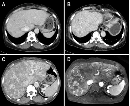
Fig. 1. (A) Three years ago, CT showed multiple, variable sized, and ill-defined low density nodule scattering through entire liver. (B) Two years ago, CT showed varia- ble sized and ill-defined low den- sity nodule increased in size and number. (C) At this time, CT showed previously noted low den- sity nodules in both lobes of the liver increased in size and number and dilated IVC and hepatic veins, inhomogenous enhancement of hepa- tic parenchyma. (D) In T2 weight- ed MRI scan, numerous nodules replaced entire liver with progress- ing centripetal enhancement.
Fig. 2. (A) Initial liver specimen showed sinusoidal dilatation and fibrous septal containing ductular proliferation and abnormal vessel (H&E, ×200). (B) Following liver specimen showed marked sinusoidal dilatation with focal sinusoidal fibrin thrombi, portal fibrosis and focal ductular proliferation (H&E, ×100).
간울혈로 확진하여 간 종괴를 배제한 증례보고도 있다.4 그 러나 심부전에 의한 간울혈에서 CT, MRI 검사로 간 종괴가 의심되어 조직검사를 시행한 증례는 없었다. 이에 저자들은 유방암 병력 및 류마티스성 심장질환으로 인한 삼첨판 역류 가 있는 심부전 환자에서 영상학적으로 간 종괴가 의심되어 간 조직검사를 시행, 간울혈의 소견을 보인 증례를 문헌 고 찰과 함께 보고하고자 한다.
증 례
64세 여자 환자가 최근 손, 발이 붓고 황달이 심해지는 증 상으로 내원하였다. 환자는 30년 전 좌측 유방암으로 완전 절제술, 방사선치료 후 완치 판정을 받았고, 4년 전 진단된 협착성 심낭염 및 삼첨판 역류에 대해 심낭막 절제술 및 삼 첨판 교체술을 시행받았다. 심초음파 검사에서, 삼첨판 역
266 The Korean Journal of Gastroenterology: Vol. 56, No. 4, 2010
류 grade 1-2, 심구혈률은 71%의 소견을 보였다. 이후 경과 관찰 중 복부탈장으로 시행한 CT에서 다발성 간 결절 소견 을 보였다(Fig. 1A). 1년 후, CT (Fig. 1B)에서 결절의 수와 크기가 증가하여 시행한 간 조직 검사 결과, 동양혈관 (sinusoid)의 확장 외의 특이 소견은 없었다(Fig. 2A). 그 후 최근 CT에서 결절의 수와 크기는 더 증가하여(Fig. 1C), MRI 촬영을 고려하였다.
내원 당시 생체 징후는 안정적이었으며 신체검사에서 상 지 및 하지의 함요 부종이 있었고 결막은 창백하였으며 공막 에 황달 소견을 보였다. 흉부 청진에서 호흡음은 정상이었으 며 심음은 불규칙하였고 grade 1의 전수축기 심잡음이 들렸 다. 검사실 소견으로 일반혈액검사에서 백혈구 4,100/mm3, 혈색소 8.9 g/dL, 혈소판 107,000/mm3, prothrombin time 71%
였다. 생화학검사에서 알부민 4.1 g/dL, 총빌리루빈 3.6 mg/dL, 직접 빌리루빈 2.0 mg/dL, γ-GT 206 U/L, AST/ALT 36/13 IU/L, 암모니아 142 ug/dL, 전해질 검사는 이상 소견이 없었으며 BUN 25.7 mg/dL, creatinine 1.67 mg/dL였다. 심초 음파 검사에서 삼천판 역류 grade 4, 조직 판막의 퇴행성 변 화, 심구혈률은 72%, 우심실의 움직임은 심각하게 떨어진 소견을 보였다. 입원 중 시행한 MRI에서 간 결절의 크기 및 수가 증가되고 T2 강조영상에서 고 신호강도를 보였다(Fig.
1D). 전이성 악성 종양 또는 혈관육종의 가능성을 배제할 수 없어, 조직 검사를 시행한 결과 fibrin thrombi, portal fi- brosis, focal ductular proliferation을 동반한 동양혈관(sinu- soid)의 확장소견을 보였다(Fig. 2B).
고 찰
이번 증례에서 처음 시행한 CT에서는 크기가 작은 저음 영의 다수의 결절이 관찰되었는데 이는 호산구성 간농양과 감별을 요한다. 호산구성 간농양의 경우 CT에서 문맥기에 가장 뚜렷해지며 동맥기에는 보이지 않는 경우가 많다. 특 징적인 소견으로는 경계가 불분명하고 미약한 저음영을 보 이며 모양이 둥글지 않다는 점이고 간혹 주위에 테두리 모 양의 조영 증강을 보일 수 있다.5 이번 환자는 30년 전 조기 유방암으로 절제술 후 완치판정을 받았다. 그러나, 조기 유 방암의 경우 재발률이 낮으나 10년에서 20년, 심지어 그 이 상의 기간까지 재발의 위험성이 존재하므로 만성 질환으로 인식되기도 한다.6 유방암 환자에서 가장 흔히 전이되는 장 기는 간, 폐, 뼈, 뇌 등이 있으며 그 중 40-50% 정도는 간으 로 전이된다.7,8 이번 증례에서 경과관찰 후 시행한 CT에서, 결절의 수와 크기가 증가하여 유방암에 의한 간전이 가능성 을 배제하기 위해 간 조직검사를 시행, 동양혈관(sinusoid)의 확장 외에는 특이 소견이 없었다.
이후, 경과 관찰 중 최근 CT에서, 간 실질에서 불균일한
조영증강(inhomogeneous enhancement)소견이 관찰되었다. 이 는 Budd-chiari syndrome 및 울혈성 심부전으로 인한 간질환 에서 관찰될 수 있으나 본 환자에서 유방암의 병력을 고려 할 때, 반드시 간암 혹은 간전이를 감별하는 것이 필요하 다.3 전이암은 CT에서 고밀도, 등밀도, 저밀도, 테두리 조영 증강을 가진 저밀도, 낭성, 혼합성, 석회화성이나 미만성 침 윤형태로 다양하게 나타난다. 대부분의 전이암은 물과 정상 간 사이의 저밀도 음영을 가진다. 전이암 병변은 저혈관성 이고 조영 증강 후 정상 간의 음영이 증가하기 때문에 종괴 의 경계가 명확해지며 문맥기 영상에서 잘 볼 수 있다.9 MRI에서는 간전이암의 T2 이완시간이 정상간보다 길기 때 문에 T2 강조영상에서 고 신호강도로 보이고 T1 강조영상 에서는 간전이암은 저신호강도를 보인다.10 이번 증례의 MRI 소견은 전이암 외에 혈관육종과도 감별이 필요한데 이유는 T2 강조영상에서 고 신호강도를 보였으나 불균일한 고 신 호강도를 보였기 때문이다. 혈관육종의 영상 소견은 비특이 적일 뿐만 아니라 흔히 혈관종과의 감별이 어려운 것으로 알려져 있다. 혈관육종은 대부분이 다발성이며 증상을 동반 하는 경우가 많다. 다발성인 경우에 주 종괴가 있는 경우와 없는 경우로 구분할 수 있고, 그 외에도 단발성인 경우와 미 만성인 경우가 있다.11-14 혈관육종의 경우 혈관종에 비해 조 영 증강의 정도가 주위 혈관보다 덜 강하고 모양이 불규칙 적이며 조영 증강이 병변의 주변부에서만 시작되지 않는 다.11 예를 들면 병변의 중간부에서 조영 증강이 시작될 수 도 있고, 병변의 전체에 균일하게 조영 증강되거나, 고리모 양으로 병변의 주변부에서 조영 증강이 시작될 수도 있다.
또한, 조영 증강 부위가 불규칙한 모양을 보이거나 다양한 모양을 보일 수도 있다. 이는 한 종괴 내에도 다양한 형태의 혈관통로를 가질 수 있기 때문으로 설명된다.11,13
심부전에 의한 간울혈의 경우 중심 정맥압이 상승하면 간 정맥으로 압력이 전해지며 간 내 동모양 공간이 충혈되어 부어 오르게 된다.15 이러한 변화는 대체로 가역적이지만 간 울혈이 지속되면 결국 간혈류가 감소하고 중심소엽성 간세 포 괴사와 섬유조직띠가 형성되어 간경변으로 진행할 수 있 다. 초기의 영상 소견은 간비대와 간정맥 확장이며 Budd-Chiari 증후군의 초기와 마찬가지의 불균일한 조영 증 강 소견을 보인다. 간의 울혈로 인해, CT에서 불균일한 조 영 증강이 보이지만 미상엽과 간문부를 포함한 중앙부와 좌 엽의 일부는 정상적인 조영 증강 패턴을 보여서 상대적으로 낮은 조영 증강을 보이는 외측과 구분된다. 이는 중앙부의 간실질이 별도의 간정맥을 통해 정맥혈을 정상적으로 배출 하는데 비해 외측은 혈류가 정체되어 있기 때문이며, 조영 후기에는 서서히 조영제가 축적되어 조영제가 빠져나간 중 앙부에 비해 오히려 강한 조영 증강을 보일 수도 있다. 또한 확장된 간정맥과 하대정맥 소견이 관찰된다.16 이번 증례는
Shin DH, et al. A Case of Hepatic Congestion Due to Right Heart Failure Mimicking Liver Tumor 267
CT에서 울혈성 심부전을 예측할 수 있는 확장된 간정맥과 하대정맥 소견이 관찰되었다.
이번 증례는 CT 소견에서 간 실질에 불균일한 조영증강 을 보였고 간울혈 혹은 Budd-Chiari 증후군의 초기에 나타나 는 미상엽과 간문부를 포함한 중앙부의 정상적인 조영 증강 패턴은 있었으나, 마치 주 종괴가 있는 다발성 종괴 및 다양 한 강도와 양상의 조영 증강 소견이 보였다. 또한, 자기공명 영상의 T1 강조영상에서 저 신호강도, T2 강조영상에서 불 균일한 고 신호강도의 경계가 불명확한 간결절 소견 등을 고려할 때 간울혈보다는 간전이, 혈관육종 등의 감별진단을 필요로 했다. 그러나 조직검사 결과 3년 전에 비해 심해진 동양혈관(sinusoid)의 확장, focal sinusoidal fibrin thrombi, portal fibrosis, focal ductular proliferation 소견을 보였다.
따라서 유방암을 진단받았던 심부전을 동반한 환자의 경 우, CT 및 MRI에서 다양한 크기의 경계가 불명확한 결절 소견을 보일 때 임상의는 간 종괴 및 전이암의 가능성을 우 선 생각해야 하지만 간 울혈도 배제하려는 노력을 해야 한 다.
참고문헌
1. Kiernan F. Philosophical transactions of the Royal Society of London 1833;123:711-770.
2. Mallory FB. Chronic passive congestion of the liver. J Med Res 1911;24:455-462.
3. Holley HC, Koslin DB, Berland LL, Stanley RJ. Inhomogen- eous enhancement of liver parenchyma secondary to passive congestion: contrast-enhanced CT. Radiology 1989;170:795- 800.
4. Nyman R, Kagevi I. Budd-Chiari syndrome misinterpreted as an infiltrating liver mass. A case report. Acta Radiol 1995;36:393-395.
5. Jang HJ, Lee WJ, Lee SJ, Kim SH, Lim HK, Lim JH. Focal eosinophilic necrosis of the liver in patients with underlying gastric or colorectal cancer: CT differentiation from meta- stasis. Korean J Radiol 2002;3:240-244.
6. Rosen PR, Groshen S, Saigo PE, Kinne DW, Hellman S. A
long-term follow-up study of survival in stage I (T1N0M0) and stage II (T1N1M0) breast carcinoma. J Clin Oncol 1989;7:355-366.
7. Marty M, Cognetti F, Maraninchi D, et al. Randomized phase II trial of the efficacy and safety of trastuzumab combined with docetaxel in patients with human epidermal growth fac- tor receptor 2-positive metastatic breast cancer administered as first-line treatment: the M77001 study group. J Clin Oncol 2005;23:4265-4274.
8. Chan S, Friedrichs K, Noel D, et al. Prospective randomized trial of docetaxel versus doxorubicin in patients with meta- static breast cancer. J Clin Oncol 1999;17:2341-2354.
9. Baron RL, Oliver JH 3rd, Dodd GD 3rd, Nalesnik M, Holbert BL, Carr B. Hepatocellular carcinoma: evaluation with biphasic, contrast-enhanced, helical CT. Radiology 1996;199:505-511.
10. Semelka R, Ascher S, Reinhold C. MRI of the abdomen and pelvis: a text-atlas. New York: Wiley-Liss, 1997.
11. Koyama T, Fletcher JG, Johnson CD, Kuo MS, Notohara K, Burgart LJ. Primary hepatic angiosarcoma: findings at CT and MR imaging. Radiology 2002;222:667-673.
12. Rademaker J, Widjaja A, Galanski M. Hepatic hemangio- sarcoma: imaging findings and differential diagnosis. Eur Radiol 2000;10:129-133.
13. Peterson MS, Baron RL, Rankin SC. Hepatic angiosarcoma:
findings on multiphasic contrast-enhanced helical CT do not mimic hepatic hemangioma. AJR Am J Roentgenol 2000;
175:165-170.
14. White PG, Adams H, Smith PM. The computed tomographic appearances of angiosarcoma of the liver. Clin Radiol 1993;
48:321-325.
15. Dunn GD, Hayes P, Breen KJ, Schenker S. The liver in con- gestive heart failure: a review. Am J Med Sci 1973;265:174- 189.
16. Van Beers B, Pringot J, Trigaux JP, Dautrebande J, Mathurin P. Hepatic heterogeneity on CT in Budd-Chiari syndrome:
correlation with regional disturbances in portal flow. Gastro- intest Radiol 1988;13:61-66.