ABSTRACT
The objective of this article is to detail the treatment for papillary thyroid microcarcinoma (PTMC). The literature presents only few contributions, with controversial results, about comparison between ‘active surveillance’ and surgery. Hemithyroidectomy is the treatment of choice for PTMC. Thyroidectomy is indicated in cases of multifocality, extrathyroid tumor growth, and familial PTMCs. Active surveillance can only be done under well-defined and controlled conditions. Collected findings and agreements with the patient must be precisely documented, also for medico-legal reasons. An observation of PTMC seems most appropriate for patients >60 years of age. In the case of observation of a PTMC, a lifelong examination of the tumor disease must be carried out, since tumor growth or metastases can still occur after 10–15 years. The follow-up periods for the ‘active surveillance’ proposed from the literature review are too short to conclude this as a real alternative.
Keywords: Papillary thyroid microcarcinoma; Surgery; Thyroid nodule
PREFACE
An increasing incidence of thyroid cancer has been observed worldwide, mainly due to an increase in papillary subtype. Papillary thyroid cancer accounts for approximately 85% of thyroid tumors in the United States and other iodine-sufficient countries (1-5).
Over the past 2 decades, the overall incidence rate has been increasing by >6% per year (1-4).
More than any other tumor entity, there was an increase in the incidence from 4.5 cases in 1982 for thyroid tumors, to 16.3 cases per 100,000 USA inhabitants in 2013 (1). In the period from 2007 to 2013, the relapse-free 5-year survival was 93.1%, the 5-year overall survival 98.1% (1).
Similar patterns of increase have been reported in Canada, Australia, and Europe (1-3).
Review Article
Received: Feb 18, 2018 Revised: Apr 2, 2018 Accepted: Apr 14, 2018 Correspondence to Gianlorenzo Dionigi
Division for Endocrine and Minimally Invasive Surgery, Department of Human Pathology in Adulthood and Childhood “G. Barresi”, University Hospital G. Martino, University of Messina, Via C. Valeria 1, Messina 98125, Italy.
E-mail: gdionigi@unime.it
Copyright © 2018. Korean Association of Thyroid and Endocrine Surgeons; KATES This is an Open Access article distributed under the terms of the Creative Commons Attribution Non-Commercial License (https://
creativecommons.org/licenses/by-nc/4.0/).
ORCID iDs Gianlorenzo Dionigi
https://orcid.org/0000-0003-0864-6087 Author Contributions
Conceptualization: Gianlorenzo Dionigi; Data curation: Giuseppe Navarra, Guido Nicola Zanghì, Francesco Freni, Bruno Galletti, Francesco Galletti, Grazia Pagano, Andrea Cogliandolo, Alberto Barbera, Salvatore Lazzara, Gianlorenzo Dionigi; Formal analysis:
Giuseppe Navarra, Guido Nicola Zanghì, Francesco Freni, Bruno Galletti, Francesco Galletti, Grazia Pagano, Andrea Cogliandolo, Alberto Barbera, Salvatore Lazzara, Gianlorenzo Dionigi; Funding acquisition:
Giuseppe Navarra, Guido Nicola Zanghì,
Giuseppe Navarra1, Guido Nicola Zanghì2, Francesco Freni3, Bruno Galletti3, Francesco Galletti3, Grazia Pagano1, Andrea Cogliandolo1, Alberto Barbera1, Salvatore Lazzara1, Gianlorenzo Dionigi 4
1 Surgical Oncology Division, Department of Human Pathology in Adulthood and Childhood “G. Barresi”, University Hospital G. Martino, University of Messina, Messina, Italy
2 Department of Surgery, Policlinico Vittorio Emanuele University Hospital - General Surgery and Oncology Unit, University of Catania, Catania, Italy
3 Division of ENT Surgery, Department of Human Pathology in Adulthood and Childhood “G. Barresi”, University Hospital G. Martino, University of Messina, Messina, Italy
4 Division of Endocrine and Minimally Invasive Surgery, Department of Human Pathology in Adulthood and Childhood “G. Barresi”, University Hospital G. Martino, University of Messina, Messina, Italy
Treatment Decision Making in
Papillary Thyroid Microcarcinoma
Francesco Freni, Bruno Galletti, Francesco Galletti, Grazia Pagano, Andrea Cogliandolo, Alberto Barbera, Salvatore Lazzara, Gianlorenzo Dionigi; Investigation: Giuseppe Navarra, Guido Nicola Zanghì, Francesco Freni, Bruno Galletti, Francesco Galletti, Grazia Pagano, Andrea Cogliandolo, Alberto Barbera, Salvatore Lazzara, Gianlorenzo Dionigi; Writing - original draft: Gianlorenzo Dionigi.
Conflict of Interest
No potential conflict of interest relevant to this article was reported.
In Germany, the incidence of thyroid carcinoma in 2012 was 4.6 in men and 10.7 in women per 100,000. The median age of onset was 56 years for men and 51 years for women (2).
From Austria, the working group of Gschwandtner et al. (3) reported a percentage increase in papillary thyroid microcarcinoma (PTMC, <10 mm) in their patient population from 3% in 1990 to 11.8% in 2008.
In Korea, thyroid carcinomas, with approximately 40,000 new cases per 51 million inhabitants (78 cases/100,000 inhabitants), represent now the most frequently diagnosed cancer (4).
By 2019, thyroid cancer is projected to become the third-most common cancer in women, with an annual age-adjusted incidence of 37 per 100,000 (1-5).
This trend is present on every continent except Africa, where detection is possibly insufficient.
The association between access to care and PTMC incidence cannot rule out a coexistent true increase in the occurrence of thyroid cancer.
In view of the increasing numbers of cases, especially of PTMC, the working group of Ito and colleagues (5) published in 2003 the results of an observational study of patients with non-surgically treated PTMC from Japan (Table 1). Between 1993 and 2001, a PTMC was confirmed in 732 patients by sonography and fine needle aspiration (FNA) cytology (5). These patients were given an ‘active surveillance,’ i.e., a well-defined follow-up instead of surgery offered. The 162 patients (22.1%) agreed with this approach. During the observation phase, a tumor progression was detected in 30% in the form of primary tumor growth or cervico- lateral lymph node (LN) metastasis. Sixty-five patients from the observation group and 570 patients who wished primarily for surgery were operated on (44% thyroidectomies, 56%
partial thyroid resections). A central LN dissection was performed in 94.9% of the operated, a lateral lymphadenectomy in 50.7%. In 50.5% of the patients histological LN metastases and in 42.8% multifocal carcinomas were detected. In 5 patients there was an infiltration of the trachea, which was partially resected. The average tumor diameter was 0.7 cm. After 8 years, 5.0% of patients experienced tumor recurrence (Table 1).
A recent Korean study describes a tumor progression in 14% of 192 patients with a median tumor diameter of 5.5 mm within an observation period of 30 months (6). In detail, the 192 patients diagnosed with PTMC under active surveillance for >1 year were included in a median 30-month follow-up. Changes in tumor size were evaluated not only using the maximal tumor diameter but also the tumor volume. The median age of patients was 51.3 Table 1. Literature review for active surveillance for papillary microcarcinomas of the thyroid gland
Study Year No. of
patients Follow-up
(mon) PTMC
unchanged (%) Tumor progress
≥3 mm (%) Surgery
(%) LN metastases
(%) Local
recurrence (%) Survival (%)
Ito et al. (5) 2003 162 48.7 70 11.1 34.6 12.3 - 100
Ito et al. (11) 2014 1,235 75 - 8 16 38 1.1 100
Kwon et al. (6) 2017 192 30.1 69 14 13 29 - 100
Tuttle et al. (7) 2017 291 25 87.6 3.8 3.4† 0 0 100
Kim et al. (30) 2018 127 26 - 19.8 - - - 100
Miyauchi et al. (31) 2018 1,211 120 - 3.5–60* - - - -
PTMC = papillary thyroid microcarcinoma; LN = lymph node.
*The age decade-specific disease progression rates at 10 years of active surveillance were 36.9% (20s), 13.5% (30s), 14.5% (40s), 5.6% (50s), 6.6% (60s), and 3.5% (70s); the respective lifetime disease progression probabilities were 60.3%, 37.1%, 27.3%, 14.9%, 9.9% and 3.5% according to the age at presentation;
†Operation due to tumor progression or patient request.
years and 145 patients (76%) were female (6). The median initial maximal tumor diameter and tumor volume were 5.5 mm and 48.8 mm3, respectively. The Authors also found that the change in tumor volume was more sensitive to detect tumor progression than the change in the maximal tumor diameter: the tumor size increased in 27 patients (14%); 23 patients showed a tumor volume increase >50% without a maximal diameter increase of ≥3 mm (6).
The other 4 patients had both an increasing tumor volume and increasing maximal tumor diameter ≥3 mm. Some PTMCs could grow significantly after a relatively short period of active surveillance: one patient (0.5%) had newly appeared cervical LN metastasis at 3 years after the initial diagnosis. There were no significant risk factors associated with increased tumor size, such as age, sex, or Hashimoto thyroiditis. Twenty-four patients (13%) underwent delayed thyroid surgery at a median of 31.2 months and seven (29%) had cervical LN metastasis on pathologic examination.
The first American study on active surveillance was published in 2017 by Tuttle et al. (7) published. Cohort study of 291 patients undergoing active surveillance for low-risk PTMC with serial tumor measurements via ultrasonography (US) at a tertiary referral center in the United States. The cumulative incidence, rate, and magnitude of the change in tumor diameter or volume, as well as associations with patient and tumor characteristics were measured. Of the 291 patients, 219 (75.3%) were women; mean (±standard deviation) age was 52 (±15) years.
During a median (range) active surveillance of 25 (6–166) months, growth in tumor diameter of 3 mm or more was observed in 11 of 291 (3.8%) patients, with a cumulative incidence of 2.5% (2 years) and 12.1% (5 years). No regional or distant metastases developed during active surveillance. In all cases, 3-dimensional measurements of tumor volume allowed for earlier identification of growth (median, 8.2 months; range, 3–46 months before increase in tumor diameter). In multivariable analysis, both younger age at diagnosis (hazard ratio [HR] per year, 0.92; 95% confidence interval [CI], 0.87–0.98; P=0.006) and risk category at presentation (HR for inappropriate, 55.17; 95% CI, 9.4–323.19; P<0.001) were independently associated with the likelihood of tumor growth. Of the tumors experiencing volume growth, kinetics demonstrated a classic exponential growth pattern, with a median doubling time of 2.2 years (range, 0.5–4.8 years; median r2=0.75; range, 0.42–0.99). The authors concluded that the rates of tumor growth during active surveillance in a USA cohort with papillary thyroid carcinomas (PTCs) measuring 1.5 cm or less were low. Serial measurement of tumor volumes may facilitate early identification of tumors that will continue to grow and thereby inform the timing of surveillance imaging and therapeutic interventions (7).
AVERAGE AGE AT FIRST DIAGNOSIS
We do not know the natural history of PTMC. Some medical definitions remain the same for many years, others change due to the progress in the diagnostic tools, which are able to distinguish markers and symptoms until then undetectable. Occult thyroid carcinoma is a general term indicating clinically different situations, whereas the incidentally detected PTMC is the most important from the clinical point of view. It is fundamental, for therapeutic management, to determine biological parameters which would define a small group of PTMC with aggressive biological behaviour.
PTMC is a prominent malignancy originating from follicular cells. This disease generally shows an indolent character, but patients demonstrating certain clinicopathological features have a dire prognosis.
Initial indications of a possibly “indolent character” of PTMC were found in autopsy studies (8).
Occult PTC is a much more common pathology than clinically evident thyroid cancer.
Autopsy studies have shown that occult PTC present in 1%–36% of patients (8,9).
Lang and colleagues (8) published in 1988 an investigation of 1.020 thyroid glands of people who had died due to non-thyroid-specific diseases. At 6.6% PTMC with a diameter between 0.4 and 10.5 mm were detected as incidental findings. Sixty-six out of 67 cases were papillary carcinomas and one was medullary carcinoma. The average age was 60.0 for men and 62.0 for women (8). The 8.6% PTMC of the thyroid gland was detected in an Austrian study with an average tumor diameter of 4.9 mm (9). The average age of these 118 patients was 67.7 years (9).
Occult/autopsy PTC appears to have no sex predilection as opposed to the clinically evident PTC, which develops more commonly in females.
Furthermore, the current study confirms that PTMC is common, but the observed increasing incidence is not mirrored by prevalence within autopsy studies and, therefore, is unlikely to reflect a true population-level increase in tumorigenesis. This strongly suggests that the current increasing incidence of PTMC most likely reflects diagnostic detection increasing over time.
The average age at first diagnosis of a PTMC is usually between the 45th and 50th year of life, not infrequently, however, these tumors are already detected from the age of 20 years (10).
PTMCs of the young patients were significantly more progressive than those of middle-aged or old patients.
In children and adolescents, but also in young adults, in the majority of cases a pronounced lymphogenous and sometimes haematogenous metastasis is found already at the time of diagnosis (10).
PTMC shows increased growth potential in younger patients (11). An increased growth potential of PTMC in younger patients was shown by another observational study by Ito et al. (11) with 1,235 patients. A size progression was reported among under-40s with a PTMC under observation, over 10 years, in 12.1% of patients vs. 4.0% in over-60s (11). In 16.1% of patients <40 years, LN metastases occurred in contrast to only 0.5% of patients ≥60 years.
In a multivariate analysis, age <40 years and tumor size ≥9 mm correlated significantly with tumor progression (11). The average follow-up period of this study was 75 months (1 to 246 months). Only 27 of 1,235 patients had their first long-term results after 15 years and showed tumor growth (>3 mm) in 50% of under-40 patients, in about 35% tumor size 12 mm and in 30% lymphoid metastasis (11). However, due to the low number of cases, these results must be evaluated with caution and the long-term results must be awaited.
The apparent slower progression of tumors in older patients is consistent with the autopsy studies, which looked primarily at people >60 years of age (12,13).
Finally, a study evaluated the influence of age on the phenotypical expression of PTMC have hitherto compared PTMC presentation either between pre-pubertal and pubertal
children or between pediatric patients and aged adults (14). The authors ascertain whether presentation of PTMC may significantly vary according to age, even within a peculiar study population covering only young patients aged less than 30 years. The main clinical, biochemical and pathologic data at PTMC diagnosis were retrospectively recorded in 2 selected cohorts including, respectively, 18 children and adolescents aged less than 18 years (group A) or 45 young adults aged between 20 and 29.8 years (group B) (14). The statistical distribution of differentiated thyroid carcinoma (DTC) cases in the different age ranges was found to progressively increase with increasing age; furthermore, the patients of group A exhibited at diagnosis a more severe clinical involvement and a higher rate of extra-regional metastases; finally, also the association with both autoimmune thyroid diseases (AITDs) and a biochemical hypothyroid pattern was more common in group A patients. In a study population younger than 30 years: 1) the risk of developing DTC increases with age, achieving its zenith during the 3rd decade of life; 2) clinical presentation is more severe in children and adolescents younger than 18 years than in the patients aged between 20 and 30; 3) in the cohort of children and adolescents DTC is more often associated with AITDs, which might play some role in conditioning the more aggressive phenotypical presentation of DTC in this patient group (14).
DIAGNOSIS
For the detection of a PTMC a meticulous sonographic examination of the thyroid gland is of crucial importance. In addition to the size of the nodule, this should also contain information about inhomogeneities of the parenchyma, an edge blurring, hypervascularization and microcalcifications.
A study investigated time-dependent changes in calcification patterns and tumor vascularity on US to clarify the natural course of PTMC. Authors examined calcification patterns and tumor vascularity for 480 lesions in 384 patients. Calcification patterns were classified as: none (A); micro (B); macro (C); or rim (D). Tumor vascularity was classified as rich or poor via color Doppler US. After a mean of 6.8 years of observation, 29 lesions (6.0%) had increased in size. Mean age for initial calcification pattern was 52.1 years for A (n=135), 54.2 years for B (n=235), 56.3 years for C (n=96), and 60.1 years for D (n=14), and the incidence rates of tumor enlargement were 9.6%, 5.5%, 3.2%, and 0%, respectively. The cumulative rate of upgrade in calcification pattern was 51.8% at 10 years. Lesions with initially rich vascularity (n=70) had significantly higher rate of tumor enlargement than those with poor vascularity (n=410); however, the majority of tumor (61.4%) with initially rich vascularity had decreased their blood supply during the follow-up. Multivariate analysis showed that strong calcification (C or D) and poor vascularity at last examination correlated significantly with non-progressive disease (15).
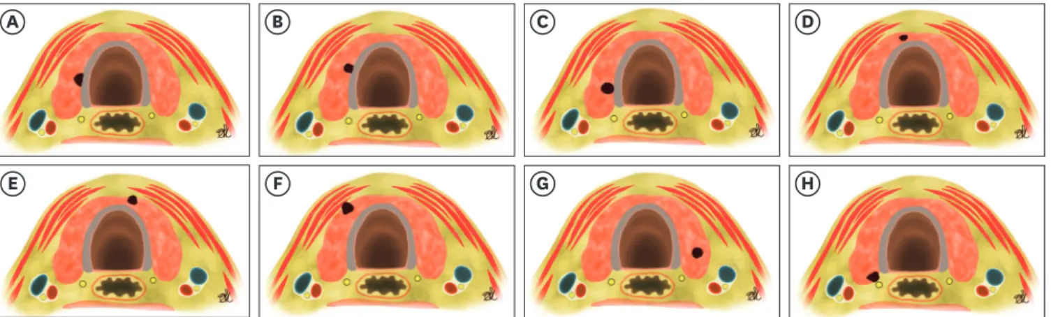
In addition to these already established criteria, Miyauchi (15) additionally nicely
recommends an exact description of the carcinoma location in relation to capsule and the trachea (Fig. 1).
Preoperative LN mapping helps in planning surgery for neck dissection and improves patient outcomes. The cervico-central and lateral LNs must be assessed and documented. In practice, an ultrasound assessment of the cervico-central LNs is limited due to their location dorsally and caudally to the thyroid gland. The cervico-lateral LNs, on the other hand, are
very easy to visualize. Nevertheless, Ito et al. (5) found that only 39% of the histologically diagnosed cervico-lateral LN metastases could be detected preoperatively sonographically in their patient collective.
A FNA of sonographically suspected thyroid nodules is usually recommended from a diameter of 10 mm (16). The Japanese and Korean papers on PTMC observation (10,11,17) have described median tumor diameters between 5.5 and 8 mm, whose puncture requires special expertise, and large multinodal struma, especially in endemic areas Thyroid volume or adiposity per magna is likely to be difficult (5,6,11).
The cytological examination of a FNA requires sufficient experience of the respective pathologist and yet cannot reliably detect subgroups such as papillary carcinoma of the follicular type. The molecular genetic detection of a BRAF (V600E) mutation can demonstrably improve the cytological diagnosis of PTMC (18). BRAF is a human gene that encodes a protein called B-Raf. The gene is also referred to as proto-oncogene B-Raf and v-Raf murine sarcoma viral oncogene homolog B, while the protein is more formally known as serine/threonine-protein kinase B-Raf (18). The B-Raf protein is involved in sending signals inside cells which are involved in directing cell growth. In 2002, it was shown to be faulty (mutated) in some human cancers (18,19). At present, researchers are working on determining the role of BRAF mutation in patients from a low-risk group and its correlations with others molecular events. Currently, BRAF mutation cannot be used as a single,
independent predictive factor. However, its usefulness in the context of other molecular and clinico-pathological risk factors cannot be excluded. They may be used to make modern prognostic scales of relapse risk and be applied to individualized diagnostic and therapeutic strategy for PTMC patients. In a meta-analysis, BRAF (V600E)-positive tumors in the definitive histopathological evaluation were found to be frequently affected by multifocal carcinomas, extrathyroid tumor growth, and LN metastases (19).
Cytologic lymphangiosis, angioinvasion or infiltration of the thyroid capsule cannot be assessed cytologically. This is only guaranteed with the greatest possible safety in the paraffin histology of the surgical specimen. Kwon et al. (6) reported extrathyroid and thus advanced
A B C D
E F G H
Fig. 1. Schematic representation of the anatomical relationship between a PTMC of the thyroid gland, the tracheal wall, the RLN and strap muscles to plan the further course of therapy (active surveillance vs. surgery). Risk of ‘tracheal’ infiltration by a PTMC: (A) angle at high risk; (B) almost right angle - middle risk;
(C) round shape tumor angle - low risk. Risk of infiltration of ‘soft tissue and muscles’ by PTMC: (D) small PTMC contained in the isthmus; (E) isthmus PTMC extending anteriorly to connective tissue; (F) lobe PTMC extending laterally to connective tissue. Risk of infiltration of ‘RLN’ by PTMC: (G) intrathyroidal - low risk;
(H) posterior PTMC - high risk (modified from reference 15).
PTMC = papillary thyroid microcarcinoma; RLN = recurrent laryngeal nerve.
tumor growth in 34% of the PTMC operated on the basis of size progression or on request of the patients, initially only observed.
OPERATIVE THERAPY
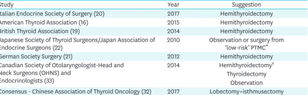
The purpose of the surgical society guidelines is to provide recommendations to assist primary care and other clinicians in the care of thyroid cancer. The guideline are developed through a collaboration between the societies and published jointly by invitation and consent in both surgeons, endocrinologist, pathologist, otorhinolaryngology (ENT), oncologist. The guidelines of the American Thyroid Association, Canadien, Chinese, British Thyroid Association, the Italian and German Society of Surgery suggested the use of hemithyroidectomy for the operative therapy of the PTMC (Table 2).
In familial PTC, which is detected in 5% of patients with PTMC, there is an indication for thyroidectomy, as these carcinomas become more multifocal and metastasise early (17,20-22).
With history of previous radiation of the neck, thyroidectomy is recommended (17,20-22).
In the Japanese Guidelines of 2010, an observation or alternatively an operation is
recommended for ‘low-risk’ PTMC (asymptomatic, without LN and distant metastases) (23).
When detecting multifocal carcinomas, a thyroidectomy is recommended (23).
Kim et al. (23) investigated in 8,676 patients (5,387 thyroidectomies, 3,289
hemithyroidectomies) with a PTMC. The average follow-up period was 65 months. The recurrence-free 5- and 10-year survival rates were 98.1% and 91.8%, respectively, for the hemithyroidectomy group and 98.5% and 97.5% for the thyroidectomy group (P<0.001) (24).
The majority of the diagnosed ‘tumor recurrences’ involved the contralateral thyroid lobes left in hemithyroidectomy. In multifocal carcinomas, thyroidectomy significantly reduced the risk of actual local recurrence. The authors therefore recommend a thyroidectomy only for multifocal carcinomas (24).
Gschwandtner et al. (3) published in 2016 comparable results with 1.2% PTMC ‘recurrences’
in the contralateral thyroid lobe after hemithyroidectomy, which were incidental findings in the operation of recurrent goiters. Tumor recurrences in the form of cervico-central LN metastases were detected at 0.4% and reoperated after thyroidectomy. Significant risk of LN
Table 2. Recommendations of various guidelines for the treatment of papillary microcarcinomas of the thyroid gland
Study Year Suggestion
Italian Endocrine Society of Surgery (20) 2017 Hemithyroidectomy
American Thyroid Association (16) 2015 Hemithyroidectomy
British Thyroid Association (19) 2014 Hemithyroidectomy
Japanese Society of Thyroid Surgeons/Japan Association of
Endocrine Surgeons (22) 2010 Observation or surgery from
‘low-risk’ PTMC*
German Society Surgery (21) 2012 Hemithyroidectomy
Canadian Society of Otolaryngologist-Head and Neck Surgeons (OHNS) and
Endocrinologists (33)
2014 Hemithyroidectomy†
Thyroidectomy Observation Consensus - Chinese Association of Thyroid Oncology (32) 2017 Lobectomy+isthmusectomy PTMC = papillary thyroid microcarcinoma.
*Low-risk PTMC: carcinomas without distant or lymph node metastases and without symptoms;
†Hemithyroidectomy (47%) or total thyroidectomy (43%) for a newly diagnosed PTMC in a low risk patient.
Observation was the preferred method for managing PTMC detected incidentally after hemithyroidectomy (76%).
recurrence was seen in young patients, in capsular infiltration of the tumor and detection of LN metastasis during primary intervention. Multifocal PTMC did not correlate with the occurrence of local recurrence in this work.
The indication for systematic cervico-central and -lateral LN dissection in the PTMC exists only in the case of pre- or intraoperative suspicion or proof of LN metastasis and not prophylactically (17,20-25).
The frequency of cervico-lateral LN involvement in PTMC was investigated in a case-control study in 196 patients with PTMC and lateral LN metastases (pN1b) compared to 199 patients without clinical evidence of LN metastases (cN0) (26). Also in the comparison group (cN0) a thyroidectomy and postoperative radioiodine therapy were obligatory. In 26 out of 199 (13.1%) cases primarily classified as cN0, a recurrent LN recurrence occurred (26). Significant risk factors for LN metastasis included age <50 years, male gender, tumor localization in the cranial portions of the thyroid, and preferably sub-capsular, and sonographically detectable microcalcifications (26).
In 2,329 patients with PTMC and routinely performed thyroidectomy and cervico-central lymphadenectomy, Oh et al. (26) had a prevalence of more than 5 affected LNs in 24% of male patients under 40 compared to only 2% of women ≥40 years.
ARGUMENTATION
The prognosis of PTMC is exceptionally favorable compared to other tumor entities.
A Literature review found 0.9%–5.7% local recurrences in studies with more than 250 patients published between 1999 and 2012 (26-28). In recent publications, 0.4%–1.7% local recurrences were treated surgically (26-28).
As risk factors for a more aggressive tumor growth and thus a higher probability for a size progression, LN metastases or a local recurrence are especially for young patients, male gender, multifocal carcinomas, a BRAF V600E mutation and LN metastasis (7,11,20,26-30).
Tumor-related deaths are reported in the literature only in 0%–0.3% (7,11,20,26-28,30).
Hay et al. (29) reported a survival rate of 99.7% after an average follow-up of 17.2 years. 85%
of these 900 PTMC patients had bilateral thyroid resection and 23% had cervico-central lymphadenectomy (31).
Thus, hemithyroidectomy, according to the guidelines of most surgical societies, is still the therapy of choice of PTMC and gives excellent results, with very low complication rates. A cure of the PTMC is only possible through surgery.
Close monitoring of PTMC as an alternative approach (‘active surveillance’) requires accurate diagnosis by FNA and should only be performed under well-defined and controlled conditions, provided that there are no clinical symptoms, such as: hoarseness, LN or distant metastases. About the 2 possibilities of active surveillance vs. surgery must be provided to the patient in detail, as this procedure is currently not established in Europe. At present, it is not clear which physicians should choose which discipline to carry out ‘active surveillance.’
In Japan, this task is performed by endocrine surgeons; in South Korea and the USA,
endocrinologists are primarily responsible. Obligate must be a high volume endocrine center.
An ‘active surveillance’ seems most suitable for patients >60 years. Of central importance for all therapeutic procedures is a good selection of the patient. While young patients <40 years are more likely to benefit from surgical therapy because of the increased risk of lymphoid metastasis, patients >60 years may also be eligible for active surveillance. In the case of observation, the patient must be fully informed and prepared to deal with, and monitor the tumor for life. This may be more of a psychological burden than an operation. The question of whether an observation or an operation affects the mental quality of life of patients more has not yet been scientifically studied. An example is from active surveillance for prostate cancer (32-34). Active surveillance is emerging as a serious alternative to radical therapy for low-risk prostate cancer (34). A review evaluated mental quality of life of patients undergoing active surveillance for prostate cancer (34). The following three are the main issues being covered in the literature on psychological aspects of active surveillance. First, the process of consultation with the physician and treatment choice in men diagnosed with low-risk prostate cancer. Second, the effect of active surveillance on physical domains and resulting anxiety and distress, and on quality of life in general. And third, the possible supportive and educational interventions for patients on active surveillance (34). At the moment of treatment choice, fear of disease progression is the main reason to reject active surveillance. Active surveillance may spare physical domains and does not cause much anxiety or distress on short term in men who choose this strategy. Once men opt for active surveillance, only a minority of them switch to radical treatment due to psychological reasons. Supportive and educational interventions should be considered (34).
Regardless of known surgical therapies or new concepts such as ‘active surveillance,’ it is still important to plan the best possible and successful treatment for each patient, taking into account their individual circumstances.
REFERENCES
1. Howlader N, Noone AM, Krapcho M, Miller D, Bishop K, Kosary CL, et al. SEER Cancer Statistics Review 1975–2014 [Internet]. Bethesda (MD): National Cancer Institute; 2017 Apr 20. Available from: https://seer.
cancer.gov/csr/1975_2014/.
2. Robert Koch-Institut. Krebs in Deutschland 2011/2012 [Internet]. Berlin: Robert Koch-Institut; 2016 Dec 10. Available from: https://www.krebsdaten.de/Krebs/DE/Content/Publikationen/Krebs_in_Deutschland/
kid_2015/krebs_in_deutschland_2015.pdf ?__blob=publicationFile.
3. Gschwandtner E, Klatte T, Swietek N, Bures C, Kober F, Ott J, et al. Increase of papillary thyroid microcarcinoma and a plea for restrictive treatment: a retrospective study of 1,391 prospective documented patients. Surgery 2016;159:503-11.
PUBMED | CROSSREF
4. Ahn HS, Kim HJ, Welch HG. Korea's thyroid-cancer “epidemic”--screening and overdiagnosis. N Engl J Med 2014;371:1765-7.
PUBMED | CROSSREF
5. Ito Y, Uruno T, Nakano K, Takamura Y, Miya A, Kobayashi K, et al. An observation trial without surgical treatment in patients with papillary microcarcinoma of the thyroid. Thyroid 2003;13:381-7.
PUBMED | CROSSREF
6. Kwon H, Oh HS, Kim M, Park S, Jeon MJ, Kim WG, et al. Active surveillance for patients with papillary thyroid microcarcinoma: a single center's experience in Korea. J Clin Endocrinol Metab 2017;102:1917-25.
PUBMED | CROSSREF
7. Tuttle RM, Fagin JA, Minkowitz G, Wong RJ, Roman B, Patel S, et al. Natural history and tumor volume kinetics of papillary thyroid cancers during active surveillance. JAMA Otolaryngol Head Neck Surg 2017;143:1015-20.
PUBMED | CROSSREF
8. Lang W, Borrusch H, Bauer L. Occult carcinomas of the thyroid. Evaluation of 1,020 sequential autopsies.
Am J Clin Pathol 1988;90:72-6.
PUBMED | CROSSREF
9. Musholt TJ, Fottner C, Weber MM, Eichhorn W, Pohlenz J, Musholt PB, et al. Detection of papillary thyroid carcinoma by analysis of BRAF and RET/PTC1 mutations in fine-needle aspiration biopsies of thyroid nodules. World J Surg 2010;34:2595-603.
PUBMED | CROSSREF
10. Machens A, Elwerr M, Thanh PN, Lorenz K, Schneider R, Dralle H. Impact of central node dissection on postoperative morbidity in pediatric patients with suspected or proven thyroid cancer. Surgery 2016;160:484-92.
PUBMED | CROSSREF
11. Ito Y, Miyauchi A, Kihara M, Higashiyama T, Kobayashi K, Miya A. Patient age is significantly related to the progression of papillary microcarcinoma of the thyroid under observation. Thyroid 2014;24:27-34.
PUBMED | CROSSREF
12. Neuhold N, Kaiser H, Kaserer K. Latent carcinoma of the thyroid in Austria: a systematic autopsy study.
Endocr Pathol 2001;12:23-31.
PUBMED | CROSSREF
13. Miyauchi A, Ito Y, Oda H. Insights into the management of papillary microcarcinoma of the thyroid.
Thyroid 2018;28:23-31.
PUBMED | CROSSREF
14. Fukuoka O, Sugitani I, Ebina A, Toda K, Kawabata K, Yamada K. Natural history of asymptomatic papillary thyroid microcarcinoma: time-dependent changes in calcification and vascularity during active surveillance. World J Surg 2016;40:529-37.
PUBMED | CROSSREF
15. Miyauchi A. Clinical trials of active surveillance of papillary microcarcinoma of the thyroid. World J Surg 2016;40:516-22.
PUBMED | CROSSREF
16. Haugen BR, Alexander EK, Bible KC, Doherty GM, Mandel SJ, Nikiforov YE, et al. 2015 American Thyroid Association management guidelines for adult patients with thyroid nodules and differentiated thyroid cancer: the American Thyroid Association guidelines task force on thyroid nodules and differentiated thyroid cancer. Thyroid 2016;26:1-133.
PUBMED | CROSSREF
17. Mohamad Yusof A, Jamal R, Muhammad R, Abdullah Suhaimi SN, Mohamed Rose I, Saidin S, et al.
Integrated characterization of microRNA and mRNA transcriptome in papillary thyroid carcinoma. Front Endocrinol (Lausanne) 2018;9:158.
PUBMED | CROSSREF
18. Li F, Chen G, Sheng C, Gusdon AM, Huang Y, Lv Z, et al. BRAFV600E mutation in papillary thyroid microcarcinoma: a meta-analysis. Endocr Relat Cancer 2015;22:159-68.
PUBMED | CROSSREF
19. Perros P, Boelaert K, Colley S, Evans C, Evans RM, Gerrard Ba G, et al. Guidelines for the management of thyroid cancer. Clin Endocrinol (Oxf ) 2014;81 Suppl 1:1-122.
PUBMED | CROSSREF
20. Lamartina L, Durante C, Lucisano G, Grani G, Bellantone R, Lombardi CP, et al. Are evidence-based guidelines reflected in clinical practice? An analysis of prospectively collected data of the Italian Thyroid Cancer Observatory. Thyroid 2017;27:1490-7.
PUBMED | CROSSREF
21. Arbeitsgemeinschaft der Wissenschaftlichen Medizinischen Fachgesellschaften. Operative Therapie maligner Schilddrüsenerkrankungen [Internet]. Berlin: Arbeitsgemeinschaft der Wissenschaftlichen Medizinischen Fachgesellschaften; 2012 Mar 15. Available online from: www.awmf.org.
22. Takami H, Ito Y, Okamoto T, Onoda N, Noguchi H, Yoshida A. Revisiting the guidelines issued by the Japanese Society of Thyroid Surgeons and Japan Association of Endocrine Surgeons: a gradual move towards consensus between Japanese and western practice in the management of thyroid carcinoma.
World J Surg 2014;38:2002-10.
PUBMED | CROSSREF
23. Kim SK, Park I, Woo JW, Lee JH, Choe JH, Kim JH, et al. Total thyroidectomy versus lobectomy in conventional papillary thyroid microcarcinoma: analysis of 8,676 patients at a single institution. Surgery 2017;161:485-92.
PUBMED | CROSSREF
24. Hou CJ, Wei R, Tang JL, Hu QH, He HF, Fan XM. Diagnostic value of ultrasound features and sex of fetuses in female patients with papillary thyroid microcarcinoma. Sci Rep 2018;8:7510.
PUBMED | CROSSREF
25. Jeon MJ, Chung MS, Kwon H, Kim M, Park S, Baek JH, et al. Features of papillary thyroid microcarcinoma associated with lateral cervical lymph node metastasis. Clin Endocrinol (Oxf ) 2017;86:845-51.
PUBMED | CROSSREF
26. Oh HS, Park S, Kim M, Kwon H, Song E, Sung TY, et al. Young age and male sex are predictors of large- volume central neck lymph node metastasis in clinical N0 papillary thyroid microcarcinomas. Thyroid 2017;27:1285-90.
PUBMED | CROSSREF
27. Leboulleux S, Tuttle RM, Pacini F, Schlumberger M. Papillary thyroid microcarcinoma: time to shift from surgery to active surveillance? Lancet Diabetes Endocrinol 2016;4:933-42.
PUBMED | CROSSREF
28. Chen Y, Sadow PM, Suh H, Lee KE, Choi JY, Suh YJ, et al. BRAF(V600E) is correlated with recurrence of papillary thyroid microcarcinoma: a systematic review, multi-institutional primary data analysis, and meta-analysis. Thyroid 2016;26:248-55.
PUBMED | CROSSREF
29. Hay ID, Hutchinson ME, Gonzalez-Losada T, McIver B, Reinalda ME, Grant CS, et al. Papillary thyroid microcarcinoma: a study of 900 cases observed in a 60-year period. Surgery 2008;144:980-7.
PUBMED | CROSSREF
30. Kim HI, Jang HW, Ahn HS, Ahn S, Park SY, Oh YL, et al. High serum TSH level is associated with progression of papillary thyroid microcarcinoma during active surveillance. J Clin Endocrinol Metab 2018;103:446-51.
PUBMED | CROSSREF
31. Miyauchi A, Kudo T, Ito Y, Oda H, Sasai H, Higashiyama T, et al. Estimation of the lifetime probability of disease progression of papillary microcarcinoma of the thyroid during active surveillance. Surgery 2018;163:48-52.
PUBMED | CROSSREF
32. Gao M, Ge M, Ji Q, Cheng R, Lu H, Guan H, et al. 2016 Chinese expert consensus and guidelines for the diagnosis and treatment of papillary thyroid microcarcinoma. Cancer Biol Med 2017;14:203-11.
PUBMED | CROSSREF
33. Merdad M, Eskander A, De Almeida J, Freeman J, Rotstein L, Ezzat S, et al. Current management of papillary thyroid microcarcinoma in Canada. J Otolaryngol Head Neck Surg 2014;43:32.
PUBMED | CROSSREF
34. van den Bergh RC, Korfage IJ, Bangma CH. Psychological aspects of active surveillance. Curr Opin Urol 2012;22:237-42.
PUBMED | CROSSREF