Introduction
Atherosclerosis is the most frequent cause of coronary and carotid artery disease. Although it rarely causes symptoms in early stages, atherosclerosis can develop at young age and progress throughout a lifetime. The development of athero- sclerotic plaques, which occurs at a late stage in atherogene- sis,1) is a progressive process and is caused by the accumulation of lipids and inflammation.2) The composition of atheroscle- rotic plaques reflects the severity of local atherosclerotic dis- ease. Advanced atherosclerotic plaques contain fibrous tissue, a necrotic lipid-rich core, calcium, and inflammatory cells.3) De- stabilization or rupture of atherosclerotic plaques can cause acute thrombosis, leading to life-threatening clinical events such as acute coronary syndrome.4)5) The possibility of rupture is related to characteristics that represent vulnerable plaques, such as a large lipid core, thin fibrous cap, or marked inflam- mation.6) Detection of atherosclerotic plaque is critical for pre- venting future cardiovascular events.
Traditional risk factors like Framingham Risk Score are not always correlated with the development of cardiovascular events.7) Researchers have sought for new imaging techniques for detection of subclinical atherosclerosis. As a result, in addi- tion to the widely used technique of carotid ultrasound, cur- rent diagnostic options include coronary artery calcium score, carotid intima-media thickness (cIMT), and carotid plaque.
This review summarizes various methods that evaluate carotid plaques using ultrasound and their role in predicting cardio- vascular disease risk.
REVIEW J Cardiovasc Ultrasound 2016;24(2):91-95
Evaluation of Carotid Plaque
The current American Society of Echocardiography (ASE) guidelines suggest standard screening methods of carotid plaques.8) Circumferential scanning ranging from anterior to posterior angles and imaging the near or far wall of the com- mon carotid artery, bulb, and internal carotid artery segments are required. If a plaque is seen in short axis view, long axis as- sessment of the plaque is used to corroborate maximum plaque size. Meanwhile, ASE and European Mannheim con- sensus defined plaque as a focal wall thickening > 50% (or 0.5 mm) of the surrounding IMT, or its cIMT > 1.5 mm.8)9) Plaque may be characterized by its presence or absence, location, thickness, number, irregularity (smooth, irregular, or ulcerat- ed), area, and echodensity (echolucent or echogenic). Carotid plaques have been evaluated by both qualitative (visual) and quantitative methods. Hollander et al.10) identified a correla- tion between the presence of carotid plaque and an increased risk of stroke and cerebral infarction (about 1.5 fold) irrespec- tive of plaque location. The presence of a carotid plaque was a stronger predictor of coronary heart disease risk than cIMT.11) This shows that qualitative method of evaluating the presence or absence of carotid plaque is important because it may indi- cate markers of generalized atherosclerosis. However, cardio- vascular risk varies widely according to the severity of the ca- rotid plaque. In our clinical cases, carotid plaques were observed in two patients. One patient had a single plaque with low cardiovascular risk (Fig. 1A) and the other patient had multiple complex plaques with recent stroke (Fig. 1B). If
Evaluation of Carotid Plaque Using Ultrasound Imaging
Tae Ho Park, MD
Department of Cardiology, Dong-A University College of Medicine, Busan, Korea
Traditional risk factors for predicting of cardiovascular disease are not always effective predictors for development of cardiovascular events. This review summarizes several newly developed noninvasive imaging techniques for evaluating carotid plaques and their role in cardiovascular disease risk.
KEY WORDS: Atherosclerosis · Carotid plaque · Cardiovascular risk.
• Received: October 15, 2015 • Revised: February 14, 2016 • Accepted: May 10, 2016
• Address for Correspondence: Tae Ho Park, Department of Cardiology, Dong-A University College of Medicine, 26 Daesingongwon-ro, Seo-gu, Busan 49201, Korea Tel: +82-51-240-2964, Fax: +82-51-240-1713, E-mail: [email protected]
• This is an Open Access article distributed under the terms of the Creative Commons Attribution Non-Commercial License (http://creativecommons.org/licenses/by-nc/3.0) which permits unrestricted non-commercial use, distribution, and reproduction in any medium, provided the original work is properly cited.
the evaluation of carotid plaque is only focused on its presence or absence, it is not possible to determine the difference in risk between the two patients.
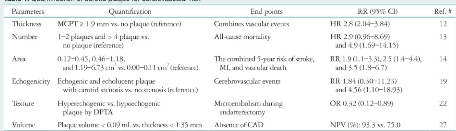
To address this issue, many studies have quantified the ca- rotid plaque to more accurately estimate cardiovascular risk (Table 1). Maximum carotid plaque thickness is associated with increased risk of vascular outcomes in a multiethnic co- hort.12) A study by Störk et al.13) looked at the relationship be- tween the total number of carotid plaques and prognosis.
They evaluated carotid burden using carotid plaque score (1 point = 1–2 plaques; 2 points = 3–4 plaques; 3 points = 5–6 plaques; and 4 points = 7–12 plaques). As compared to pa- tients with no plaque, the risk of mortality increased 2.9-fold when one to two plaques were present and 4.9-fold when greater than four plaques were present. These findings demon- strated a graded relationship between the number of carotid plaques and mortality. Carotid plaque area, determined by the sum cross-sectional area of all carotid plaques, has also been identified as an independent predictor of future cardiovascular
risk.14) Echolucent plaques are lipid-rich, whereas echogenic plaques are rich of fibrous tissue and calcification. Plaque echogenicity can be graded from 1 to 4 based on visual analy- sis with gradient progressing, from echolucent (defined as a plaque appearing black) to predominant echolucent, predomi- nant echogenic, and, finally, echogenic (defined as a plaque ap- pearing white or almost white).15) Honda et al.16) defined an echolucent plaque as less than -13.4 dB by using integrated backscatter (IBS) analysis. They demonstrated that echolucent carotid plaque with low IBS value was a significant and inde- pendent predictor of future coronary events in patients with stable angina. Echolucent plaque was defined by use of the grey-scale median (GSM), a computerized measurement of plaque echogenicity. el-Barghouty et al.17) have contended that a GSM of less than 32 corresponds to a high risk plaque. Ad- ditional studies have further confirmed echolucent plaques as predictor of cerebrovascular events.18)19) Prati et al.20) studied whether plaque score was an independent predictor of cerebro- vascular ischemic events. The score was based on degree of ste-
Table 1. Quantification of carotid plaque for cardiovascular risk
Parameters Quantification End points RR (95% CI) Ref. #
Thickness MCPT ≥ 1.9 mm vs. no plaque (reference) Combines vascular events HR 2.8 (2.04–3.84) 12 Number 1–2 plaques and > 4 plaque vs.
no plaque (reference) All-cause mortality HR 2.9 (0.96–8.69)
and 4.9 (1.69–14.15) 13 Area 0.12–0.45, 0.46–1.18,
and 1.19–6.73 cm2 vs. 0.00–0.11 cm2 (reference) The combined 5-year risk of stroke,
MI, and vascular death RR 1.9 (1.1–3.3), 2.5 (1.4–4.4),
and 3.5 (1.8–6.7) 14
Echogenicity Echogenic and echolucent plaque
with carotid stenosis vs. no stenosis (reference) Cerebrovascular events RR 1.84 (0.30–11.23)
and 4.56 (1.10–18.93) 19 Texture Hyperechogenic vs. hypoechogenic
plaque by DPTA Microembolism during
endarterectomy OR 0.32 (0.12–0.89) 22
Volume Plaque volume < 0.09 mL vs. thickness < 1.35 mm Absence of CAD NPV (%): 93.3 vs. 75.0 27 MCPT: maximum carotid plaque thickness, MI: myocardial infarction, DPTA: detailed plaque texture analysis, CAD: coronary artery disease, NPV: negative predictive value, CI: confidence interval, HR: hazard ratio, OR: odds ratio, RR: relative risk
Fig. 1. Carotid ultrasound showed carotid plaques in two patients, one patient has an asymptomatic single plaque with low cardiovascular risk (A), and the other patient has multiple complex plaques with recent stroke (B). The image (A) shows a small amount of smooth homogenous plaque (arrow) located on the far wall of the proximal CCA. In contrast, the image (B) demonstrates moderate to large amount of heterogeneous multiple plaques (arrows) with focal calcification and irregular surface, in mid CCA. CCA: common carotid artery.
B A
nosis and plaque morphology; (degree of stenosis = 0 if lumen obstruction < 40%, = 1 if 40%), (echogenicity = 1 for low echogenicity or echolucent plaque, = 2 for intermediate, and = 3 for hyper-echogenicity), (texture = 0 if homogeneous, and = 1 if heterogeneous), and (surface characteristics = 0 if smooth, = 1 if irregular). A total plaque risk score was calculated from each subject examined, which ranged from 1 to 6. The most pow- erful predictor of cardiovascular events during their follow-up was total plaque risk score > 4.
Recently, several new ultrasound technologies have been de- veloped for the evaluation of carotid plaques. A study by Lal et al.21) analyzed plaque texture by pixel distribution analysis and histology. Madycki et al.22) found that pixel distribution analy- sis predicted the risk of perioperative complications more pre- cisely than standard GSM analysis. Another evaluation tech- nique relies on contrast-enhanced ultrasound imaging of the carotid vasa vasorum.23) Coli et al.24) demonstrated a strong correlation between histologic density and the degree of con- trast-agent enhancement by ultrasound imaging. Contrast en- hanced ultrasound may also be used to detect subclinical ath- erosclerosis in the carotid arteries, serving as an additional method for the detection of carotid plaque ulceration.25)26)
The final technique quantifies carotid plaque using a three- dimensional (3D) measurement of plaque volume. 3D mea- surement demonstrated a higher negative predictive value and sensitivity for coronary artery disease (CAD) than two-dimen- sional (2D) measurement. In particular, total 3D plaque vol- umes less than 0.09 mL could predict the absence of CAD.27) 3D plaque volume is significantly more sensitive to changes with therapy than measurements of cIMT or total plaque area.28) Sillesen et al.29) found that carotid plaque volume was more closely correlated with coronary atherosclerosis than cIMT, abdominal aortic diameter, or ankle-brachial index in a large number of patients (n = 6101).
Carotid Plaque as a Predictor of Subclinical Atherosclerosis
Traditional cardiovascular disease risk factors such as old age, gender, smoking, cholesterol levels, blood pressure, and diabetes have long been used to predict cardiovascular risk, but this method has its limitations. Imaging modalities such as carotid ultrasound are superior means of predicting cardio- vascular risk.30) Measurement of cIMT has been widely used to predict cardiovascular risk, but it may not be useful for risk stratification in the general population. A meta-analysis by Den Ruijter et al.31) demonstrated that, for the general popu- lation, the added predictive value of including common cIMT measurement to the Framingham Risk Score was small and not significant. However, in individuals of intermediate risk, the added value was 3.2% in men and 3.9% in women.
Despite such limited evidence of cardiovascular risk predic- tion, recent guidelines recommend measuring cIMT in as- ymptomatic patients of intermediate cardiovascular risk. The
ASE consensus statement recommends that common cIMT measurement should always be supplemented by thorough scanning of extracranial carotid arteries for carotid plaques for higher diagnostic accuracy of subclinical vascular diseases.8) Although cIMT is a well-known surrogate marker for sub- clinical atherosclerosis, thickening of cIMT does not necessari- ly represent subclinical atherosclerosis. cIMT is primarily a re- sult of the hypertensive thickening of smooth muscles in the arterial media layer, rather than the subintimal changes which are indicative of atherosclerosis. Compared with cIMT, pres- ence of carotid plaque may be more representative for athero- sclerosis. Atherosclerosis Risk In Communities (ARIC) trial showed that the addition of plaque to cIMT resulted in a net reclassification improvement of 9.9% in the overall popula- tion and 21.7% in the intermediate risk group.11) Carotid plaque, compared with cIMT, was shown to predict CAD events more accurately in a meta-analaysis.32) Ultrasound as- sessment of carotid plaque was found to have a higher diag- nostic accuracy than cIMT for the prediction of future myo- cardial infarction. In addition, the absence of carotid plaque provided greater assurance, with low event rate (4.0%) of 10- year myocardial infarction. The ARIC trial, which evaluated the presence or absence of carotid plaque without quantifica- tion, demonstrated that including plaque with cIMT signifi- cantly improved risk prediction of coronary heart disease.11)
Subsequent studies used quantified methods to measure plaques and evaluated risk prediction using quantified plaques.
In earlier studies, plaque quantification included the thickness, number, surface, area, and texture of plaques which were de- tailed by 2D ultrasound. These studies showed significantly improved risk prediction compared to cIMT and traditional risk factors. Studies investigating the evaluation of coronary heart disease based on carotid plaque score also demonstrated its efficacy as a significant predictor for CAD.
Recent developments in new technologies for evaluating ca- rotid plaques show promise in improving our ability to pre- dict the risk of cardiovascular disease. 3D ultrasound shows par- ticular prognostic promise. 3D measurement of carotid plaque volume is more sensitive to the presence of carotid plaque be- cause it creates simultaneous visualization of carotid plaque us- ing all 3 planes, decreasing the odds of missing any plaques present. It also allows for a more accurate assessment of total plaque volume and enhances the ability to observe changes in plaque burden compared to 2D techniques. Further research is required to confirm this, but present evidence suggests that 3D plaque volume measurement will be a stronger predictor of cardiovascular events in patients.
Ultrasound is used for initial evaluation of carotid plaque.
The advantage of ultrasound is its low cost and that it is rela- tively safe as an imaging modality. The presence of carotid plaque on ultrasound is a better predictor of future cardiovas- cular events compared to cIMT. The disadvantage of ultra- sound is that it is dependent on the operator’s skill and image
quality. The limitation of using carotid plaque in clinical prac- tice is that plaque quantification, such as morphology and vol- ume, has not been well studied yet.
Conclusion
Quantification of carotid plaques is an important step in as- sessing subclinical atherosclerosis for the prediction of future coronary heart disease. While some studies suggest a plaque scoring method for improved assessment, there is no consen- sus classification system for carotid plaque severity. In the fu- ture, 3D ultrasound is expected to be a more reliable means of evaluating plaque burden and predicting cardiovascular risk.
References
1. Hegele RA. The pathogenesis of atherosclerosis. Clin Chim Acta 1996;
246:21-38.
2. Vaya J. The association between biomarkers in the blood and carotid plaque composition-focusing on oxidized lipids, oxysterols and plaque status. Bio- chem Pharmacol 2013;86:15-8.
3. Kragel AH, Reddy SG, Wittes JT, Roberts WC. Morphometric anal- ysis of the composition of atherosclerotic plaques in the four major epicardial coronary arteries in acute myocardial infarction and in sudden coronary death. Circulation 1989;80:1747-56.
4. Schaar JA, Muller JE, Falk E, Virmani R, Fuster V, Serruys PW, Colombo A, Stefanadis C, Ward Casscells S, Moreno PR, Maseri A, van der Steen AF. Terminology for high-risk and vulnerable coronary artery plaques. Report of a meeting on the vulnerable plaque, June 17 and 18, 2003, Santorini, Greece. Eur Heart J 2004;25:1077-82.
5. Naghavi M, Libby P, Falk E, Casscells SW, Litovsky S, Rumberger J, Badimon JJ, Stefanadis C, Moreno P, Pasterkamp G, Fayad Z, Stone PH, Waxman S, Raggi P, Madjid M, Zarrabi A, Burke A, Yuan C, Fitzgerald PJ, Siscovick DS, de Korte CL, Aikawa M, Juhani Airak- sinen KE, Assmann G, Becker CR, Chesebro JH, Farb A, Galis ZS, Jack- son C, Jang IK, Koenig W, Lodder RA, March K, Demirovic J, Navab M, Priori SG, Rekhter MD, Bahr R, Grundy SM, Mehran R, Colombo A, Boerwinkle E, Ballantyne C, Insull W Jr, Schwartz RS, Vogel R, Serruys PW, Hansson GK, Faxon DP, Kaul S, Drexler H, Greenland P, Muller JE, Virmani R, Ridker PM, Zipes DP, Shah PK, Willerson JT. From vulnerable plaque to vulnerable patient: a call for new definitions and risk assessment strategies: part I. Circulation 2003;108:1664-72.
6. Shah PK. Mechanisms of plaque vulnerability and rupture. J Am Coll Cardiol 2003;41(4 Suppl S):15S-22S.
7. Schlendorf KH, Nasir K, Blumental BS. Limitations of the Framing- ham risk score are now much clearer. Prev Med 2009;48:115-6.
8. Stein JH, Korcarz CE, Hurst RT, Lonn E, Kendall CB, Mohler ER, Najjar SS, Rembold CM, Post WS; American Society of Echocar- diography Carotid Intima-Media Thickness Task Force. Use of carot- id ultrasound to identify subclinical vascular disease and evaluate cardio- vascular disease risk: a consensus statement from the American Society of Echocardiography Carotid Intima-Media Thickness Task Force. Endorsed by the Society for Vascular Medicine. J Am Soc Echocardiogr 2008;21:93- 111; quiz 189-90.
9. Touboul PJ, Hennerici MG, Meairs S, Adams H, Amarenco P, Bornstein N, Csiba L, Desvarieux M, Ebrahim S, Fatar M, Hernandez Hernandez R, Jaff M, Kownator S, Prati P, Rundek T, Sitzer M, Schmin- ke U, Tardif JC, Taylor A, Vicaut E, Woo KS, Zannad F, Zureik M.
Mannheim carotid intima-media thickness consensus (2004-2006). An update on behalf of the Advisory Board of the 3rd and 4th Watching the Risk Symposium, 13th and 15th European Stroke Conferences, Mannheim,
Germany, 2004, and Brussels, Belgium, 2006. Cerebrovasc Dis 2007;23:
75-80.
10. Hollander M, Bots ML, Del Sol AI, Koudstaal PJ, Witteman JC, Grobbee DE, Hofman A, Breteler MM. Carotid plaques increase the risk of stroke and subtypes of cerebral infarction in asymptomatic elderly: the Rot- terdam study. Circulation 2002;105:2872-7.
11. Nambi V, Chambless L, Folsom AR, He M, Hu Y, Mosley T, Vol- cik K, Boerwinkle E, Ballantyne CM. Carotid intima-media thickness and presence or absence of plaque improves prediction of coronary heart disease risk: the ARIC (Atherosclerosis Risk In Communities) study. J Am Coll Cardiol 2010;55:1600-7.
12. Rundek T, Arif H, Boden-Albala B, Elkind MS, Paik MC, Sacco RL. Carotid plaque, a subclinical precursor of vascular events: the Northern Manhattan Study. Neurology 2008;70:1200-7.
13. Störk S, van den Beld AW, von Schacky C, Angermann CE, Lam- berts SW, Grobbee DE, Bots ML. Carotid artery plaque burden, stiffness, and mortality risk in elderly men: a prospective, population-based cohort study. Circulation 2004;110:344-8.
14. Spence JD, Eliasziw M, DiCicco M, Hackam DG, Galil R, Lohm- ann T. Carotid plaque area: a tool for targeting and evaluating vascular preventive therapy. Stroke 2002;33:2916-22.
15. Mathiesen EB, Bønaa KH, Joakimsen O. Low levels of high-density li- poprotein cholesterol are associated with echolucent carotid artery plaques: the tromsø study. Stroke 2001;32:1960-5.
16. Honda O, Sugiyama S, Kugiyama K, Fukushima H, Nakamura S, Koide S, Kojima S, Hirai N, Kawano H, Soejima H, Sakamoto T, Yo- shimura M, Ogawa H. Echolucent carotid plaques predict future coronary events in patients with coronary artery disease. J Am Coll Cardiol 2004;
43:1177-84.
17. el-Barghouty N, Nicolaides A, Bahal V, Geroulakos G, Androulakis A. The identification of the high risk carotid plaque. Eur J Vasc Endovasc Surg 1996;11:470-8.
18. Kitta Y, Obata JE, Takano H, Nakamura T, Kodama Y, Fujioka D, Saito Y, Kawabata K, Mende A, Kobayashi T, Kugiyama K. Echolu- cent carotid plaques predict in-stent restenosis after bare metal stenting in native coronary arteries. Atherosclerosis 2008;197:177-82.
19. Mathiesen EB, Bønaa KH, Joakimsen O. Echolucent plaques are associ- ated with high risk of ischemic cerebrovascular events in carotid stenosis: the tromsø study. Circulation 2001;103:2171-5.
20. Prati P, Tosetto A, Casaroli M, Bignamini A, Canciani L, Bornstein N, Prati G, Touboul PJ. Carotid plaque morphology improves stroke risk prediction: usefulness of a new ultrasonographic score. Cerebrovasc Dis 2011;31:300-4.
21. Lal BK, Hobson RW 2nd, Pappas PJ, Kubicka R, Hameed M, Chakhtoura EY, Jamil Z, Padberg FT Jr, Haser PB, Durán WN.
Pixel distribution analysis of B-mode ultrasound scan images predicts histo- logic features of atherosclerotic carotid plaques. J Vasc Surg 2002;35:1210-7.
22. Madycki G, Staszkiewicz W, Gabrusiewicz A. Carotid plaque texture analysis can predict the incidence of silent brain infarcts among patients un- dergoing carotid endarterectomy. Eur J Vasc Endovasc Surg 2006;31:373- 80.
23. Feinstein SB. Contrast ultrasound imaging of the carotid artery vasa vaso- rum and atherosclerotic plaque neovascularization. J Am Coll Cardiol 2006;48:236-43.
24. Coli S, Magnoni M, Sangiorgi G, Marrocco-Trischitta MM, Meli- surgo G, Mauriello A, Spagnoli L, Chiesa R, Cianflone D, Maseri A.
Contrast-enhanced ultrasound imaging of intraplaque neovascularization in carotid arteries: correlation with histology and plaque echogenicity. J Am Coll Cardiol 2008;52:223-30.
25. van den Oord SC, ten Kate GL, Akkus Z, Renaud G, Sijbrands EJ, ten Cate FJ, van der Lugt A, Bosch JG, de Jong N, van der Steen AF, Schinkel AF. Assessment of subclinical atherosclerosis using contrast-en-
hanced ultrasound. Eur Heart J Cardiovasc Imaging 2013;14:56-61.
26. ten Kate GL, van Dijk AC, van den Oord SC, Hussain B, Verhagen HJ, Sijbrands EJ, van der Steen AF, van der Lugt A, Schinkel AF.
Usefulness of contrast-enhanced ultrasound for detection of carotid plaque ulceration in patients with symptomatic carotid atherosclerosis. Am J Cardi- ol 2013;112:292-8.
27. Johri AM, Chitty DW, Matangi M, Malik P, Mousavi P, Day A, Gravett M, Simpson C. Can carotid bulb plaque assessment rule out sig- nificant coronary artery disease? A comparison of plaque quantification by two- and three-dimensional ultrasound. J Am Soc Echocardiogr 2013;
26:86-95.
28. Spence JD. Technology insight: ultrasound measurement of carotid plaque-- patient management, genetic research, and therapy evaluation. Nat Clin Pract Neurol 2006;2:611-9.
29. Sillesen H, Muntendam P, Adourian A, Entrekin R, Garcia M, Falk E, Fuster V. Carotid plaque burden as a measure of subclinical atheroscle- rosis: comparison with other tests for subclinical arterial disease in the High
Risk Plaque BioImage study. JACC Cardiovasc Imaging 2012;5:681-9.
30. del Sol AI, Moons KG, Hollander M, Hofman A, Koudstaal PJ, Grobbee DE, Breteler MM, Witteman JC, Bots ML. Is carotid inti- ma-media thickness useful in cardiovascular disease risk assessment? The Rotterdam Study. Stroke 2001;32:1532-8.
31. Den Ruijter HM, Peters SA, Anderson TJ, Britton AR, Dekker JM, Eijkemans MJ, Engström G, Evans GW, de Graaf J, Grobbee DE, Hedblad B, Hofman A, Holewijn S, Ikeda A, Kavousi M, Kitagawa K, Kitamura A, Koffijberg H, Lonn EM, Lorenz MW, Mathiesen EB, Nijpels G, Okazaki S, O'Leary DH, Polak JF, Price JF, Robertson C, Rembold CM, Rosvall M, Rundek T, Salonen JT, Sitzer M, Stehouwer CD, Witteman JC, Moons KG, Bots ML.
Common carotid intima-media thickness measurements in cardiovascular risk prediction: a meta-analysis. JAMA 2012;308:796-803.
32. Inaba Y, Chen JA, Bergmann SR. Carotid plaque, compared with carot- id intima-media thickness, more accurately predicts coronary artery disease events: a meta-analysis. Atherosclerosis 2012;220:128-33.