• 검색 결과가 없습니다.

Inflammatory myofibroblastic tumors arising from pancreas head and peri-splenic area mimicking a malignancy

N/A
N/A
Protected

Academic year: 2021

Share "Inflammatory myofibroblastic tumors arising from pancreas head and peri-splenic area mimicking a malignancy"

Copied!
6
0
0

로드 중.... (전체 텍스트 보기)

전체 글

(1)

Inflammatory myofibroblastic tumors arising from

pancreas head and peri-splenic area mimicking a malignancy

Eun Jeong Jang

1

, Kwan Woo Kim

1

, Sung Hwa Kang

1

, Min Gyoung Pak

2

, and Song Hee Han

2

Departments of

1

Surgery and

2

Pathology, Dong-A University Hospital, Dong-A University College of Medicine, Busan, Korea

Inflammatory myofibroblastic tumors (IMTs) are a rare chronic inflammatory disease with unclear pathogenesis and pathological features that are not those of a malignant tumor. It is difficult to differentially diagnose them without surgical excision because of their unpredictable clinical behavior, which ranges from benign to locally invasive aggressiveness.

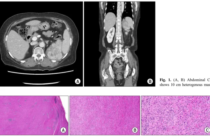
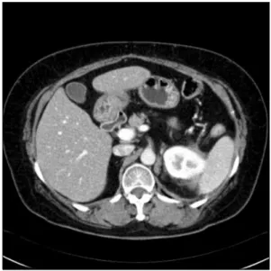
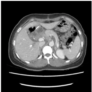
We report two cases of IMTs that were diagnosed after surgery. In one case, the IMT originated in peri-splenic area in a 63-year-old female patient. The other case involved a 48-year-old female patient who suffered from an IMT of the head of the pancreas. Both of these cases did not require further treatment based on histological findings, and there has been no evidence of recurrence or metastasis so far. These cases show that the primary choice for the exact diagnosis and proper treatment of IMTs is complete surgical resection. (Ann Hepatobiliary Pancreat Surg 2021;25:287-292)

Key Words: Inflammatory myofibroblastic tumor; Pancreas; Retroperitoneum; Case report

Received: January 10, 2021; Revised: March 4, 2021; Accepted: March 4, 2021 Corresponding author: Kwan Woo Kim

Department of Surgery, Dong-A University College of Medicine, 26 Daesingongwon-ro, Seo-gu, Busan 49201, Korea Tel: +82-51-240-5146, Fax: +82-51-247-9316, E-mail: [email protected]

Copyright Ⓒ 2021 by The Korean Association of Hepato-Biliary-Pancreatic Surgery

This is an Open Access article distributed under the terms of the Creative Commons Attribution Non-Commercial License (http://creativecommons.org/

licenses/by-nc/4.0) which permits unrestricted non-commercial use, distribution, and reproduction in any medium, provided the original work is properly cited.

Annals of Hepato-Biliary-Pancreatic Surgery ∙ pISSN: 2508-5778ㆍeISSN: 2508-5859

INTRODUCTION

Inflammatory myofibroblastic tumors (IMTs) have been reported to occur in multiple anatomical locations, includ- ing the lungs, bladder, spleen, breast, pancreas, liver, co- lon, spermatic code, prostate, and soft tissue.

1

An IMT is a fibroblastic neoplasm composed of spindle cells accom- panied by an inflammatory infiltrate of plasma cells, lym- phocytes, or eosinophils.

2

The incidence of IMTs is ap- proximately 0.04-0.7%, and clinical data have shown that they have a 25% rate of local recurrence and up to a 5%

rate of distant metastasis.

3

The symptoms of IMT are ambiguous and generally re- lated to obstructive symptoms caused by the large-sized mass.

2

Additionally, when a mass is found in the pan- creas, IMTs are most commonly mistaken for pancreatic cancer according to their clinical and radiological charac- teristics. Therefore, the initial diagnosis of IMTs is diffi- cult without extensive histological examination. Usually, they are diagnosed by histological evaluations after surgery.

In this study, we report two cases of IMTs that were diagnosed after surgery. The first case arose in peri-splen- ic area of a 63-year-old female patient who underwent complete mass excision. The second case occurred in a 48-year-old female patient who underwent pylorus-pre- serving pancreaticoduodenectomy (PPPD) due to a pan- creas-head mass.

CASE

Case 1

A 63-year-old female patient with diabetes mellitus and

hypertension had been suffering from fever and sweating,

which were controlled easily for several months. After

visiting a local clinic, she was referred to our hospital for

the evaluation of an abdominal mass >10 cm in size,

which was strongly suspicious for lymphoma, leiomyo-

sarcoma, or malignant fibrous histiocytoma in the left up-

per quadrant. Laboratory findings showed a slight increase

in the white blood cell count (14.93; normal range: 3-9.3

(2)

Fig. 1. (A, B) Abdominal CT shows 10 cm heterogenous mass.

Fig. 2. The histopathological evaluation shows a well-circumscribed whitish mass characterized by the proliferation of spindle cells mixed with inflammatory cells such as lymphocytes and plasma cells, compatible with Inflammatory myofibroblastic tumor.

×10 magnification (A), ×100 magnification (B), ×400 magnification (C).

10

3

/ul) and C-reactive protein (CRP) (1.47; normal range:

0-0.5 mg/dl). However, no particular abnormality was noticed. Moreover, a tumor marker test showed normal results.

Enhanced abdominal computed tomography (CT) showed a 10-cm heterogeneous mass extending in the peri-splenic area (Fig. 1). No metastatic lesions were observed in the enhanced chest CT and abdominal CT scans. We did not perform any pre-operative biopsies due to the patient’s re- fusal and the clinical judgment that complete surgical ex- cision was necessary for a definite pathologic diagnosis.

The patient underwent radical excision of the retroperito- neal mass. During the operation, a huge mass was found that strongly adhered to the posterior portion of the spleen, upper pole of the left kidney, and the diaphragm. There- fore, diaphragmatic excision was performed simultaneously.

The patient was discharged on the 9th day after surgery without any peri- or post-operative complications.

Upon histological examination, the mass was diagnosed as an IMT associated with frequent IgG4-positive cell infiltration. Grossly, it was a grayish, encapsulated mass

with dimensions of 13 cm×9.5 cm. Immunochemical stud- ies showed negative results for C-kit, S100, and P53, along with positive results for anaplastic lymphoma kinase (ALK), smooth muscle actin (SMA), vimentin, desmin, CD68, and phosphorylated histone H3 (PHH3) (Fig. 2). As of two years after surgery, the patient has had no local recurrence or metastasis without further treatment, which was determined through multidisciplinary opinions. Fig. 3 shows the CT findings from the latest follow-up.

Case 2

A 48-year-old female patient was referred to our hospi- tal after regular health examinations revealed the presence of hyperbilirubinemia and an abdominal mass. She had a medical history of hypertension. The initial laboratory find- ings were as follows: alanine transaminase (AST), 178 (nor- mal range: 0-35 U/L); aspartate transaminase (ALT), 77 (normal range: 0-35 U/L); alkaline phosphatase (ALP), 1,295 (normal range: 30-120 U/L); total bilirubin, 3.9 (normal range: 0.2-1.2 mg/dl); direct bilirubin, 2.2 (normal range:

0-0.4 mg/dl), alpha-fetoprotein (AFP), 6.58 (normal range:

(3)

Fig. 4. Initial abdominal CT (A, B), MRI (C) and PET (D) images show 7.5 cm mass in the head of pancreas.

Fig. 3. Two years after surgery, CT shows no local recurrence and metastasis.

0-7 ng/ml); carcinoembryonic antigen (CEA), 2.34 (normal range: 0-5 ng/ml); carbohydrate antigen 19-9 (CA19-9), 235 (normal range: 0-27 U/ml).

CT and magnetic resonance imaging (MRI) findings re-

vealed a 7.5-cm mass with obstructive jaundice in the head of the pancreas, which was suspected to be either a solid pseudopapillary tumor or retroperitoneal solitary fibrous tumor (Fig. 4A-C). Moreover, a huge hyper- metabolic mass was observed in the head of the pancreas without any metastatic lesions using positron emission to- mography (PET) (Fig. 4D). The patient underwent pylo- rus-preserving pancreaticoduodenectomy (PPPD) for accu- rate pathologic diagnosis and complete surgical excision without pre-operative biopsy. During the operation, there was no evidence of distal organ or lymph node metastasis.

The patient was discharged routinely after 12 days with- out any surgical or medical complications.

After the operation, the final pathological diagnosis was IMT. The tumor was relatively well defined with di- mensions of 8.9 cm×5.7 cm and involving the duodenal wall. A total of 23 lymph nodes were retrieved during sur- gery, but all were negative. The tumor was composed of fascicles of plump spindle cells with oval nuclei and small nucleoli (Fig. 5A). There was no necrosis or hemorrhage.

Inflammatory cells (mostly plasma cells) were either seen

(4)

Fig. 5. (A) The tumor is composed of fascicles of plump spindle cells with oval nuclei and small nucleoli (arrows). Inflammatory cells, mostly plasma cells are seen either as small aggregates or more uniformly dispersed (dot-lined circle) (H-E stain, ×400 magnification). (B) The tumor cells show focal immunohistochemical positivity for smooth muscle actin (SMA, ×400 magnification). (C) Immunohistochemical staining for IgG4, a marker of IgG4-positive plasma cell shows only one positive IgG4-positive cell per high power field (dot-lined circle) (IgG4, ×400 magnification). (D) Ki-67 proliferation index is about 5% (Ki-67, ×400 magnification).

Fig. 6. After 6 month, no evidence of recurrence is observed in the CT.

as small aggregates or were more uniformly dispersed.

The immunohistochemical staining test results were negative for ALK, S-100, CD23, CD34, and CD99, but the results were focally positive for only SMA (Fig. 5B).

Only three IgG-positive plasma cells per high-power field were recognized (Fig. 5C), so IgG4-related pancreatitis was ruled out. The tumor showed low mitotic activity, and the Ki-67 proliferation index was about 5% (Fig. 5D). As with the first case, no adjuvant treatment was provided to the patient after surgical excision of the tumor based on histological findings, and no evidence of recurrence was observed in the CT scan at the 6-month follow-up (Fig. 6). As far, about 1 year after surgery, the patient has been well without treatment and will be screened ev- ery 6 months.

DISCUSSION

Also known as inflammatory pseudotumors, IMTs refer to a heterogeneous group of lesions occurring in various organs that are histologically characterized by fibroblastic and myofibroblastic proliferation with inflammatory infil- trate.

4

The current World Health Organization’s (WHO’s) classification for this rare tumor entity is a fibroblastic sarcoma or myofibroblastoma, which is a distinctive neo- plasm of intermediate biological potential that may be ma-

lignant or aggressive.

3

We have reported 2 cases of large IMTs occurring in different locations: one in the retro- peritoneum and the other in the pancreas head.

Generally, retroperitoneal tumors grow slowly and are

usually presented as a solid abdominal palpable mass ac-

companied by fever, pain, or other symptoms.

5

A differ-

ential diagnosis must be made to rule out benign fiber

mesenteric tumors (such as mesenteric fibromatosis and

sclerosing mesenteritis), lymphomas, soft tissue sarcomas,

(5)

and metastatic malignancies.

6

Because of its nonspecific symptoms and imaging findings, definitive diagnosis of IMT relies on histological evaluation. However, it is often difficult to obtain enough tissue using an ultrasonography- guided biopsy or a trans-endoscopic biopsy to reveal a de- finitive histological diagnosis.

6

Additionally, although IMT may be detected during preoperative biopsy, surgical excision is mostly the treatment of choice for malignancy as there is no clear basis for conservative treatment.

1,7-9

There have been recent reports of spontaneous re- gression of IMTs,

10-12

which also indicated that surgical resection could be necessary for accurate diagnosis and treatment. The medical treatment is not clearly defined due to a lack of strong evidence in cases of large tumors, and palliative treatment cannot be recommended as a standard regimen. Moreover, in cases of a large IMTs, surgical treatment is required to relieve obstructive symp- toms caused by the large mass and to exclude malignancy.

This is why we performed complete surgical resection without pre-operative biopsy in both cases.

The prognosis of IMTs is favorable, and the 5-year sur- vival rate is 91%.

3

Although metastasis is a rare event in IMTs,

13

recurrence rate is approximately 25% related to location, resectability and multinodularity.

8

So far, neither of our cases have had recurrence or distant metastasis.

However, long-term periodic follow-up could be neces- sary because of the possibility of local recurrence or dis- tant metastasis of IMTs.

CONCLUSION

IMTs are a rare chronic inflammatory disease of un- clear pathogenesis, and their pathological features are not those of a malignant tumor. They are very difficult to dif- ferentially diagnose from malignant diseases before com- plete surgical excision, especially in the pancreas.

Therefore, according to the current literature and our ex- perience, the primary choice for the exact diagnosis and proper treatment of IMTs is complete surgical resection.

ETHICAL STATEMENT

This study was approved by the ethics committee of Dong-A University Hospital (IRB No. DAUH IRB-20-090).

The requirement for informed consent from patients was

waived due to the retrospective nature of study.

ACKNOWLEDGEMENTS

This paper was not supported by any research fund.

CONFLICT OF INTEREST

The authors have no potential conflicts of interest to disclose.

ORCID

Eun Jeong Jang: https://orcid.org/0000-0002-8993-7769 Kwan Woo Kim: https://orcid.org/0000-0002-7432-6949 Sung Hwa Kang: https://orcid.org/0000-0002-5614-5968 Min Gyoung Pak: https://orcid.org/0000-0001-6142-4569 Song Hee Han: https://orcid.org/0000-0002-4564-7014

AUTHOR CONTRIBUTIONS

Conceptualization: KWK. Data curation: EJJ. Formal analysis: EJJ, KWK, SHK, MGP, SHH. Funding acquis- ition: None. Methodology: EJJ, KWK. Writing - original draft: EJJ, SHK, MGP. Writing - review & editing: EJJ, KWK, SHH.

REFERENCES

1. Kovach SJ, Fischer AC, Katzman PJ, Salloum RM, Ettinghausen SE, Madeb R, et al. Inflammatory myofibroblastic tumors. J Surg Oncol 2006;94:385-391.

2. Thompson LDR. Inflammatory myofibroblastic tumor. Ear Nose Throat J 2019. doi: 10.1177/0145561319890165. [in press]

3. Ding D, Bu X, Tian F. Inflammatory myofibroblastic tumor in the head of the pancreas with anorexia and vomiting in a 69- year-old man: a case report. Oncol Lett 2016;12:1546-1550.

4. Battal M, Kartal K, Tuncel D, Bostanci O. Inflammatory myofi- broblastic pancreas tumor: a case report. Clin Case Rep 2016;4:

1122-1124.

5. Tao YL, Wang ZJ, Han JG, Wei P. Inflammatory myofibro- blastic tumor successfully treated with chemotherapy and non- steroidals: a case report. World J Gastroenterol 2012;18:7100- 7103.

6. Poves I, Alonso S, Jimeno M, Bessa X, Burdío F, Grande L.

Retroperitoneal inflammatory pseudotumor presenting as a pan- creatic mass. JOP 2012;13:308-311.

7. Zanchi C, Giurici N, Martelossi S, Cheli M, Sonzogni A, Alberti D. Myofibroblastic tumor of the pancreatic head: recurrent cholangitis. J Pediatr Gastroenterol Nutr 2015;61:e28-e29.

8. Panda D, Mukhopadhyay D, Datta C, Chattopadhyay BK,

Chatterjee U, Shinde R. Inflammatory myofibroblastic tumor

(6)

arising in the pancreatic head: a rare case report. Indian J Surg 2015;77:538-540.

9. Sim A, Lee MW, Nguyen GK. Inflammatory myofibroblastic tu- mour of the pancreas. Can J Surg 2008;51:E23-E24.

10. Matsubayashi H, Uesaka K, Sasaki K, Shimada S, Takada K, Ishiwatari H, et al. A pancreatic inflammatory myofibroblastic tumor with spontaneous remission: a case report with a literature review. Diagnostics (Basel) 2019;9:150.

11. Yamaguchi J, Sakamoto Y, Sano T, Shimada K, Kosuge T.

Spontaneous regression of inflammatory pseudotumor of the liv- er: report of three cases. Surg Today 2007;37:525-529.

12. Zhao JJ, Ling JQ, Fang Y, Gao XD, Shu P, Shen KT, et al.

Intra-abdominal inflammatory myofibroblastic tumor: sponta- neous regression. World J Gastroenterol 2014;20:13625-13631.

13. Coffin CM, Hornick JL, Fletcher CD. Inflammatory myofibro-

blastic tumor: comparison of clinicopathologic, histologic, and

immunohistochemical features including ALK expression in

atypical and aggressive cases. Am J Surg Pathol 2007;31:509-520.

수치

Fig. 1. (A, B) Abdominal CT  shows 10 cm heterogenous mass.
Fig. 4. Initial abdominal CT (A,  B), MRI (C) and PET (D)  images show 7.5 cm mass in  the head of pancreas.
Fig. 6. After 6 month, no evidence of recurrence is observed  in the CT.

참조

관련 문서