LPS
로 유도된 대식세포에 대한 와송 핵산추출물의 AP-1과 IRF3 전사인자의 억제에 의한 전염증성 사이토카인의 감소 효과
이형선*
중원대학교임상병리학과
Received: February 26, 2020 / Revised: March 30, 2020 / Accepted: March 31, 2020
서 론
염증은손상과감염그리고치유과정에대한숙주의보호 반응이다. 하지만이러한염증반응이과도하고지속적으로 일어나만성화되면암, 자가면역질환, 동맥경화등다양한
질병을유발하는것으로알려져있다[1]. 염증반응을조절하
는세포중대식세포는다양한미생물감염에대항한숙주 방어시스템에서매우중요한세포이다[2]. 대식세포는다양 한자극으로활성화되는데특히그람음성균의세포벽성분 인 lipopolysaccharide (LPS)가 세포막 수용체인 toll-like receptor (TLR) 4에결합하면 MyD88과 TRIF 의존성경로 가시작된다[3]. 활성화된두경로는 nuclear factor-κB (NF-
κB), activator protein-1 (AP-1), interferon regulatory factor 3 (IRF3)와같은전사인자를핵내로이동시킨다[4].
전사인자의핵내로이동은염증자극에의해일어나고결과 적으로 interleukin-1β (IL-1β), interleukin-2 (IL-2), interleukin-6 (IL-6), interferon gamma-induced protein 10 (IP-10) 및 tumor necrosis factor (TNF) 와같은전염증 성 사이토카인과 inducible nitric oxide synthase (iNOS) 및 cyclooxygenase-2 (COX-2)와같은 염증성인자들의생 성을촉진한다[5−7]. 흔히상용되는염증성질환의치료제 로는 non-steroidal anti-inflammatory drugs (NSAIDs)와 glucocorticosteroids 등이있다. 이들의다양한부작용때문 에현재는부작용이적은천연물질로부터염증을억제할수
있는물질탐색에대한연구가증가하고있다[8].
와송(Orostachys japonicus)은다년생초본식물로전통의 약에서다양한치료에사용되어왔다[9]. 와송은 triterpenoid 류인 friedelin, epi-friedianol, glutanone, glutinol과 sterol Orostachys japonicus Hexane Fraction Attenuates Pro-inflammatory Cytokines in LPS-activated Macrophage Cells by
Suppression of AP-1 and IRF3 Transcription Factors Hyeong-Seon Lee*
Department of Biomedical Laboratory Science, Jungwon University, Goesan 28024, Republic of Korea
Orostachys japonicus (O. japonicus) is known as a medicinal plant for the treatment of various symptoms.
This study investigated the anti-inflammatory effect of the hexane fraction from O. japonicus (OJH) on the LPS-stimulated response in RAW 264.7 macrophage cells. This study was conducted to confirm the effect of cell cytotoxicity and production of reactive oxygen species (ROS) in OJH-treated macrophage cells. Addi- tionally, pro-inflammatory cytokines and transcription factors were determined using RT-PCR and west- ern blotting assay. OJH showed no change in lactate dehydrogenase (LDH) levels and exhibited reduced ROS levels in LPS-induced inflammatory cells. Moreover, OJH significantly suppressed the mRNA levels of pro-inflammatory cytokines, including IL-1β, IL-2, IL-6, TNF-α, and IP-10. Furthermore, OJH effectively inhibited the protein levels of AP-1 (p-c-Jun and p-c-Fos) and p-IRF3 in a dose-dependent manner. In con- clusion, our results demonstrate that OJH exhibits strong anti-inflammatory activities via regulation of inflammatory factors.
Keywords: Orostachys japonicus, hexane fraction, pro-inflammatory cytokine, AP-1, IRF3
*Corresponding author
Tel: +82-43-830-8861, Fax: +82-43-830-8679 E-mail: [email protected]
© 2020, The Korean Society for Microbiology and Biotechnology
계열물질인 β-sitosterol, campesterol 등과 aromatic acid 인 4-hydroxy benzoic acid, 3,4-dihydroxybenzoic acid와 tannin 계열 물질인 gallic acid와 flavonoid인 kaemferol,
quercetin 등의 다양한식물 화합물이있다고보고되었다
[10, 11]. 본연구에서는 와송을건조 및분쇄하여에탄올
(EtOH), 핵산(Hex), 디클로로메탄(DCM), 에틸아세테이트 (EtOAc), 부탄올(BuOH), 물(H2O) 순으로분획하여성분을 추출하였다. 본연구방식으로추출된와송의유기용매분획 물은정등(2011)에의해항염증활성이평가되었고[12], 그 중 DCM과 Hex 분획물로 NF-κB 전사인자를매개로한염증 성 인자인 inducible nitric oxide synthase (iNOS)와 cyclooxygenase-2 (COX-2)의탁월한억제활성에대해본연 구자가선행연구하였다[13, 14]. 전체 6가지분획물중 DCM 과 Hex 분획물이항염증활성이탁월하였다. 여러연구가진 행중인 DCM 분획물을제외하고그중핵산추출물(OJH)을 이용하여항염증활성실험을진행하였다. 와송핵산추출물 을 전처리하여 LPS로 염증을 자극한 대식세포 주(RAW
264.7 cells)를이용하여항염증관련사이토카인에미치는
영향을확인하였다. 본연구에서는이전연구에서한번도확 인한적없는 AP-1과 IRF3 전사인자관련경로를연구하고 염증성사이토카인에어떠한영향을미치는지조사하고자 한다.
재료 및 방법
와송 추출물 제조
농장에서재배하여건조한와송을경남밀양농장에서직 접구입하였다. 건와송을갈아서가루로만들고에탄올에 침지하여 95℃에서 3시간 3회반복추출하여상층만을여과 (Whatman, NO. 1)하였다. 여과한에탄올추출물은회전식 감압농축기(Laborata 4000, Heidolph, Germany)로최대한
농축하고물을이용하여완전히용해시켜핵산, 디클로로메 탄, 에틸아세테이트, 부탄올, 물순으로계층분획한후농 축하여유기용매를제거하였다. 세포실험을사용하기위해 dimethyl sulfoxide (DMSO)에농도 100 mg/ml로제조하여 사용하였다(Fig. 1). 본연구의재료추출방법은인제대학교 이동석교수님연구실에서제공받았으면본연구자가동일 한방법으로추출및분리하여공동으로연구를수행중이다.
세포배양 및 실험처치
RAW 264.7는 murine macrophage cell line으로한국 세포주은행에서 분양받아사용하였다. 1% 100 units/ml penicillin-streptomycin과 10% fetal bovine serum (FBS) 가 함유된 Dulbecco’s Modified Eagle Medium (DMEM, Gibco, USA) 배지를사용하여 37℃, 5% CO2조건에서배양 하였고, 2−3일간격으로계대배양하여세포를유지하였다. 모든실험은세포수(5 × 10⁵/ml)를일정하게준비하여부착 시킨후 OJH를농도별(0, 25, 50, 100μg/ml)로 2시간전처 리하고 LPS (1μg/ml)로조건에맞추어염증을유도하였다.
세포독성 평가(LDH assay)
OJH의 세포독성은 Cyto Tox 96 non-radioactive cytotoxicity assay kit (Promega, USA)를 이용하여 lactic dehydrogenase (LDH) 를측정하였다. RAW 264.7 세포를 5 × 10⁵/ml로 96 well plate에분주하였다. 하루동안안정화 하여 OJH를 2시간동안농도별로전처리하고 LPS (1μg/ml) 를 24시간처리후 3,000 rpm으로 5분간원심분리하여상층 배양배지를얻었다. 50μl 상층액과 reconstituted substrate mix를 96 well plate에옮겨담아 30분간반응시킨후 stop solution을 넣어 FilterMax F5 Multi-Mode microplate reader (Molecular Devices, USA) 490 nm에서흡광도를측정 하였다.
Fig. 1. Fractionated Orostachys japonicus with organic solvents. O. japonicus powder extracted in 95% EtOH for 3 h. EtOH extract is concentrated using rotary evaporator to dryness. The concentrate was suspended in water and fractioned with following sequence of organic solvents: hexane, DCM, EtOAc, BuOH, and H2O. Each solvent fraction was concentrated by rotary evaporator to dryness.
활성산소 측정(DCF-DA assay)
ROS를측정하기위해 2'7'–dichlorofluorescein diacetate (DCF-DA) 형광시약을 이용하였다. RAW 264.7 세포를 5 × 10⁵/ml로 96 black well plate에분주하고, 하루동안안 정화하여 20μM DCF-DA (Sigma, USA) 시약으로 1시간동 안전처리하였다. 형광시약을제거하고 1 × PBS로 2번씻 어내고 OJH를 2시간동안농도별로전처리하고 LPS (1μg/
ml)를 2시간처리후 1 × PBS로 2번씻어내고 485/20 nm와 528/20 nm에서형광값을측정하였다.
Reverse transcription-polymerase chain reaction (RT- PCR)
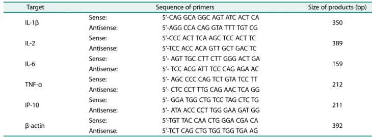
RAW 264.7 세포를 6 well plate에 2 ml씩 (5 × 10⁵/ml) 분주하여하루동안안정화시킨후시료를농도별로전처 리하고 LPS (1μg/ml)를 18시간처리하여염증반응을유도 하였다. 배지를완전히제거하고 1 ml의 TRIzol 용액을이 용하여세포를회수하여클로로포름및이소프로판올로총 RNA를 분리하였다. RNA 1μg을 주형으로 AccuPower Reverse Transcription PreMix tube for the cDNA synthesis (Bioneer, Korea)를이용하여 cDNA를합성하고목적유전자 의 oligo primer를이용하여 PCR을수행하였다. PCR에사용 된 primer는 Table 1과 같으며 PCR은 AccuPower PCR PreMix (Bioneer)를이용하여 95℃에서 5분간 denaturation하 고 95℃에서 30초, 각 primer 온도에서 30초, 72℃에서 30초 25-30 cycle을 반복 후 72℃에서 10분간 extension하였다. PCR product는 1.5% agarose gel에서 전기 영동하여 ethidium bromide (EtBr)로 염색하여 Davinch-ChemiTM imaging system (Davinch-K, Korea)에서결과를확인하 였다.
단백질 Immune blot 측정
RAW 264.7 세포를 6 well plate에 2 ml씩 (5 × 10⁵/ml) 분주하여하루동안안정화시킨후시료를농도별로전처 리하고 LPS (1μg/ml)를 1.5시간처리하여염증반응을유도 하였다. 배지를저거하고 1× PBS로 2번씻어내고 100μl cell lysate buffer (Cell signaling Technology, USA)를 분주하 여스크래퍼로세포를회수하여얼음에 30분간반응시켰다. 세포추출물을 14,000 rpm에서 10분간원심분리하여상층을 얻어 bicinchoninic acid (BCA) protein determination kit (Thermo scientific, USA)를이용하여단백질농도를측정하였 다. 40−50μg 단백질샘플을 8−12% sodium dodecyl sulfate polyacrylamide gel electrophoresis (SDS-PAGE) gel에 전기 영동하여 polyvinylidene fluoride (PVDF) membrane (Bio-rad, USA)으로옮겨목적단백질에특이적이 p-c-Jun, p- c-Fos, p-IRF3 1차 항체(Cell signaling Technology, USA)와 anti-rabbit HRP 2차 항체에 반응시켜 ECL 용액(Santa Cruz Biotechnology, USA)을이용하여발색하였다. 통계 처리
실험은모든과정을최소 3회이상반복실시하였으며, 실 험결과는평균 ± 표준편차로나타내었다. 각실험결과의유 의성검정은 Student’s t-test로비교하였으며 p 값이 0.05 미 만일때통계적의의가있다고판단했다.
결과 및 고찰
와송 추출물의 제조와 세포 독성 확인
자연건조된와송으로부터에탄올추출물을얻고유기용 매의극성을이용하여용매분획을실시하였다. 농축감압기 (Laborata 4000, Heidolph)를이용하여용매를제거한건조
Table 1. Primer sequence design for RT-PCR.
Target Sequence of primers Size of products (bp)
IL-1β Sense: 5’-CAG GCA GGC AGT ATC ACT CA
Antisense: 5’-AGG CCA CAG GTA TTT TGT CG 350
IL-2 Sense: 5’-CCC ACT TCA AGC TCC ACT TC
Antisense: 5’-TCC ACC ACA GTT GCT GAC TC 389
IL-6 Sense: 5’- AGT TGC CTT CTT GGG ACT GA
Antisense: 5’- TCC ACG ATT TCC CAG AGA AC 159
TNF-α Sense: 5’- AGC CCC CAG TCT GTA TCC TT
Antisense: 5’- CTC CCT TTG CAG AAC TCA GG 212
IP-10 Sense: 5’- GGA TGG CTG TCC TAG CTC TG
Antisense: 5’- ATA ACC CCT TGG GAA GAT GG 211
β-actin Sense: 5’-TGT TAC CAA CTG GGA CGA CA
Antisense: 5’-TCT CAG CTG TGG TGG TGA AG 392
상태의 hexane, DCM, EtOAc, BuOH, 그리고 H2O 추출물 의각각수율은 3.38 g, 2.81 g, 6.08 g, 13.36 g, 6.22 g이었다
(Fig. 1). 세포막의손상또는괴사가일어나면세포막이나
핵내에 LDH의활성이증가하고세포밖으로용출되는현
상이일어난다[15]. 배양배지를이용하여세포밖으로유출
된 LDH의양을측정하여세포독성여부를확인할수있다. 항염증활성을보인 hexane 추출물을 RAW 264.7 세포에농 도별(0, 25, 50, 100, 200μg/ml)로 처리하고 동시에 LPS (1μg/ml)를처리하여 24시간배양후세포독성을측정하였 다(Fig. 2A). 각농도에서 LDH 활성율은대조군과비교하여 수치적 차이가 없는 것으로 확인되었다. 따라서 와송의
hexane 추출물을실험한농도내에서는독성이없는것으로
생각되어이를바탕으로실험을진행하였다.
ROS 생성 억제 효과
ROS의생성은세포를공격하여핵산및지질을파괴하고
여러단백효소의기능을저해한다. 그결과노화촉진, 암 및염증등의유발로다양한질병을발생시키는것으로보고 되어 있다[16]. ROS의 종류는 superoxide (O2·), hydrogen peroxide (H2O2), hydroxyl radical (.OH) 등이있다. 세포내
ROS가존재할 경우 DCF-DA와반응하여높은 형광을띈
DCF로산화됨으로써 ROS 생성량을측정할수있다[17]. 대 식세포주에 LPS로염증을유발하여와송 hexane 추출물이
LPS에의해생성된 ROS에대한억제능이있는지확인한결
과농도의존적으로 ROS의형성이저하되었다(Fig. 2B). 특 히, 50μg/ml 이상에서는 LPS 단독처리에대비통계적으로 유의성있는감소(p < 0.05)를보였으며정상과매우비슷한 수준으로생성이억제되는것을확인할수있었다. 이러한
결과는와송 hexane 추출물이활성산소의제거능을가지고
있음을보여준다. 염증으로유발된산화스트레스에대해대 항하는능력이있을것으로생각된다.
염증성 사이토카인 억제 효과
면역세포는염증반응이일어나면 IL-1β, IL-2, IL-6, TNF-
α, IP-10과같은전염증성사이토카인이생성되어증가하는
양상을보인다[18]. 이와같은염증매개인자의과다발현
은전신성염증반응, 만성염증, 동맥경화, 암성장촉진등 다양한질병의기전에관여한다[19, 20]. IP-10은 IRF3 전사 인자의활성화에의해생성되는데식세포의화학주성을유
도하여염증반응에관여하는것으로알려져있다[21]. 따라
서염증에의해생성된사이토카인의조절은염증반응에의 해영향을받는다양한질병의조절가능성을시사한다. 본 Fig. 2. Effect of hexane fraction on cell cytotoxicity and ROS formation in LPS-stimulated cells. (A) RAW 264.7 cells were incubated in the presence of hexane fraction and LPS for 24 h. Cell cytotoxicity was determined by LDH assay. (B) Cells were first incubated with 50 μM DCF-DA for 1 h, and then incubated with hexane fraction and LPS for 12 h. *significantly different from LPS only at p < 0.05.
Fig. 3. Effect of hexane fraction on mRNA levels of pro- inflammatory cytokines in LPS-stimulated cells. RAW 264.7 cells were pretreated with hexane fraction for 2 h and stimulated with LPS for 18 h. The mRNA expression of IL-1β, IL-2, IL-6, TNF- α, IP-10, and β-actin were analyzed by RT-PCR. Target products were separated on 1.5% agarose gel and detected by Davinch- ChemiTM imaging system (Davinch-K, Korea).
실험에서는 RAW 264. 7 대식세포주에와송 hexane 추출 물을 2시간농도별로전처리하고 LPS로염증을자극하여 전염증성사이토카인의생성과관련하여 mRNA 수준에서 측정하여확인하였다(Fig. 3). LPS 단독처리에의해사이토 카인의발현이강하게나타났으나, 와송 hexane 추출물전 처리에의해 IL-1β, IL-2, IL-6, IP-10의발현이농도의존적
으로 확연한 감소 양상을 보인다. 그러나 TNF-α는 와송
hexane 추출물처리에도불구하고 mRNA의양이감소하지
않았으며오히려약간씩증가하는듯한결과를보여준다. 따 라서와송 hexane 추출물은 IL-1β, IL-2, IL-6, IP-10의발현 억제를통해항염증효과를나타냄을확인할수있었다.
핵 내 전사인자 신호 전달 감소
LPS는대식세포의막수용체를자극하여세포내 MyD88 과 TRIF 의존성경로를활성화시킨다. 두경로는 NF-κB,
AP-1, IRF3와같은전사인자를핵내로이동시켜염증관련
사이토카인인자의생성을유도한다[22]. AP-1은 c-Jun와 c- Fos 두가지단백질로구성된이형이량체의염증, 스트레스, 성장인자, 감염에의해자극을받는핵내전사인자로 MyD88 의존성경로에의해활성화된다[23]. 또한 IRF3 전사인자 는염증반응에서 TRIF 의존성경로에의해활성화되어 IFN 과 IP-10과같은사이토카인의생성에관여한다[24]. 염증자 극에의해생성된사이토카인의조절은상위단백질인 AP-
1과 IRF3의전사인자활성억제를확인함으로써가능하다.
와송 hexane 추출물을 2시간농도별로전처리하고 LPS로 염증을자극하여핵내전사인자의인산화발현억제와관 련하여단백질수준에서측정하여확인하였다(Fig. 4). 그결 과, AP-1 구성단백질인 c-Jun과 c-Fos의인산화의발현이 와송 hexane 추출물전처리에의해 LPS 염증자극에대항
하여농도의존적으로감소하였다. 또한, IRF3의인산화발 현도와송 hexane 추출물에농도에따라감소하여 100μg/
ml에서는 탁월한 효과를 보인다. 이러한 결과들은 와송
hexane 추출물이염증자극에의한전사인자의인산화억제
로핵내이동을제한하여염증성매개사이토카인의생성 을억제하는것으로판단된다.
요 약
본연구는와송에유기용매를활용하여순차적으로추출 하여항염증활성에대한가능성을평가하기하기위해수 행되었다. 대식세포에 와송 hexane 추출물을전처리하고
LPS로염증을자극하여염증과관련한세포내신호전달경
로에미치는 영향을확인하고자하였다. 대식세포에와송 hexane 추출물은 LPS 자극에의해세포독성이나타나지않 았고, ROS의생성을억제하는것으로확인되었다. 또한, IL- 1β, IL-2, IL-6, IP-10과같은전염증성사이토카인의분비를
mRNA 수준에서확인한결과탁월하게억제하였다. 이러한
전염증성사이토카인의생성억제는상위전사인자인 AP-1
과 IRF3의조절을통해이루어지므로이들을단백질수준에
서발현량을확인하였다. 그결과 c-Jun, c-Fos, IRF-3의인 산화억제로핵내전사활성이제한되었을것으로생각된다. 이들결과를종합해볼때, 와송 hexane 추출물은염증반응 을저해하는효과가있는것으로나타나다양한염증성질 환의예방및개선에유용하게활용할수있을것으로생각 된다.
Acknowledgment
This study was received by Basic Science Research Program through the National Research Foundation of Korea (NRF) funded by the Ministry of Education (No. NRF-2017R1D1A1B03035934).
Conflict of Interest
The authors have no financial conflicts of interest to declare.
References
1. Kim JK, Jun JG. 2015. Licochalcone B exhibits anti-inflammatory effects via modulation of NF-κB and AP-1. Biomed. Sci. Lett. 21:
218-226.
2. Gilroy D, De Maeyer R. 2015. New insights into the resolution of inflammation. Semin. Immunol. 27: 161-168.
3. O’Neill LA. 2006. How toll-like receptors signal: what we know and what we don’t know. Curr. Opin. Immunol. 18: 3-9.
4. Fitzgerald KA, McWhirter SM, Faia KL, Rowe DC, Latz E, Golen- Fig. 4. Effect of hexane fraction on protein levels of transcrip-
tion factors in LPS-stimulated cells. RAW 264.7 cells were pre- treated with hexane fraction for 2 h and stimulated with LPS for 1.5 h. The protein expression of p-c-Jun, p-c-Fos, p-IRF3, and β- actin were analyzed by western blotting. The result was visualized using ECL solution and detected by Davinch-ChemiTM imaging system (Davinch-K, Korea).
bock DT, et al. 2003. IKK epsilon and TBK1 are essential compo- nents of the IRF3 signaling pathway. Nat. Immunol. 4: 491-496.
5. Jeong JB, Hong SC, Jeong HJ, Koo JS. 2012. Anti-inflammatory effects of ethyl acetate fraction from Cnidium officinale Makino on LPS-stimulated RAW 264.7 and THP-1 cells. Korean J. Plant Res. 25: 299-307.
6. Guha M, Mackman N. 2001. LPS induction of gene expression in human monocytes. Cell Signal. 13: 85-94.
7. Yeom MJ, Choi BH, Han DO, Lee HJ, Shim IS. 2007. In vitro inhibi- tion of pro-inflammatory mediator mRNA expression by Neph- rite in lipopolysaccharide-induced mouse macrophage cells.
Korean J. Oriental Physiol. Pathol. 18: 1622-1627.
8. Smale ST. 2010. Selective transcription in response to an inflam- matory stimulus. Cell 140: 833-844.
9. Kim SG, Choi JW, Park HJ, Lee SM, Jung HJ. 2009. Anti-hyperlipid- emic effects of the flavonoid-rich fraction from the methanol extrect of Orostachy japonicus in rats. Korean J. Pharmacogn. 40:
51-58.
10. Park HJ, Young HS, Kim JO, Rhee SH, Choi JS. 1991. A study on the chemical constituents of Orostachys japonicus A. Berger.
Korean J. Pharmacogn. 22: 78-84.
11. Park JG, Park JC, Hur JM, Park SJ, Choi DR, Shin DY, et al. 2000.
Phenolic compounds from Orostachys japonicus having anti- HIV-1 protease activity. Nat. Prod. Sci. 6: 117-121.
12. Jeong JH, Ryu DS, Suk DH, Lee DS. 2011. Anti-inflammatory effects of ethanol extract from Orostachys japonicus on modula- tion of signal pathways in LPS-stimulated RAW 264.7 cells. BMB Rep. 44: 399-404.
13. Lee HS, Ryu DS, Lee GS, Lee DS. 2012. Anti-inflammatory effects of dichloromethane fraction from Orostachys japonicus in RAW 264. 7 cells: Suppression of NF-κB activation and MAPK signal- ing. J. Ethnopharmacol. 140: 271-276.
14. Lee HS, Bilehal D, Lee GS, Ryu DS, Kim HK, Suk DH, et al. 2013.
Anti-inflammatory effect of hexane fraction from Orostachys japonicus in RAW 264.7 cells by suppression of NF-κB and PI3K- Akt signaling. J. Funct. Foods. 5: 1217-1225.
15. Kim MJ, Chung YC, Kim SS, Lim CK, Park KJ, Choi YH, et al. 2019.
Anti-inflammatory effect of Sechium edule extract in LPS-stimu- lated RAW 264.7 cells via p-JNK and p-p38 down-regulation.
KSBB J. 34: 99-106.
16. Jeong YJ, Nam MK, Kang KJ. 2011. The effect of Angelica keiskei ethanol extract on proliferation, apoptosis and ROS accumula- tion in human breast cancer MDA-MB-231 cells. J. East Asian Soc.
Diet Life. 21: 24-30.
17. Lee YH, Ho JN, Dong MS, Park JH, Kim HK, Hong BS, et al. 2005.
Transfected HepG2 cells for evaluation of catechin effects on alcohol-induced CYP2E1 cytotoxicity. J. Microbiol. Biotechnol. 15:
1310-1316.
18. Kim YS, Joung NY, Ryu BS, Park PJ, Jeong JH. 2016. Anti-inflam- matory activities of exracts from fermented Taraxacum platycar- pum D. leaves using Hericium erinaceum mycelia. J. Korean Soc.
Food Sci. Nutr. 45: 20-26.
19. Choi YJ, Park MH, Kim MH, Jung KI. 2018. Antioxidnat and anti- inflammatory effects of Mulberry (Morus alba L.) fermented liq- uid in LPS-induced RAW 264.7 cells. J. Korean Soc. Food Sci. Nutr.
47: 995-1005.
20. Lee YB, Ham YM, Yoon SA, Oh DJ, Song SM, Hong IC, et al. 2017.
Antioxidant and anti-inflammatory activities of crud extract and solvent fractions of Allium hookeri. J. Korean Soc. Food Sci. Nutr.
46: 18-25.
21. Zeng X, Moore TA, Newstead MW, Deng JC, Lukacs NW, Standi- ford TJ. 2005. IP-10 mediates selective mononuclear cell accu- mulation and activation in response to intrapulmonary transgenic expression and during adenovirus-induced pulmo- nary inflammation. J. Interferon Cytokine Res. 25: 103-112.
22. Kim HY, Han AR, Kil YS, Seo EK, Jin CH. 2019. Anti-inflammatory effects of Catalpalactone isolated from Catalpa ovate in LPS- induced RAW 264.7 cells. Molecules 24: 1236.
23. Kim JK, Jun JG. 2015. Licochalcone B exhibits anti-inflammatory effects via modulation of NF-κB and AP-1. Biomed. Sci. Lett. 21:
218-226.
24. Sato S, Sugiyama M, Yamamoto M, Watanabe Y, Kawai T, Takeda K, et al. 2003. Toll/IL-1 receptor domain-containing adaptor inducing IFN-β (TRIF) associates with TNF receptor-associated factor 6 and TANK-Binding kinase 1, and activates two distinct transcription factors, NF-κB and IFN-Regulatory factor-3, in the toll-like receptor signaling. J. Immunol. 171: 4304-4310.