Volume 10, Number 1, April, 2007
경직성 뇌성마비 환자에서 근육내 Ubiquitin 및 Neural Cell Adhesion Molecule의 발현
연세대학교 의과대학 정형외과학교실, 분당제생병원 정형외과*
정성훈*∙한수봉∙공선영∙김현우
= Abstract =
Expression of Ubiquitin and Neural Cell Adhesion Molecule in the Muscles of Spastic Cerebral Palsy Patients
Sung Hoon Jung, M.D.*, Soo Bong Hahn, M.D., Sun Young Kong, M.S., Hyun Woo Kim, M.D.
Department of Orthopaedic Surgery, Yonsei University College of Medicine, Seoul, Korea, Department of Othopaedic Surgery, Boondang Jesaeng General Hospital, Sungnam, Korea*
Purpose: This study was performed to examine the histopathologic changes of muscles and the expression patterns of ubiquitin and N-CAM (neural cell adhesion molecule) in accordance with cerebral palsy patient’s spasticity.
Materials and Methods: We studied thirteen specimens from seven patients with spastic cerebral palsy, five patients suspected to have neuromuscular diseases, and one normal person. We performed the routine histolog- ic procedures, the reverse transcriptional polymerase chain reaction (RT-PCR), and immunostaining.
Results: There were no disease-specific abnormalities related with the degree of spasticity on histopatholog- ic evaluation. However, in the cerebral palsy patients, the degree of spasticity seems to have positive correla- tions with the expression of ubiquitin gene and negative correlations with the expression of N-CAM gene. On the other hand, in the immunostaining procedures, the reactions to ubiquitin protein were all negative and reac- tions to N-CAM protein were strongly positive only in two hereditary motor sensory neuropathy patients.
Conclusion: The results of our study seem to be caused by multiple mechanisms. If more studies about the changes after the transcription of ubiquitin and N-CAM genes are performed, these results can be applied to the research and treatment of cerebral palsy on molecular biologic aspects.
Key Words: Cerebral palsy, Spasticity, Ubiquitin, Neural cell adhesion molecule
※ 통신저자: 김 현 우
서울특별시 서대문구 성산로 250 연세대학교 의과대학 정형외과학교실
TEL: 02) 2228-2187 FAX: 02) 363-1139 E-mail: [email protected]
� 본 연구는 연세대학교 교수연구비로 이루어졌음.
서 론
뇌성마비 환자에서의 뇌 병변 자체는 진행되지 않고 더 이상의 활성적인 변화가 없으나 이로 인 한 상, 하지의 변형은 성장하면서 점점 진행되고 변화한다. 경직성이란 근육을 수동적으로 늘일 때 근육의 신전 반사가 항진됨으로써 근육의 긴장성 이 증가되는 것이며 이에 대한 정확한 병태생리학 적인 기전은 밝혀져 있지 않다20). 또한 현재까지 의 경직성 뇌성마비에 관한 연구는 중추성 뇌병변 이라는 관점 때문에 신경학적 측면에서의 병리 현 상에 초점이 맞추어져 왔으며 이환된 근육 자체에 서의 조직병리학적, 생화학적 연구는 아직 미미한 실정으로 과거 연구 결과도 일관되지 않았다2,4,7-
12,15-19,22,24)
.
정형외과적으로 이들 환자에서의 근 경직은 매 우 중요한 임상적 의미를 지니는데, 특히 근육의 구축에 대해 흔히 시행되는 근, 건연장술 시 근육 탄력성의 저하 등으로 인해 만족할만한 교정을 얻 지 못하는 경우도 많다. 본 연구에서는 뇌성마비 환자에서 경직도에 따른 근육에서의 조직병리학적 변화를 관찰하고, 최근 탈신경 표식자 및 근 위축 에 관련되어 있는 것으로 알려진 ubiquitin과
neural cell adhesion molecule (N-CAM)의 발현 변화를 관찰하고자 하였다.
대상 및 방법 1. 연구 대상
중증의 첨내반족 변형에 대해 수술적 교정을 시 행받았던 환자 12명과 하지의 압궤손상으로 인해 슬관절 이하 절단술을 시행받은 정상 성인 환자 1 명을 대상으로 하였다. 수술 전 사전 동의 후 장 무지 외전근에 대한 근 생검을 실시하였는데 경직 성 사지 뇌성마비(spastic CP quadriplegia) 환 자 7명, 유전성 운동 및 감각 신경병증(heredi- tary motor sensory neuropathy, type I) 환 자 3명, 원인 불명의 보행 이상 환자 1명, 피부근 염(dermatomyositis) 환자 1명이었다. 뇌성마비 환자에서의 근육의 경직도는 술 전 3명의 정형외 과 의사에 의해 측정되었는데 modified Ash- worth’s scale (MAS)을 이용하여 1등급에서 4 등급까지로 나누었다. 모든 뇌성마비 환자에서 정 상보다 증가된 근 경직이 촉지되었으며, 3명의 환 자에서 1등급의 경직도를 보였고 나머지 4명에서
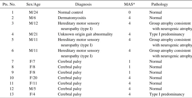
Table 1. Summary of Patients
Pts. No. Sex/Age Diagnosis MAS* Pathology
Pts. 01 M/24 Normal control 0 Normal
Pts. 02 M/60 Dermatomyositis 4 Normal
Pts. 03 M/12 Hereditary motor sensory 4 Group atrophy consistent
neuropathy (type I) with neurogenic atrophy
Pts. 04 M/21 Unknown origin gait abnormality 4 Type I predominancy
Pts. 05 M/11 Hereditary motor sensory 4 Group atrophy consistent
neuropathy (type I) with neurogenic atrophy
0Pts. 6 M/11 Hereditary motor sensory 4 Group atrophy consistent
neuropathy (type I) with neurogenic atrophy
Pts. 07 F/70 Cerebral palsy 1 Normal
Pts. 08 F/80 Cerebral palsy 1 Normal
Pts. 09 F/80 Cerebral palsy 1 Normal
Pts. 10 F/20 Cerebral palsy 4 Normal
Pts. 11 F/11 Cerebral palsy 4 Normal
Pts. 12 M/50 Cerebral palsy 4 Normal
Pts. 13 F/40 Cerebral palsy 4 Type I predominancy
*: modified Ashworth’s score
는 4등급이었다. 남자가 7명, 여자가 6명이었고 나이는 4세에서 24세까지로 다양하였으며 평균 나이는 11.4세였다(Table 1).
2. 조직학적 검사
통상적인 방법으로 haematoxylin and eosin 염색, 효소조직화학 염색(enzyme histochem- istry) 및 Gomori trichrome 염색을 시행하였으 며, 면역조직화학 염색(immunohistochem- istry)을 위해 3% hydrogen peroxide (H2O2) 로 조직내 내인성 peroxidase를 불활성시킨 후 blocking serum으로 실온에서 10분간 반응시켰 다. Anti-torpedo dystrophin 일차항체, ubiq- uitin 일차항체(DAKO, Denmark), N-CAM 일차 항체(Santa cruz, California, USA)로 37�C에서 2시간 반응시켰다. Avidin-biotin (Zymed Histo Plus kit, San Francisco, CA, USA) 방법을 이용하여 면역조직화학 염색을 수 행하였으며 blocking serum에서 일차항체 단계 를 제외한 각 단계에서 PBS 용액으로 수세하였 다. 3,3’-diaminobenzidine (DAB, Vector laboratories, Inc., Burlingame, CA, USA) 으로 발색하고 Mayer’s hematoxilin으로 대조 염색을 수행한 후 70, 90, 100% 에틸알코올에서 탈수하고 봉입하여 광학현미경하에서 관찰하였다.
3. 유전자 발현
근육 조직의 RNA 분리는 Trizol� (Invitro- gen, Carlsbad, Canada)를 이용한 acid guanidium thiocyanate-phenol chloroform
extraction 방법으로 시행하였다. 추출된 RNA 1 μl에 DEPC water를 첨가하여 50배 희석시킨 후 Spectrophotometry (λ=260 nm)로 흡광도 를 측정하였다. 측정된 값으로 전체 RNA양을 측 정하였고, DEPC water를 첨가하여 농도가 0.5 μg/μl가 되도록 하였다.
AccuPower�-RT PreMix (Bioneer, Korea) kit 20 μl에 정량된 total RNA 5 μg을 첨가하고 deoxyribonucleotide triphosphate (dNTP, Promega, USA) 1 μl를 최종적으로 첨가한 후 Thermo Hybrid PCR reactor (USA)를 이용 72�C에서 5분간 반응시켰다. AccuPower�-PCR premix tube (Bioneer, Korea)에 제작된 cDNA 1 μl와 primer 10 pmole를 넣고, 전체 부피가 20 μl가 되도록 증류수를 첨가하였다. 각 유전자에 대해 사용된 primer는 참고문헌과 Gene Bank를 이용하여 제작되었으며 염기서열 은 Table 2와 같다. Ubiquitin과 N-CAM의 경 우 94�C에서 1분 유지 후 94�C 5초, 54�C 5초 와 72�C 30초 조건으로 27 cycle을 시행하였고 72�C에서 2분간 안정시켰다. GAPDH의 경우 각각의 primer와 동일한 조건으로 수행하였다.
이후, 2% Agarose gel에 RT-PCR 결과로 얻어 진 cDNA 3 μl를 well에 넣고 100 volt에서 약 15분간 전기영동시켰다. DNA ladder를 함께 전 기영동하여 ubiquitin과 N-CAM의 경우 각각 120 bp, 140 bp에서 유전자 발현을 확인하였고 GAP DH의 경우 226 bp에서 유전자 발현을 확 인하였다. 확인된 두 유전자 발현을 ultraviolet 디지털 카메라로 촬영 후 Image-pro� 프로그램 을 이용하여 GAPDH 유전자의 발현을 기준으로 상대적인 발현 정도를 측정하여 비교하였다.
Table 2. Primer of ubiquitin, N-CAM and GAPDH
Ubiquitin Forward 5’- GGG TGT CAG TAT CTT TAA CGG C- 3’
Reverse 5’- CAG ACG GGC AAA GAT ACA CA- 3’
N-CAM Forward 5’- CGT AGA AAA CAT TGC ACA GA- 3’
Reverse 5’- CTA TGA ACA TGC ACG CAT AC- 3’
GAPDH Forward 5’- GAA GGT GAA GGT CGG AGT- 3’
Reverse 5’- GAA GAT GGT GAT GGG ATT TC- 3’
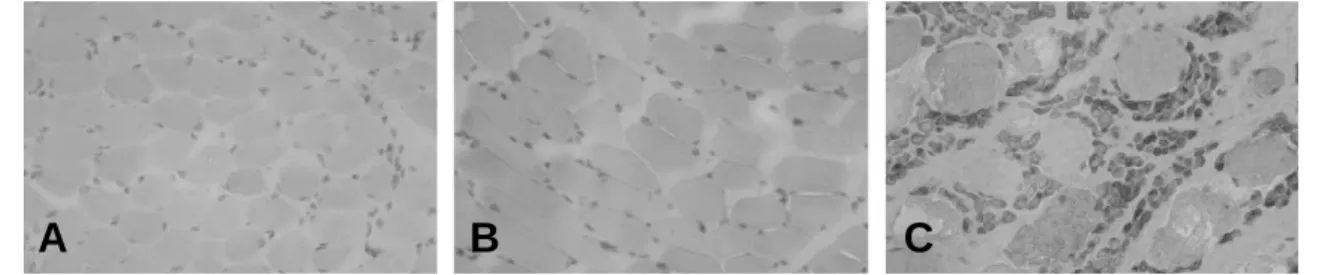
결 과 1. 조직병리학적 소견
뇌성마비 환자 7명 중 6명은 근육세포가 정상 크기의 다각형 모양으로 구성되어 있었고 핵은 근 섬유막하에 존재하며 효소조직화학 염색(ATPase pH 9.4, pH 4.3, pH 4.6, NAPH-TR)에서 제 1형 근섬유와 제 2형 근섬유가 정상 비율로 구 성되어 있었다(Fig. 1A). 근 섬유형 군화(fiber type grouping)나 근 섬유형 우세(fiber type predominency), 특정 근섬유의 위축이나 비대가 없는 정상 조직 소견을 보였으며 1명에서만 제 1 형 근섬유 우세를 보였다(Fig. 1B). Dystrophin 면역 염색에서도 근 섬유막을 따라 정상적인 면역 활성도를 보였으며 utrophin 증가 반응도 없었 다. 또한 미토콘드리아 근 질환을 관찰할 수 있는 Gomori trichrome 염색에서는 ragged red fiber나 nemaline 간체는 없었다. 유전성 운동 및 감각 신경병증 환자 3명에서는 신경원성 근 위
축에 합당한 소견인 소집합 위축(small grouped atrophy)이 관찰되었다(Fig. 1C). 원인 미상의 보행 이상 환자에서는 제 1형 근 섬유형 우세 소 견이 관찰되었다.
2. 조직화학적 소견
모든 환자들에서 ubiquitin 단백에 대한 염색 은 음성반응을 보였다(Fig. 2A-C). 한편 N- CAM 단백에 대한 면역화학염색 소견 상 뇌성마 비 환자에서는 염색이 되지 않았으며 근 위축이 심했던 유전성 운동 및 감각 신경병증 환자 3명 모두에서 위축된 근섬유의 세포질에서 강한 N- CAM 발현이 관찰되었다(Fig. 3A-C).
3. 유전자 발현
1) Ubiquitin 유전자 발현
생검한 근육의 일부를 이용하여 전체 RNA를 적출한 후 역전사 중합효소 연쇄반응을 시행한 결
Fig. 1. Histopathological findings (×20). (A) Normal muscle cells were noted in patient 7, who was a cerebral palsy patient (H-E staining). (B) Type I fiber was dominant in patient 13, a cerebral palsy patient (ATPase pH 9.4 staining). (C) Small grouped atrophy was noted in patient 6, a hereditary motor sensory neuropathy patient (H-E staining).
Fig. 2. The immunochemical staining of ubiquitin protein (×20). (A) Negative reaction was noted in patient 1, a con- trol group. (B) This figure is a negative control of patient 7, and you can compare it with other stained figures.
(C) Negative reaction was noted in patient 13, a cerebral palsy patient.
A B C
A B C
과, 근 경직도가 낮았던(MAS 1등급) 뇌성마비 환자 7, 환자 8, 환자 9에서는 ubiquitin 유전자 발현이 다른 환자에 비해 낮았으나 경직도가 높았 던 4등급 뇌성마비 환자들에서는 ubiquitin 유전 자 발현이 다른 환자들에 비해 증가한 소견이 관 찰되었다(Fig. 4). 유전성 운동 및 감각 신경병 증 환자 3명 및 제 1형 근 섬유형 우세를 보인 원 인 불명의 보행 이상 환자에서는 대조군과 비슷하 거나 높은 발현을 보였다.
2) N-CAM 유전자 발현
뇌성마비 환자에서는 환자 12를 제외하고 모두 발현되었다(Fig. 5). 또한 경직도가 낮았던 환자 들에서 경직도가 높았던 환자 10, 11, 12, 13보
다 상대적으로 강한 발현이 관찰되었으며 피부근 염 환자, 원인 미상의 보행 장애 환자 및 3명의 유전성 운동 및 감각 신경병증 환자에서는 모두 강하게 발현되었다.
고 찰
일반적으로 정상 골격근은 잘 조화된 합성과 분 해 과정을 통해 항상 근육의 안정성과 항상성을 유지하고 있다. 정상적인 생체 반응으로써 금식을 하게 되면 근육 단백질의 분해가 일어나 아미노산 이 생성되며 간에서의 포도당신생(gluconeogen- esis)에 쓰이게 된다. 이러한 외부 환경의 변화에 대한 생체의 유익한 반응이 있는 반면 신경이 단
Fig. 3. The immunochemical staining of N-CAM protein (×20). (A) Negative reaction was noted in patient 1, a con- trol group. (B) This figure is a negative control of patient 13, and you can compare it with other stained fig- ures. (C) Strong positive reaction was noted in patient 5, a hereditary motor sensory neuropathy patient.
Fig. 4. Ubiquitin gene expression. In the cerebral palsy patients, there seemed to be positive correlations between the expression of ubiquitin gene and the degree of spasticity. 1: normal control, 2: dermatomyositis, 3: hereditary motor sensory neuropathy, 4: unknown origin gait abnormality, 5, 6: hereditary motor sensory neuropathy, 7, 8, 9, 10, 11, 12, 13: cerebral palsy.
A B C
절되는 경우와 같은 병적인 환경에서는 생체의 정 상적인 조절 범위를 벗어나게 되어 근 단백 분해 가 가속화되고 단백질 합성보다 분해가 더 많아져 근 소모가 일어나게 된다13,14). 또한 근 소모가 지 속될 경우에는 비가역적 단계인 근 위축으로 진행 하게 된다.
경직성 뇌성마비 환자는 골격근의 비정상적인 긴장상태로 인한 근육 구축, 관절 변형 및 탈구 등 근, 골격계 이상으로 인해 정형외과적 수술이 적응되는 경우가 흔하다. 본 연구에서는 수술 전 신체 문진 및 수술 시에 근육 탄력성을 기준으로 하여 근육 경직도에 따라서 환자를 선택하여 근건 이전술 및 연장술시 근 생검을 시행하였다. 역전 사 중합효소 연쇄반응에서 ubiquitin 유전자 발 현은 경직도 1등급이었던 환자 3명에서 다른 환 자에 비해 낮은 유전자 발현을 보였고, 경직도 4 등급이었던 환자들에서는 높은 유전자 발현이 관 찰되었다. 따라서 근육의 경직도와 ubiquitin 유 전자 발현과의 상관관계를 유추해 볼 수 있다.
Castle4) 등은 경직성 뇌성마비 환자에 있어서 근 육의 조직학적 변화를 기술하면서 경직성 뇌성마 비 환자는 지속적인 긴장성 근육 활성화를 보이므 로 제 1형 근섬유의 비대가 보여야 하나 오히려 위축을 보였는데 그에 대한 가설로 경직성의 관절
고정역할을 제시하였다. Booth2) 등도 경직성이 골격근에 미치는 영향은 고정이나 탈신경, 건절제 술이 골격근에 미치는 영향과 유사하다고 하였다.
Ubiquitin은 고정, 탈신경, 근 무부하(muscle unloading)와 같은 불용성 상태(disuse condi- tion)에서 활성화되는데 경직성이 이러한 불용성 상태를 유발했다고 할 수 있겠다. 또한 경직성 뇌 성마비에서 경직도가 심해질수록 근 병변성 (myopathic) 조직변화가 보이는데 이러한 질환 의 만성화가 근육의 구축을 심화시키면서 관절 고 정상태를 유발시켰다고 생각할 수 있다4). 경직도 가 1등급이었던 환자 7, 환자 8, 환자 9에서 대 조군보다 더 낮은 ubiquitin의 발현을 보였는데 이는 ubiquitin이 아닌 다른 경로를 통한 근 소 모가 더 활성화되었기 때문이라고 유추해 볼 수 있겠다.
한편, N-CAM 유전자 발현은 정상인과 뇌성마 비 환자 1명을 제외한 모든 환자들에서 발현이 증 가되었다. 특이할 만 한 것은 뇌성마비 환자에서 경직도가 낮았던 환자 군이 경직도가 높았던 환자 군에 비해 상대적으로 강한 발현을 보였다는 점이 다. N-CAM은 대부분의 말초신경이나 중추신경 의 표면에 있는 막성 당단백으로써 태아 발생시에 관상근세포(myotube)의 표면에서 나타나지만 성 Fig. 5. N-CAM gene expression. In the cerebral palsy patients, there seemed to be negative correlations between the expression of N-CAM gene and the degree of spasticity. 1: normal control, 2: dermatomyositis, 3: hereditary motor sensory neuropathy, 4: unknown origin gait abnormality, 5, 6: hereditary motor sensory neuropathy, 7, 8, 9, 10, 11, 12, 13: cerebral palsy.
장하면서 없어지게 되어 성인이 되면 완전히 사라 지게 된다5). 그러나 성인 근육에 탈신경화가 일어 나게 되면 다시 근육에 나타나게 되는데 여기서 N-CAM이 재신경지배에 중요한 역할을 한다5). N-CAM은 근섬유 사이의 간질조직에서도 나타나 며 신경 종판이 있었던 자리에 집중적으로 분포함 으로써 재신경이 지배될 자리를 유도한다고 알려 져 있다5). 재신경지배가 완전히 끝나게 되면 N- CAM도 사라지게 된다. N-CAM은 정상적인 근 육에는 없으며 다양한 신경근육성 질환에서 발현 이 증가 되는 것으로 알려져 있는데 대표적 질환 은 척수성 근육 위축증(spinal muscular atro- phy), 다발성 근염(polymyositis), 진행성 근이 영양증(progressive muscular dystrophy) 등 이다23). 탈신경 및 재신경지배는 연속적이면서 서 로 중복되는 과정으로 볼 수 있는데 이런 과정에 서 N-CAM 유전자 발현이 가장 많이 나타나는 시점은 탈신경 과정과 재신경지배가 중복되는 기 간이고 재신경지배가 진행되면서 서서히 N-CAM 도 사라지게 된다. 뇌성마비 환자에서 N-CAM 발현 기전은 탈신경-재신경지배 시에 일어나는 기 전과 일치하지는 않지만 비정상적인 상부 신경계 통으로부터의 근세포 반응이라는 점에서 볼 때 경 직성이 심한 경우를 탈신경-재신경지배 과정에서 N-CAM 유전자가 발현되는 과정의 말기 과정으 로 볼 수 있으며, 여기서 뇌성마비 환자의 경직도 와 N-CAM 유전자 발현 강도의 역상관관계를 유 추해 볼 수 있겠다. 또한 과도한 지속적인 긴장성 자극에 대한 근세포의 탈감작 및 탈진(exhaus- tion)으로 경직도가 심한 경우에는 N-CAM 유전 자 발현이 감소했다고 유추할 수 있다. 한편, 면 역화학 염색에서 ubiquitin 단백은 모든 환자에 있어서 염색이 되지 않았으며 N-CAM 단백은 유 전성 운동 및 감각 신경병증 환자 3예에서만 염색 이 되었다. 이는 전사 후(post-translational) 단백질 합성과정에서의 장애라고 유추해 볼 수 있 으며 이에 대한 추가적인 연구가 필요하리라 사료 된다. 또한 본 연구 자체가 환자를 대상으로 한 생검이었기 때문에 충분한 근육을 얻을 수 없었고 면역 염색을 실시한 냉동 절편이 매우 적은 부위 이기 때문에 실험 오차가 있을 수 있다는 점을 고 려해 볼 수 있겠다. 따라서 추가적인 연구에서는
western blot을 이용한 실험 방법을 택하는 것이 더 정확한 연구 결과을 얻는 데 도움이 될 것으로 사료된다.
조직학적 변화는 경직성 뇌성마비 환자 7명 중 1명이 제 1형 근섬유 우세를 보였으며 다른 환자 에서는 정상 소견을 보였다. Castle4) 등의 보고 에서도 약 반수에서만 다양한 소견의 조직학적 변 화가 보인다고 했다. 제 1형 근섬유형 우세는 뇌 성마비 환자에서 흔히 보이는 조직학적 소견으로 써 긴장성 근섬유(tonic muscle)인 제 1형 근섬 유의 지속적인 활성에 의한 소견으로 볼 수 있다.
Seter21) 등은 동물 실험에서 지속적인 고주파 자 극(high frequency stimulation)에 의해서 제 2형 근섬유가 제 1형 근섬유로 변환 되는 것을 증 명하였다. 따라서 경직성 뇌성마비 환자에서도 경 직성으로 인한 지속적인 근육 활성화에 의해 유사 한 과정이 생겼다고 추측해 볼 수 있다. Ito9) 등 은 경직성 뇌성마비 환자들의 근육 생검에서 질병 특이적인 이상소견은 없었으나 근섬유형 분포에 있어서 변화를 관찰하였는데 이전 연구 결과와 유 사한 제 1형 근섬유형 우세와 제 2B형 근섬유형 결핍 양상을 확인하였다. 유전성 운동 및 감각 신 경병증 환자 3명에서는 신경원성 근 위축에 합당 한 소견인 소집합 위축(small grouped atro- phy)을 보였다. 근육은 정상적으로 한 종류의 근 섬유형끼리 모여 있지 않고 서양바둑판 모양으로 서로 다른 근섬유형이 섞여 있으나 비정상적인 경 우에 한 종류의 근섬유형끼리 모여 있는 소견을 근섬유형 군화(fiber type grouping)라 부른다.
이 소견은 신경원성 근위축의 대표적인 조직학적 소견으로 탈신경 후 나타나는 신경 재지배에 의한 이차적인 변화로 생각된다. Booth2) 등은 뇌성마 비 환자에서 경직도에 따른 조직학적 변화를 보고 하였는데 경직도가 높은 환자일수록 교원질의 양 이 많으며 근섬유의 원형화와 섬유간 간격의 증가 를 보인다고 하였다. 본 연구에서는 경직도에 따 른 특이적인 변화를 보인 예는 없었다.
이상에서 본 바와 같이 본 연구에서도 다양한 결과의 조직학적인 소견과 ubiquitin 및 N- CAM 유전자 발현을 보였다. 이는 여러 가지 요 인들이 복합적으로 작용하여 뇌성마비 환자의 골 격근에 영향을 미치기 때문이라고 유추할 수 있
다. 가능한 변수로는 경직도, 보행가능 여부, 변 형된 관절 및 근육에 대한 물리치료 기간 및 효율 성, 일상생활의 활동성 등이 있을 수 있으며 이에 대한 추가적인 연구가 더 필요하다고 판단된다.
결 론
본 연구에서는 뇌성마비 환자의 경직도에 따른 조직병리학적 변화 및 ubiquitin과 N-CAM 유 전자 발현 양상을 관찰하고자 하였다. 경직이 심 한 환자에서 ubiquitin 유전자 발현은 증가하였 고 N-CAM 유전자 발현은 감소하는 양상을 보였 다. 이러한 소견은 향후 뇌성마비 환자에 있어서 분자생물학적 연구 및 치료에 적용이 될 수 있으 리라 사료된다.
REFERENCES
01) Auclair D, Garrel DR, Chaouki ZA and Fer- land LH: Activation of the Ubiquitin pathway in rat skeletal muscle by catabolic doses of glu- cocorticoids. Am J Physiol, 272: C1007-16, 1997.
02) Booth CM, Cortina MJ and Theologis TN:
Collagen accumulation in muscles of children with cerebral palsy and correlation with severity of spasticity. Dev Med Child Neurol, 43: 314- 320, 2001.
03) Calore EE, Sesso A, Puga FR, Cavaliere MJ, Calore NMP and Weg R: Early expression of Ubiquitin in myofibers of rats in organophos- phate intoxication. Ecotoxicol Environ Saf, 43:
187-194, 1999.
04) Castle ME, Reyman TA and Schneider M:
Pathology of spastic muscle in cerebral palsy.
Clin Orthop, 142: 223-232, 1979.
05) Covault J and Sanes JR: Neural cell adhesion molecule (N-CAM) accunulates in denervated and paralyzed skeletal muscle. Proc Natl Acad Sci USA, 82: 4544-4548, 1985.
06) Curtis JW, Joshua MVM, Dan DH and Per- Olof H: Sepsis up regulate the gene expression of multiple ubiquitin ligases in skeletal muscle.
Int J Biochem Cell Biol, 35: 298-705, 2003.
07) Fang CH, Tiao G, James JH, Ogle CK, Fischer JE and Hasselgren PO: Burn injury stimulates multiple proteolytic pathways in skeletal muscle, including the ubiquitin-energy-dependent path- way. J Am Coll Surg, 180: 161-170, 1995.
08) Furuno K, Goodman MN and Goldberg AL:
Role of different proteolytic systems in the degra- dation of muscle proteins during denervation atrophy. J Biol Chem, 265: 8550-8557, 1990.
09) Ito J, Araki A, Tanaka H, Tasaki T, Cho K and Yamazaki R: Muscle histopathology in spastic cerebral palsy. Brain Dev, 18: 299-303, 1996.
10) Jackman RW and Kandarin SC: The molecu- lar basis of skeletal muscle atrophy. Am J Physi- ol, 287: 834-843, 2004.
11) Jagoe RT and Goldberg AL: What do we really know about the ubiquitin-proteosome pathway in muscle atrophy? Curr Opin Clin Nutr Metab Care, 4: 183-190, 2001.
12) Kumamoto T, Fujimoto S, Ito T, Horinouchi H, Ueyama H and Tsuda T: Proteasome expression in the skeletal muscles of patients with muscular dystrophy. Acta Neuropathol, 100(6): 595-602, 2000.
13) Lecker SH, Solomon V, Mitch WE and Gold- berg AL: Muscle protein breakdown and the critical role of the ubiquitin-proteosome pathway in normal and disease states. J Nutr, 129: 227S- 237S, 1999.
14) Lecker SH, Thomas J, Alexander G, et al.:
Multiple types of skeletal muscle atrophy involves a common program of changes in gene expression. FASEB J, 18: 39-51, 2004.
15) Llovera M, Garcia-Martinez C, Agell N, Marzabel M, Lopez-Sariano FJ and Argiles JM: Ubiquitin gene expression is increased in skeletal muscle of tumor-bearing rats. FEBS Lett, 338: 311-318, 1994.
16) Mansoor O, Beaufrere B, Boirie Y, et al.:
Increased mRNA levels for components of the
lysosomal, Ca-activated, and ATP-ubiquitin- dependent proteolytic pathways in skeletal mus- cle from head trauma patients. Proc Natl Acad Sci USA, 93: 2714-2718, 1996.
17) Marbini A, Ferrari A, Cioni G, Bellanova MF, Fusco C and Gemignani F: Immunohistochem- ical study of muscle biopsy in children with cere- bral palsy. Brain Dev, 24(2): 63-66, 2002.
18) Micth WE and Goldberg AL: Mechanisms of disease: Mechanisms of muscle wasting- The role of the ubiquitin-proteosome pathway. N Engl J Med, 335(25): 1897-1905, 1996.
19) Mitch WE, Medina R, Grieber S, et al.: Meta- bolic acidosis stimulates muscle protein degrada- tion by activating the adenosine triphosphate- dependent pathway involving ubiquitin and pro- teasomes. J Clin Invest, 93: 2127-2133, 1994.
20) Mutch L, Alberman E, Hagberg B, Kodama K and Perat MV: Cerebral palsy epidemiology;
where are we now and where are we going? Dev
Med Child Neurol, 34: 547-551, 1992.
21) Sreter FA, Pinker K, Jolesz F and Mabuchi K: Fast to slow transformation of fast muscles in response to long-term phasic stimulation. Exp Neurol, 75: 95-102, 1982.
22) Tiao G, Hobler S, Wang JJ, et al.: Sepsis is associated with increased mRNAs of the Ubiqui- tin-proteasome proteolytic pathway in human skeletal muscle. J Clin Invest, 99: 163-168, 1997.
23) Walsh FS, Moore SE and Lake BD: Cell adhe- sion molecule N-CAM is expressed by denervat- ed myofibres in Wernig-Hoffman and Kugel- berg-Welander type spinal muscular atrophies. J Neurol Neurosurg Psychiatry, 50: 439-442, 1987.
24) Williams A, Sun X, Fischer JE and Hassel- gren PO: The expression of genes in the ubiqui- tin-proteasome proteolytic pathway is increased in skeletal muscle from patients with cancer.
Surgery, 126(4): 744-750, 1999.