J. of Korean Orthopaedic Research Society V o l u m e 5 , N u m b e r 1 , A p r i l , 2 0 0 2
골시멘트에 함유된 BaSO
4가 쥐의 골모세포에 미치는 영향
가톨릭대학교 의과대학 정형외과학교실, 가톨릭대학교 의과대학 약리학교실*
김용식・우영균・정진화・양성철 권순용・이은정*・이권행*
= Abstract =
Effect of BaSO
4in Cement on Rat Osteoblast
Yong-Sik Kim, M.D., Young-Kyun Woo, M.D., Jin-Wha Chung, M.D., Seong-Chul Yang, M.D., Soon-Yong Kwon, M.D., Eun-Jung Lee, M.D.*, Kweon-Haeng Lee, M.D.*
Department of Orthopedic Surgery, Department of Pharmacology*, College of Medicine, Catholic University of Korea
Purpose : We hypothesize that the presence of barium sulfate debris plays an influential role to deteriorate the balance of bone remodelling around prosthesis via cytotoxic mechanism to osteoblast.
Materials and Methods : Osteoblasts were obtained from the neonatal rat calvarium, and SiO2, TiO2, PMMA and BaSO4 particles were prepared for the evaluation of particle induced cytotoxicity to osteoblast.
Osteoblasts were grown in DMEM and then were seeded into 6 well culture plates. 1.0wt% solution of each particle was added to culture medium to generate a final concentration of 0.1wt%, and 0.005wt% of various particles in each well, respectively. The measurement of intracellular calcium was conducted using various agonists of calcium. The cell viability assay for osteoblast was performed with MTT reduction assay and the mineralization of the matrix was checked by Von Kossa staining. ELISA kit was used to quantify the level of osteocalcin in osteoblast.
Results : BaSO4significantly lowered the cell viability. All particles except TiO2increased [Ca2+]i transient- ly, and the rank of differential cytosolic [Ca2+]i was in order as follows; SiO2, BaSO4, and PMMA. The mineral- ization was significantly prohibited in SiO2 and BaSO4(0.1wt%), however the PMMA showed no definite inhibitory effect on bone mineralization. PMMA(0.1wt%) and BaSO4(0.1wt%) showed significantly inhibitory effect on osteocalcin production.
C o n c l u s i o n : In higher concentration, BaSO4has a cytotoxic effect on osteoblast and inhibitory effect of osteocalcin production as well as mineralization of osteoblast. Also, this study has shown that the concentration of intracellular calcium is strongly influenced by exposure to BaSO4particles in vitro. The effect of BaSO4on osteoblast observed in this study could have implications for the role of BaSO4particles on osteoblast function at aseptic loosening of cemented total joint arthroplasty.
Key Words : Bone Cement, Barium sulfate (BaSO4), Ostoblast
※ 통신저자 : 권 순 용
서울시 영등포구 여의도동 6 2번지 가톨릭의과대학 성모병원 정형외과학교실 Tel : 02) 3779-1192, Fax : 02) 783-0252 E-mail : [email protected]
서 론
고관절 인공 관절 전치환술에서 생기는 마모 미 립자(wear particle)는 관절주위 골용해 (periprosthetic osteolysis)의 주된 원인 중 한 가지로 제시되어 왔으며, 이는 인공 관절 삽입물 주변을 싸고 있는 섬유막에서 발견되고, 성분으로 는 금속, 골 시멘트, 폴리에틸렌, 그리고 세라믹 등이 있다1 - 5 ). 이 마모 미립자들은 생물학적으로 대 식세포, 섬유아세포, 림프구, 거대세포 등으로부터 염증 물질을 분비하게 하여 세포의 식작용( p h a g o- c y t o s is)과 그에 따른 골 흡수를 유발한다6 - 9 ).
이들 중 골 시멘트(bone cement) 미립자는 인 공 삽입물의 적절한 삽입에도 불구하고 골시멘트 성 인공 관절 주변에서 삽입 후 즉시 발견되는 것 으로 보고되고 있고, 또한 골 시멘트에 포함된 성 분 중 방사선 비투과성 조영제인 B a S O4( b a r i u m s u l f a t e )에 대해서, Sabokbar등은, 대식세포-파 골세포 분화를 촉진함으로써 무균성 인공 관절 해 리에서의 골용해에 기여하며, 임상적 비교실험에 서 이러한 B a S O4를 함유한 P M M A ( p o l y- m e t h y l m e t h a c r y l a t e )가 Z r O2( z i r c o n i u m d i o x i d e )를 함유한 P M M A보다 골 흡수를 더 잘 유발한다고 보고하였다1 0 - 1 3 )
. 또한, 실험동물에게 B a S O4를 피하 주사하여 육아종성 염증반응 (granulomatous inflammatory reaction)을 유발하였다는 보고도 있었다1 4 ).
현재까지 P M M A에 대한 생물학적 독성기전은 실험을 통하여 많이 연구되어왔으나, 과연 이러한 실험 결과들이 PMMA particle 단독에 의한 것 인지, 아니면 방사선 비투과성 조영제를 포함한 여러 첨가제와 같이 작용을 하는 지는 아직 명확 하지 않고, 실험 방법도 파골세포의 활동도에 대 한 효소 측정 및 염증반응의 측정 등이 대부분이 었으며, 골모세포에 관한 연구는 알려진바 없다.
이에, 저자들은 인공 관절 주위의 세포들이 골 형성과 연관된 골모세포, 골수 유래 세포( m a r- row derived cells), 간엽세포 등으로 구성되어 있다는 사실을 고려하여, BaSO4의 골모세포에 대한 영향을 실험적으로 연구하였으며, SiO2, T i O2, BaSO4 그리고 PMMA particle들을 비
교 분석함으로써, BaSO4가 골모세포에 미치는 세포 독성 기전을 규명하고자 하였다.
연구재료 및 방법 1. Particles
평균 1.6 ㎛ 크기의 S i O2p a r t i c l e ( M i n - U - S i l 5 ; U.S. Silica Co, Clarks-town, WV, USA)과 1
㎛ 보다 작은 크기의 T i O2(Sigma Chemical Co.
St. Louis, MO, USA)를 준비하였다. 또한, 직경 1 ㎛에서 10 ㎛정도의 MMA-styrene copoly- mer 입자로 구성된 PMMA p o w d e r와 골시멘트 CMV (Depuy, Leeds,U K )에서 얻어진 s u b m i- c r o n에서 10 ㎛ 정도 크기의 B a S O4를 준비하였다.
2. Cell Culture and Particle Exposure
골모세포는 neonatal rat의 c a l v a r i u m에서 채취하였으며, Osteoblast의 분리에 효소( c o l l a- g e n a s e )를 이용하였으며, 얻어진 세포들을 1 0 % fetal bovine serum (FBS), non-essential amino acid (0.10 mM), L-glutamine (4 mM), penicillin (100 U/ml), strep-tomycin (100 ㎍/ml), fungizone (0.25 ㎍/ml) (Biowhit- taker, Walkersville, MD)가 함유된 D u l b e c- c o’s Modified Eagle’s Medium(DMEM)에서 배양하였다. 배양 과정 중 3 7℃ 및 5% CO2를 유 지하였으며, trypsin-EDTA (1:250 trypsin, 200 ㎍/ml EDTA; Biowhittaker, Walk- ersville, MD) 혼합액을 이용하여 5분 동안 t r y p s i n화 한 후에, DMEM으로 두 차례 세척하 였으며, 부유 한 후 DMEM 배지에서 교반하였 다. 마지막으로 골모세포들을 w e l l당 5 0 0 , 0 0 0개 의 밀도로 5개의 시험관 배지에 각각 나누었다.
대조군 시험관에는 아무런 입자도 첨가하지 않 았으며, SiO2, TiO2, PMMA 그리고 B a S O4
p a r t i c l e들을 a u t o c l a v e를 이용하여 소독한 후, 골모세포들이 들어있는 DMEM 시험관 배지에 넣어 입자와 배지의 질량 비율이 1 . 0 w t %가 되게 stock solution을 만들었다. 그리고, 분자들의 응집을 최소화하기 위하여 세포 배양액에 넣기 전
에 3 0분 동안 초음파 처리하였다. 입자들을 배양 표면에 잘 부착시키기 위해 3 7℃에서 4시간 동안 배양하였으며, 1.0wt% particle solution을 배 지에 첨가하여 입자마다 각각 0 . 1 w t % , 0 . 0 0 5 w t %의 농도를 형성하도록 하여 대조군 시 험관 2개를 포함한 총 1 0개의 시험관 배지를 만 들었다.
3. M T T ( 3 - ( 4 , 5 - d i m e t h y l t h i a z o l - 2 - y l ) - 2 , 5 - diphenyltetrazolium bromide) Reduc- tion Assay
위의 방법으로 배양된 골모세포들의 생존도를 측정하기 위해 MTT reduction assay를 이용하
였다1 5 ). MTT는 살아있는 세포의 마이토콘드리아
에 있는 succinate-tetrazolium reductase s y s t e m을 통해 f o r m a z a n으로 분해된다. 그러므 로, 각각의 시험관 배지에 DMEM 1ml당 M T T 0.5 mg/ml를 가한 후 4시간, 24시간, 72시간 동안 배양하였고, 마지막으로 40 mM HCL-iso- propanol 100 ㎕를 이용하여 배지에 남은 M T T 를 씻어낸 후, microplate reader(MR700, Dynatech, Chantilly, VA, USA)를 이용하여 f o r m a z a n의 농도를 측정하였다.
4. Intracellular Ca2 + A s s a y
C a2 + sensitive indicator인 F u r a - 2 / A M (Sigma Chemicals, Poole, UK)를 이용하여 각 배지들의 intracellular calcium level을 측 정하였다.
5. Calcium Phosphate Mineral Deposit
50 ㎍/ml ascorbic acid와 10mM ß- g l y c- erol phosphate을 포함한 DMEM 배지를 3주간 배양 Von Kossa staining 을 이용하여 무기질 침착 정도를 측정하였다.
6. Production of Osteocalcin
상기와 같은 방법으로 배지를 배양한 후 E L I S A kit(Biomedial technologies Inc., Stoughton, M A )를 이용하여 osteocalcin 의 농도를 측정하 였다.
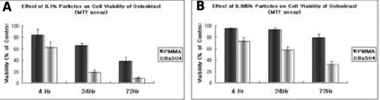
결 과 1. MTT Reduction Assay
B a S O4를 포함한 배지에서는 배양시간이 경과 함에 따라 그리고, 농도가 증가함에 따라 세포 생 존도가 낮아지는 현상을 보였다. 또한 P M M A에 대한 세포 생존도의 비교실험에서도 시간의 경과 와 농도의 증가에 따라 세포증식 능에서 의미 있 는 차이를 보였다(p<0.05)(Fig. 1).
2. Intracellular Ca2+ Assay
세포 내 칼슘이온 농도에 영향을 주는 다양한 입자를 이용한 비교측정 결과, TiO2를 제외한모 든 p a r t i c l e들이 intracellular calcium을 일시
Fig. 1. Effects of 0.1wt%(A) and 0.005wt%(B) particles on cell viability using MTT reduction assay. BaSO4particles decreased more cell viability with time and concentration dependent manner as compared with PMMA particles.
A B
Fig. 2. Intracellular calcium assay of osteoblast with various particles.The rank of differential cytosolic [Ca2+]i was SiO2, BaSO4and PMMA(A) in order. BaSO4shows more significant transient increase of intracellular calcium level as compared with PMMA. (▲) is the point at which particles were added to ostoblasts(B)
Fig. 3. Intracellular calcium assay of control(A,C) and BaSO4(B,D) Each 9 images of stippled cluster are the calcium speckled into the cytoplasm of osteoblasts(A,B) and was modified into graph (C,D). When BaSO4is added to osteoblast(B,D), transient increase of intracellular calcium level is observed.
Fig. 4. The mineralization was significantly prohibited in 0.1wt% BaSO4. (p<0.05)
A B
A B
C D
Fig. 5. Both PMMA and BaSO4decreased significantly osteocalcin release from osteoblasts. (p<0.05)
적으로 증가시켰으며, 그 정도는 S i O2, BaSO4, P M M A의 순서로 나타났다(Fig. 2). PMMA와 B a S O4 와의 비교실험에서 B a S O4 첨가시의 세포 내 칼슘의 증가 정도가 P M M A에 대하여, 급격 하게 증가하는 경향을 보였다(p<0.05)(Fig. 3).
3. Calcium Phosphate Mineral Deposit
0.1wt% PMMA및 B a S O4 입자를 이용한 3주 간의 배양 후, Von Kossa 염색법에서 무기질 침 착( m i n e r a l i z a t i o n )은 대조군에 비하여 P M M A 와 B a S O4에서 감소를 보였다(p<0.05)(Fig. 4).
4. Production of Osteocalcin
골모세포로부터 유리되는 o s t e o c a l c i n은 P M M A와 B a S O4에서 대조군과 비교하여 감소하 였다(p<0.05)(Fig. 5).
고 찰
고관절 인공 관절 치환술에서 골시멘트를 이용 할 때 방사선학적 추시 검사를 위해 비투과성 물 질인 B a S O4나 Z r O2는 P M M A에 흔히 혼합되 며, 이 물질들은 조직학적으로 구체(sphere) 모 양의 PMMA 표면에 작은 과립의 형태로 분포한 다. 관절주위 골용해가 있는 경우, 이들 방사선 비투과 물질들이 높은 농도로 발견된다.
L a z a r u s등은 쥐의 슬관절에 B a S O4 가 포함된 PMMA particle과 그렇지 않은 PMMA parti- c l e을 주사하여 추시 관찰한 결과, BaSO4 가 포 함된 PMMA particle이 골흡수를 더 잘 유도하 였다고 보고하였다1 6 ). 이와 같은 P M M A의 세포 독성 및 생물학적 영향에 대한 실험적 연구는 상 당히 많이 있었으나, 이런 연구들이 단독으로 PMMA 자체가 원인인지, 아니면 방사선 비투과 성 물질 등 다른 첨가물을 함유한 P M M A가 원 인인지에 대한 연구는 부족하였다. 이에 S a b o k- b a r등은 실험적으로 방사선 비투과성 물질이 첨 가된 PMMA particle을 mononuclear phago- c y t e에 첨가하면, PMMA 자체만 첨가할 때와 비교하여 대식세포-파골세포의 분화와 파골세포와
관련된 효소들이 증가한다고 하였다1 2 ). 이는 결국 B a S O4 와 Z r O2 모두 파골세포 분화를 촉진하고 골흡수를 유도하지만, 특히 B a S O4가 포함된 PMMA particle에서 골흡수가 더욱 의미 있게 증가하였다고 하였으며, 그 기전으로 B a S O4가 포함된 PMMA particle에 접촉한 단핵세포가 파골세포로 분화되면서 골 용해를 유발한다고 주 장하였다.
인공관절이 삽입되어있는 골은 지속적인 재형성 과정이 일어나게 되며, 이와 연관된 가장 주된 세 포군은 골 모세포, 골세포, 파골세포 등의 상호 균형적이 관계에 의한다. 최근 인공관절해리의 병 리기전에 대한 다수의 연구들 중에서 p a r t i c l e과 골모세포와의 생물학적 영향에 대한 보고들은 새 로운 측면에서의 인공관절해리를 해석하고자 하였 다1 7 - 1 9 ).
따라서 저자들은 barium sulfate의 인공관절 해리와 연관된 세포들에 대한 생물학적 영향을 골 모세포를 대상으로, 이미 세포독성효과가 극명하 다고 밝혀진 S i O2와 세포 독성효과가 별무한 T i O2를 각각 대조군으로 설정하여 비교분석하고 자 하였다1 5 , 1 8 ).
본 실험을 통하여 시멘트형 인공고관절 치환술 에 흔히 사용되는 방사선 조영제 barium sul- f a t e는 높은 농도인 경우에 골모세포에 대하여 세 포 내의 급격한 calcium 증가를 통한 세포 독성 의 효과를 보였고, 이와 함께 세포 증식의 감소, 무기질 침착 감소 및 osteocalcin 세포 내 생산의 감소가 각각 관찰되었다. 따라서 본 연구를 통하 여 관찰된 barium sulfate에 대한 골모세포의 생물학적 효과는 인공관절의 무균성 해리의 병리 기전과 관계 있는 인자로 고려될 수 있을 것이다.
결 론
시멘트형 인공관절 치환술에서 P M M A에 방사 선 비투과성 물질로 사용되는 B a S O4는 대식세 포-파골세포의 분화를 촉진하여 골흡수 및 인공 관절 해리를 유도한다고 보고되고 있는 바, 본 연 구에서는 B a S O4가 직접적으로 골 형성에 관여하 는 골모세포의 영향을 연구함으로써, 실험적으로 (in vitro) BaSO4가 골모세포의 기능을 감소시
켜 골의 재형성 과정에 불균형을 초래함으로써 이 러한 골흡수 및 인공 관절 해리에 기여한다고 추 정할 수 있었다. 향후, 골시멘트 내에 포함된 B a S O4가 생물학적 측면에서 골의 재형성과정에 어떠한 영향을 미치는가에 대한 추가적인 동물 실 험을 통한 연구가 필요할 것으로 요구된다.
R E F E R E N C E S
01 ) Goldring SR, Schiller AL, Roelke M : The synovial- like membrane at the bone-cement interface in loose total hip replacements and its proposed role in bone l y s i s . J Bone Joint Surg Am, 65(5): 575-584, 1983.
02 ) Goldring SR, Jasty M, Roelke MS : Formation of a synovial-like membrane at the bone-cement interface.
Its role in bone resorption and implant loosening after total hip replacement. Arthritis Rheum, 29(7): 836-842, 1 9 8 6 .
03 ) Maguire JK Jr, Coscia MF, Lynch MH : Foreign body reaction to polymeric debris following total hip arthroplasty. Clin Orthop, 216: 213-223, 1987.
04 ) Goodman SB, Chin RC, Chiou SS : A clinical-patho- logic-biochemical study of the membrane surrounding loosened and nonloosened total hip arthroplasties. C l i n O r t h o p , 244: 182-187, 1989.
05 ) Willert HG, Bertram H, Buchhorn GH : Osteolysis in alloarthroplasty of the hip. The role of bone cement fragmentation. Clin Orthop, 258: 108-121, 1990.
06 ) Murray DW, Rushton N : Macrophages stimulate bone resorption when they phagocytose particles. J Bone Joint Surg Br, 72(6): 988-992, 1990.
07 ) Amstutz HC, Campbell P, Kossovsky N, Clark IC : Mechanism and clinical significance of wear debris- induced osteolysis. Clin Orthop, 276: 7-18, 1992.
08 ) Jiranek WA, Machado M, Tasty M : Production of cytokines around loosened cemented acetabular com- ponents. Analysis with immunohistochemical tech- niques and in situ hybridization. J Bone Joint Surg Am, 75(6): 863-879, 1993.
09 ) J, Rubash HE, Kim KJ, Iwaki Y : The characteriza- tion of cytokines in the interface tissue obtained from failed cementless total hip arthroplasty with and without
femoral osteolysis. Clin Orthop,300: 304-312. 1994.
1 0 ) Isaac GH, Atkinson JR, Dowson D : The causes of femoral head roughening in explanted Charnley hip prostheses. Eng Med,16(3): 167-73, 1987.
1 1 ) Caravia L, Dowson D, Fisher J, Jobbins B : The influence of bone and bone cement debris on counter- face roughness in sliding wear tests of ultra-high molec- ular weight polyethylene on stainless steel. Proc Inst Mech Eng [H], 204(1): 65-70, 1990.
1 2 ) Sabokbar A, Fujikawa Y, Murray DW, Athanasou N A : Radio-opaque agents in bone cement increase bone resorption. J Bone Joint Surg Br, 79(1): 129-134, 1 9 9 7 .
1 3 ) Quinn J, Joyner C, Triffitt JT, Athanasou NA : Polymethylmethacrylate-induced inflammatory macrophages resorb bone. J Bone Joint Surg Br, 7 4 ( 5 ) : 652-8, 1992.
1 4 ) Adams DO : The gramulomatous inflammatory response: a review. Am J Pathol, 54: 65-70, 1976.
1 5 ) Yamashoji S, Nishimoto F, Usuda M, Kubota H, Isshiki K : Application of the chemiluminescent assay to cytotoxicity test: Detection of menadione-catalyzed H2O2 production by viable cells. Anal. Biochem, 2 0 7 : 2 5 5 - 2 6 0 .
1 6 ) Lazarus MD, Cuckler JM, Schumacher HR, Ducheyene P, Baker DG : Comparison of the inflam- matory response to particulate polymethylmethacrylate debris with and without barium sulfate. J Orthop Res, 12(4): 532-541, 1994.
1 7 ) Takei H, Pioletti DP, Kwon SY and Sung KL : Combined effect of titanium particles and TNF-alpha on the production of IL-6 by osteoblast-like cells. J Bio - med Mater Res. 2000 Nov;52(2):382-7.
1 8 ) Kwon SY, Takei H, Pioletti DP, Lin T, Ma QJ, Ake- son WH, Wood DJ and Sung KL : Titanium particles inhibit osteoblast adhesion to fibronectin-coated sub- strates. J Orthop Res. 2000 Mar;1 8 ( 2 ) : 2 0 3 - 1 1 . 1 9 ) Pioletti DP, Takei H, Kwon SY, Wood D and Sung
KL : The cytotoxic effect of titanium particles phagocy- tosed by osteoblasts. J Biomed Mater Res. 1999 Sep 5 ; 4 6 ( 3 ) : 3 9 9 - 4 0 7 .
![Fig. 2. Intracellular calcium assay of osteoblast with various particles.The rank of differential cytosolic [Ca 2+ ]i was SiO 2 , BaSO 4 and PMMA(A) in order](https://thumb-ap.123doks.com/thumbv2/123dokinfo/5375187.200706/4.894.54.703.58.908/intracellular-calcium-assay-osteoblast-various-particles-differential-cytosolic.webp)
