ISSN: 2233-601X (Print) ISSN: 2093-6516 (Online)
Received: October 15, 2018, Revised: November 7, 2018, Accepted: November 12, 2018, Published online: April 5, 2019
Corresponding author: Do Kyun Kang, Department of Thoracic and Cardiovascular Surgery, Inje University Haeundae Paik Hospital, Inje University College of Medicine, 875 Haeun-daero, Haeundae-gu, Busan 48108, Korea
(Tel) 82-51-797-3135 (Fax) 82-51-797-3135 (E-mail) [email protected]
© The Korean Society for Thoracic and Cardiovascular Surgery. 2019. All right reserved.
This is an open access article distributed under the terms of the Creative Commons Attribution Non-Commercial License (http://creativecommons.org/
licenses/by-nc/4.0) which permits unrestricted non-commercial use, distribution, and reproduction in any medium, provided the original work is properly cited.
A Prospective Randomized Trial Comparing Manual Needle Aspiration to Closed Thoracostomy as an Initial Treatment for the First Episode of Primary Spontaneous Pneumothorax
In Ha Kim, M.D., Do Kyun Kang, M.D., Ho-Ki Min, M.D., Youn-Ho Hwang, M.D.
Department of Thoracic and Cardiovascular Surgery, Haeundae Paik Hospital, Inje University College of Medicine, Busan, Korea
Background: Variation exists in the initial treatment for the first episode of primary spontaneous pneumo- thorax (PSP), and no definitive consensus exists due to a lack of high-quality evidence. This study examined the outcomes of needle aspiration and closed thoracostomy in first episodes of PSP requiring intervention.
Methods: This study was a randomized, prospective, single-center trial conducted between December 2015 and August 2016. Patients of all ages with a documented first episode of PSP who were unilaterally affected, hemodynamically stable, and had a pneumothorax measuring over 25% in size were included. Patients with underlying lung disease, severe comorbidities, bilateral pneumothorax, tension pneumothorax, recurrent pneu- mothorax, traumatic pneumothorax, and pregnancy were excluded. Patients were randomly assigned to the needle aspiration or closed thoracostomy group using a random number table. Results: Forty patients with a first episode of PSP were recruited, and 21 and 19 patients were included in the needle aspiration group and the closed thoracostomy group, respectively. The hospital stay of each group was 2.1±1.8 days and 5.4±3.6 days, respectively (p<0.01). However, no significant differences were found in the success rate of in- itial treatment or the 1-month and 1-year recurrence rates. Conclusion: Needle aspiration is a favorable ini- tial treatment in patients experiencing a first episode of PSP.
Key words: 1. Primary spontaneous pneumothorax 2. Chest aspiration
3. Thoracostomy
Introduction
Primary spontaneous pneumothorax (PSP) is de- fined as spontaneously occurring pneumothorax in a patient without underlying pulmonary disease [1,2].
The pathophysiology of PSP is heterogeneous, and in- volves the rupture of bullae or blebs, which are em- physema-like changes of the lung parenchyma caused by distal airway inflammation and obstruction [1,3-5].
Treatment options for PSP include observation with
O
2inhalation, manual needle aspiration (NA), closed thoracostomy (CT), and surgical intervention [6]. In PSP patients with a small pneumothorax (size ≤25%), the general consensus is that observation with O2 in- halation is most appropriate [6,7]. However, there is no clear guideline for PSP patients with a large pneumothorax (size >25), and variations exist in in- ternational guidelines and clinical practice [8,9].
These discrepancies arise from a lack of high-quality evidence obtained from a variety of prospective stud-
https://doi.org/10.5090/kjtcs.2019.52.2.85
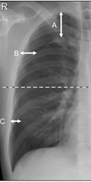
Fig. 1. Quantification of pneumothorax size on chest radiograph using the Collins method. Size of pneumothorax is 4.2+{4.7×(A+
B+C)}.
ies [6,10]. Furthermore, in Korea, variation exists in the initial treatment of first episodes of PSP, and no definitive consensus has emerged. Herein, we report the results of a randomized prospective study con- ducted at our center for first episodes of large PSP requiring intervention.
Methods
1) Study design and population
This study was a randomized, prospective, and sin- gle-center trial conducted between December 2015 and August 2016. Patients of all ages with a docu- mented first episode of PSP who were unilaterally af- fected, hemodynamically stable, and had a pneumo- thorax measuring over 25% in size were included.
Patients with underlying lung disease (e.g., chronic obstructive pulmonary disease, interstitial lung dis- ease, emphysematous lung, pulmonary tuberculosis, malignancy, and infectious lung disease), severe co- morbidities, bilateral pneumothorax, tension pneumo- thorax, recurrent pneumothorax, traumatic pneumo- thorax, or pregnancy were excluded. The size of the pneumothorax was measured using the Collins meth- od (Fig. 1) [11]. All patients provided informed consent. Patients were randomly assigned to the NA or CT group using a random number table. This study received the Institutional Review Board appro-
val of Inje University Haeundae Paik Hospital, Inje University College of Medicine (IRB approval no., 129792-2015-116).
2) Manual needle aspiration group
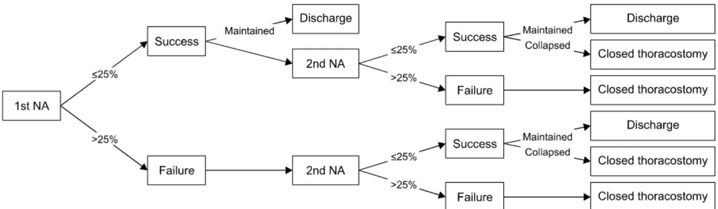
The treatment protocol for the NA group is shown in Fig. 2. Patients were placed in the semi-Fowler position and skin disinfection was performed. After local anesthesia using 2% lidocaine, a 16-gauge intra- venous angio-catheter with a diameter of 1.7 mm was introduced through the second intercostal space, on the midclavicular line. When the catheter entered the pleural space, it was placed at the apex of the thoracic cavity and connected to a 3-way valve and syringe. After confirming the functionality of the catheter with a syringe, the air in the pleural cavity was suctioned using a mechanical suction system.
After sufficient mechanical suction, manual aspiration was performed until resistance was felt. When no more air was evacuated, the catheter was removed and the procedure was terminated. Chest radiog- raphy was performed immediately after the proce- dure and 12 hours later to determine the results of NA.
NA was considered successful when the pneumo- thorax measured ≤25% on chest radiography taken immediately after the procedure and when no signs of worsening were observed on follow-up chest ra- diography taken 12 hours later. We defined initial treatment success as achieving successful NA within 2 attempts. If NA was successful, the patient was discharged. If the first attempt to perform NA failed, a second attempt was made immediately. If the sec- ond attempt to performed NA failed, CT was performed.
3) Closed thoracostomy group
Under local anesthesia using 2% lidocaine, CT was
performed using a 12-French (F) catheter (Argyle
Suture Rib Trocar Catheter; Covidien, Mansfield, MA,
USA) through the fourth intercostal space, on the an-
terior axillary line. The catheter was connected to an
underwater seal drainage system with a negative
pressure of −10 to −15 mm H
2O. When complete
lung expansion on chest radiography was confirmed
and there was no air leakage, the chest tube was
removed. If no signs of worsening were observed on
chest radiography taken 12 hours after removal of
Fig. 2. Protocol of needle aspiration for a first episode of primary spontaneous pneumothorax. NA, needle aspiration; ≤25%, size of pneumothorax is same or less than 25% as estimated by Collins method on immediate follow-up chest radiography after NA; >25%, size of pneumothorax is greater than 25% on immediate follow-up chest radiography after NA; maintained, lung expansion is maintained on chest radiography 12 hours after needle aspiration; collapsed, worsening of lung expansion on chest radiography 12 hours after needle aspiration.
Table 1. Patient characteristics
Characteristic Needle aspiration (N=21) Closed thoracostomy (N=19) p-value
Age (yr) 24.0±10.9 24.8±11.5 0.83
Sex (male:female) 20:1 17:2 0.44
Body mass index (kg/m
2) 20.2±2.8 20.7±3.3 0.55
Size of pneumothorax (%) 56.0±23.3 65.6±22.1 0.23
Affected side (right:left) 11:10 10:9 0.57
Current smoker 5 3 0.44
Values are presented as mean±standard deviation or number.
the chest tube, the patient was discharged. If there was persistent air leakage (>5 days), a surgical in- tervention was performed. Successful CT was defined as removal of the chest tube within 5 days.
4) Statistical analysis
All statistical analyses were performed using PASW SPSS statistics ver. 18.0 (SPSS Inc., Chicago, IL, USA).
Baseline characteristics and information about the hospital stay were compared using the Student t-test, and the categorical variables were assessed with the chi-square test or the Fisher exact test. All p-values
<0.05 were considered to indicate statistical significance.
Results
Forty patients with a first episode of PSP were re- cruited between December 2015 and August 2016.
There were 21 patients in the NA group and 19 pa-
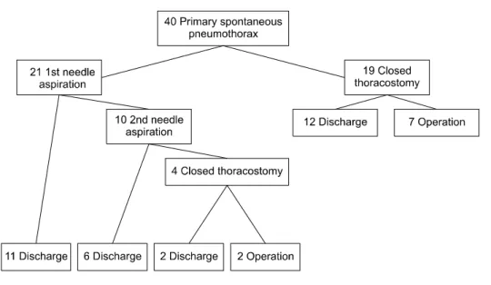
tients in the CT group. The characteristics of the pa- tients in each group are shown in Table 1. There were no statistically significant differences in age, sex, body mass index, size of pneumothorax, the af- fected side, or smoking status. Fig. 3 shows the flow chart of study allocation and overall outcomes.
Table 2 shows information about patients’ hospital stay, the success rate of initial treatment, the 1-month recurrence rate, the 1-year recurrence rate, and the size of pneumothorax in both groups. The hospital stay of the patients in the NA group was shorter than that of the patients in the CT group.
This difference was statistically significant (p<0.01).
The success rate of initial treatment in the NA group and CT group was 81.0% and 63.2%, respectively.
However, this difference was not statistically significant. There was no significant difference in the 1-month or 1-year recurrence rate between the NA and CT groups.
We also conducted a subgroup analysis according
Fig. 3. Flow chart of study alloca- tion and outcomes.
Table 2. Results comparing needle aspiration to closed thoracostomy
Variable Needle aspiration (N=21) Closed thoracostomy (N=19) p-value
Initial treatment success rate 17/21 (81.0) 12/19 (63.2) 0.16
Success, first attempt 11/21 (52.4)
Success, second attempt 6/10 (60.0)
1-Month recurrence rate 3/21 (14.3) 1/19 (5.3) 0.34
Success 3/17 (17.7) 1/12 (8.3) 0.44
Failure 0 0 0
1-Year recurrence rate 4/21 (19.0) 3/19 (15.7) 0.79
Success 4/17 (23.5) 3/12 (25.0) 0.93
Failure 0 0 0
Hospital stay 2.1±1.8 5.4±3.6 <0.01
Success 1.4±0.5 3.3±1.4 <0.01
Failure 5.5±1.3 8.9±3.4 0.05
Size of pneumothorax
Success, first attempt 48.5±22.1 58.4±19.6
Success, first attempt 55.9±19.9
Failure 77.2±22.7 77.0±22.3
Values are presented as number (%) or mean±standard deviation. The category of ‘success’ refers to patients in whom the intervention succeeded, while ‘failure’ refers to the patients in which the intervention failed.
to whether treatment succeeded or failed. In the NA group, the success rate of the first attempt was 52.4% (11 of 21) and that of the second attempt was 60.0% (6 of 10). There was no difference in the recurrence rate between these 2 subgroups, and the hospital stay of each of these subgroups was 1.4±0.5 days and 3.3±1.4 days, respectively (p<0.01). In the NA group, a statistically significant difference was found in the size of the pneumothorax among sub- groups in which the first round of treatment suc- ceeded, the second round of treatment succeeded, or
treatment failed (p=0.04).
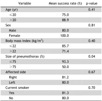
Table 3 shows the success rate according to multi- ple clinicopathologic factors in the NA group.
Pneumothorax size was identified as an independent
variable that affected the initial treatment success
rate. The success rate was significantly higher in pa-
tients in whom the size of the pneumothorax was
less than 75%.
Table 3. Results of subgroup analysis of the patients who re- ceived needle aspiration