ISSN: 2233-601X (Print) ISSN: 2093-6516 (Online)
− 207 −
†
This study was presented at the 275 monthly conference of Seoul and Gyeonggi-do.
Received: August 29, 2016, Revised: October 27, 2016, Accepted: October 28, 2016, Published online: June 5, 2017
Corresponding author: Jeong-Jun Park, Department of Thoracic and Cardiovascular Surgery, Asan Medical Center, University of Ulsan College of Medicine, 88 Olympic-ro 43-gil, Songpa-gu, Seoul 05505, Korea
(Tel) 82-2-3010-3575 (Fax) 82-2-3010-6811 (E-mail) [email protected]
© The Korean Society for Thoracic and Cardiovascular Surgery. 2017. All right reserved.
This is an open access article distributed under the terms of the Creative Commons Attribution Non-Commercial License (http://creativecommons.org/
licenses/by-nc/4.0) which permits unrestricted non-commercial use, distribution, and reproduction in any medium, provided the original work is properly cited.
Fontan Revision with Y-Graft in a Patient with Unilateral Pulmonary Arteriovenous Malformation
Jeong-woo Lee, M.D. 1 , Jeong-Jun Park, M.D., Ph.D. 1 , Hyun Woo Goo, M.D., Ph.D. 2 , Jae Kon Ko, M.D., Ph.D. 3
Departments of
1Thoracic and Cardiovascular Surgery,
2Radiology, and
3Pediatric Cardiology, Asan Medical Center, University of Ulsan College of Medicine
The extracardiac conduit Fontan procedure is the last surgical step in the treatment of patients with a func- tional single ventricle. An acquired pulmonary arteriovenous malformation may appear perioperatively or postoperatively due to an uneven hepatic flow distribution. Here we report a case of a bifurcated Y-graft Fontan operation in a 15-year-old male patient with a unilateral pulmonary arteriovenous malformation after an extracardiac conduit Fontan operation.
Key words: 1. Congenital heart defects 2. Fontan
3. Conduits 4. Vascular disease 5. Y-graft
Case report
A 15-year-old male patient with a history of com- plete atrioventricular septal defect, pulmonary atre- sia, right isomerism, asplenia, and separate hepatic venous drainage was treated with a left modified Blalock -Taussig shunt at 50 days after birth, a bidir- ectional cavopulmonary shunt at 21 months, and an external cardiac conduit Fontan operation at 5 years of age. His oxygen saturation was maintained at ap- proximately 93%–95% after the Fontan procedure.
During follow-up, mild desaturation at about 90%–
93% was observed. In the cardiac catheterization, there were no anomalous findings regarding ven- tricular function or systemic venous pressure; how- ever, preferential flow was observed. Blood from the
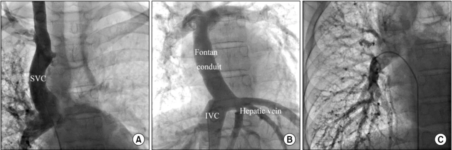
superior vena cava showed a tendency to flow to- ward the right lung, while blood from the inferior vena cava (IVC) showed a tendency to flow toward the left lung. The right lung had a diffuse pulmonary arteriovenous malformation (pAVM) (Fig. 1). A lung perfusion scan (LPS) was performed by a 99mTc al- bumin-aggregated (99mTc-MAA) injection in the low- er limb and the shunt fraction was reported as 15%.
Given the diagnosis of a unilateral pAVM, a Fontan revision was planned. A bifurcated Y-graft was used for the redistribution of the hepatic blood flow.
Under general anesthesia, a redo median sternotomy and adhesiolysis were performed. After aortic/bicaval cannulation, the operation was performed under car- diopulmonary bypass. In the operative findings, the previous Fontan graft was offset to the left side, and
Korean J Thorac Cardiovasc Surg 2017;50:207-210 □ CASE REPORT □
https://doi.org/10.5090/kjtcs.2017.50.3.207
Jeong-woo Lee, et al
− 208 −
Fig. 1. A selective angiogram showing that (A) the right lung received almost all blood flow from the SVC, (B) while the blood from the IVC mainly flowed to the left lung. (C) The right lung displays a reticulonodular density representing a diffuse pulmonary arteriovenous malformation. SVC, superior vena cava; IVC, inferior vena cava.
Fig. 2. The Y-graft conduit connected to the left pulmonary ar- tery and the lower branch of the RPA (arrow). SVC, superior vena cava; RPA, right pulmonary artery.
the previous bidirectional cavopulmonary shunt site was very wide. The previous Fontan conduit was re- moved, and a 22×11×11-mm Y-graft conduit was connected to the left pulmonary artery and the right pulmonary artery lower branch. The IVC side was connected to the previous remnant graft. At the pre- vious anastomosis site of the graft to the pulmonary artery, a direct closure and angioplasty using GORE- TEX Vascular Grafts (W. L. Gore & Associates Inc., Newark , NJ, USA) had been performed (Fig. 2). The total cardiopulmonary bypass time was 100 minutes.
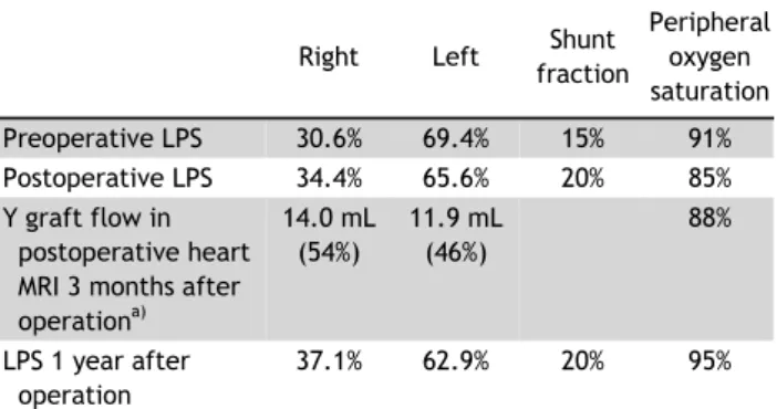
The patient was transferred to the general ward on postoperative day 1 and discharged on post- operative day 8 without any complications. In the postoperative LPS, performed by using a right foot vein, the shunt fraction was found to have increased by 20% as compared to the preoperative LPS. Three months after surgery, the patient underwent a mag- netic resonance imaging (MRI) scan of the heart, and oxygen saturation was still 88%. In the MRI quantifi- cation, the total blood flow per strok e volume of the Y-graft was 25.9 mL. The blood flow volumes in the right and left limbs of the Y-graft were 14.0 mL (54%) and 11.9 mL (46%), respectively. Considering that this patient had a preoperative preferential flow of the Fontan graft to the left lung, greater IVC flow, including hepatic venous return, was directed to the right lung postoperatively. One year after the Fontan revision, LPS was performed again. It showed an in- creased blood flow to the right lung and oxygen sat- uration amounting to 95%, considerably greater than
the results of the previous test (Table 1).
Discussion
There are various factors that cause pAVMs, but
these do not account for the details of how pAVMs
are formed. A congenital heart defect, such as a left
isomerism or exclusion of the hepatic vein blood
flow to the pulmonary circulation, could be a risk
factor for a pAVM. Even after the completion of the
Fontan operation in patients with a functional single
ventricle (FSV), an unbalanced distribution of the
hepatic flow might result in a pAVM.
Fontan Revision with Y-Graft
− 209 −
Table 1. Changes in preoperative and postoperative blood flow, shunt fraction, and oxygen saturation
Right Left Shunt fraction
Peripheral oxygen saturation
Preoperative LPS 30.6% 69.4% 15% 91%
Postoperative LPS 34.4% 65.6% 20% 85%
Y graft flow in postoperative heart MRI 3 months after operation
a)14.0 mL (54%)
11.9 mL (46%)
88%
LPS 1 year after operation
37.1% 62.9% 20% 95%
LPS, lung perfusion scan, using Technetium 99mTc albumin aggregated injection to the lower limb; MRI, magnetic resonance imaging.
a)