저작자표시 2.0 대한민국 이용자는 아래의 조건을 따르는 경우에 한하여 자유롭게 l 이 저작물을 복제, 배포, 전송, 전시, 공연 및 방송할 수 있습니다. l 이차적 저작물을 작성할 수 있습니다. l 이 저작물을 영리 목적으로 이용할 수 있습니다. 다음과 같은 조건을 따라야 합니다: l 귀하는, 이 저작물의 재이용이나 배포의 경우, 이 저작물에 적용된 이용허락조건 을 명확하게 나타내어야 합니다. l 저작권자로부터 별도의 허가를 받으면 이러한 조건들은 적용되지 않습니다. 저작권법에 따른 이용자의 권리는 위의 내용에 의하여 영향을 받지 않습니다. 이것은 이용허락규약(Legal Code)을 이해하기 쉽게 요약한 것입니다. Disclaimer 저작자표시. 귀하는 원저작자를 표시하여야 합니다.
Functional Role of Hepatitis B Virus
Core Protein in Viral Replication
by
Jaesung Jung
Major in Molecular Medicine
Department of Biomedical Sciences
The Graduate School, Ajou University
Functional Role of Hepatitis B Virus
Core Protein in Viral Replication
by
Jaesung Jung
A Dissertation Submitted to The Graduate School of
Ajou University in Partial Fulfillment of the Requirements for the
Degree of Doctor of Philosophy of Biomedical Sciences
Supervised by
Kyongmin Kim, Ph.D.
Major in Molecular Medicine
Department of Biomedical Sciences
The Graduate School, Ajou University
This certifies that the dissertation
of Jaesung Jung is approved.
SUPERVISORY COMMITTEE
Sun Park
Soon Bong Hwang
Myoung Hee Ahn
Ho-Joon Shin
Kyongmin Kim
The Graduate School, Ajou University
December, 5th, 2012
i
-ABSTRACT-
Functional Role of Hepatitis B Virus Core Protein in Viral
Replication
PART I
Arginine-Rich
167RRRSQSPRR
175Domain in C-Terminus of Core
is Critical for HBV Replication
To investigate the contributions of carboxyl-terminal nucleic acid binding domain of HBV core protein for hepatitis B virus (HBV) replication, chimeric HBV core proteins were generated by substituting varying lengths of the carboxyl-terminus of duck hepatitis B virus (DHBV) core protein for the corresponding regions of HBV core protein. All chimeric core proteins formed core particles. A chimeric core protein with 221–262 amino acids of DHBV core protein, in place of 146–185 amino acids of the HBV core protein, supported HBV pregenomic RNA (pgRNA) encapsidation and DNA synthesis: 40% amino acid sequence identity or 45% homology in the nucleic-acid binding domain of HBV core protein was sufficient for pgRNA encapsidation and DNA synthesis, although we predominantly detected spliced DNA. A chimeric core protein with 221–241 and 251–262 amino acids of DHBV core, in place of HBV C 146–166 and 176–185 amino acids, respectively, could
ii
rescue full-length DNA synthesis. However, a reciprocal core chimera with 242–250 of DHBV core (242RAGSPLPRS250) introduced in place of 167–175 of HBV core
(167RRRSQSPRR175) significantly decreased pgRNA encapsidation and DNA synthesis, and
full-length DNA was not detected, demonstrating that the arginine-rich 167RRRSQSPRR175
domain may be critical for efficient viral replication. Five amino acids differing between viral species (underlined above) were tested for replication rescue; R169 and R175 were found to be important.
Key words: Hepatitis B virus, Core protein, Chimeric core, Carboxyl-terminal domain of core protein, Encapsidation, Hepatitis B virus replication
iii
-ABSTRACT-
PART II
Phosphorylation of the C-Terminal Domain of HBV Core Protein
modulates Genome Replication
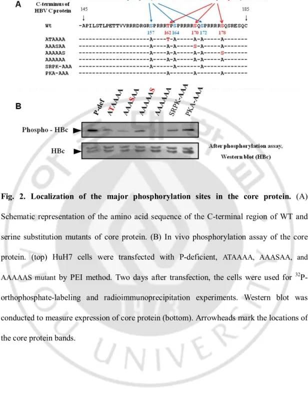
Phosphorylation of hepatitis B virus (HBV) core protein at Ser157, Ser164, and Ser172 residues by host serine/arginine protein-specific kinases (SRPK) or protein kinase C (PKC) has been demonstrated to modulate HBV replication. Also, three additional amino acid residues, Thr162, Ser170, and Ser178, of HBV core protein have been suggested as the putative protein kinase A (PKA) phosphorylation sites with the conserved RRXS/T motif. The in vivo phosphorylaiton assay reveals that Thr 162, Ser170, or Ser178 can be phosphorylated. In order to elucidate importance of these residues for HBV replication, each was mutated to Ala to mimic nonphosphorylated Ser or to Glu to mimic phosphorylated Ser. Thr 162 to Ala (T162A) mutation decreased replicative intermediate DNA significantly. To further investigate the importance of Thr 162 in conjunction with Ser170Ala and/or Ser178Ala mutations, more core protein mutants were constructed. In the presence of T162A mutation, the HBV DNA synthesis was decreased more dramatically, indicating that Thr 162 residue may be important for HBV DNA synthesis. Taken together, our results indicate that
iv
the putative PKA phosphorylation sites, Thr 162, Ser170, or Ser178, is phosphorylated and can modulate DNA replication possibly through phosphorylation and dephosphorylation.
Key words: hepatitis B virus, phosphorylation of HBV core protein, cAMP depdent protein kinase
v
TABLE OF CONTENTS
ABSTRACT ... i
TABLE OF CONTENTS ... v
LIST OF FIGURES ... viii
PART I
I. INTRODUCTION ... 1II. MATERIALS AND METHODS ... 5
A. DNA construction ... 5
B. Cell culture, transfection, and isolation of core particles ... 6
C. RNase protection analysis... 7
D. RNA encapsidation assay and Southern blotting ... 8
E. SDS-PAGE and Western blotting ... 8
F. PCR to detect HBV DNA from spliced RNA ... 9
III. RESULTS ... 10
A. Chimeric core protein expression and core particle formation ... 10
vi
C. HBV DNA is synthesized in core particles by the HD221-262 C variant ... 19
D. Core particle formation and RNA encapsidation by additional chimeric C variant ... 21
E. Full-length HBV DNA is synthesized in core particles by the HDHD C variants ... 27
F. Residues R169 and R175 are important for HBV replication ... 31
IV. DISCUSSION ... 37 V. CONCLUSION ... 44 REFERENCES ... 45 국문요약 ... 50
PART II
I. INTRODUCTION ... 53II. MATERIALS AND METHODS ... 57
A. DNA construction ... 57
B. Cell culture and transfection ... 57
C. siRNA transfection ... 58
D. Isolation of core particles and western blot ... 58
E. In vivo phosphorylation assay ... 59
F. Southern blotting ... 60
vii
H. Endogenous polymerase assay... 60
I. Immunofluorscence assay ... 61
J. Primer extension assay ... 61
III. RESULTS ... 63
A. Amino acids sequence alignment of HBV core proteins with the related hepadnaviruses ... 63
B. In vivo phosphorylation of core protein... 65
C. Core protein expression and core particle formation by phosphorylation site mutant core proteins ... 68
D. PgRNA encapsidation, DNA synthesis, and endogenous polymerase activity by the nonphosphorylated and phosphorylated core protein mutants. ... 72
E. Distribution of nonphosphorylated and phosphorylated core protein mutants. ... 79
F. Individual roles of putative PKA phosphorylation sites of the core protein in pgRNA encapsidation and DNA synthesis. ... 81
G. Over expression and knockdown of PKAα on HBV replication... 87
IV. DISCUSSION ... 90
V. CONCLUSION ... 94
REFERENCES ... 95
viii
LIST OF TABLES
PART II
Table.1. PgRNA encapsidation, minus-DNA elongation, and replicative intermediate DNAs by phosphorylation mutants
...
86ix
LIST OF FIGURES
PART I
Fig. 1. Core protein expression and core particle assembly by chimeric core
protein variants
...
13 Fig. 2. HBV pgRNA encapsidation in core particles with chimeric core protein variants....
17 Fig. 3. HBV DNA synthesis in core particles with chimeric core protein variants...
20 Fig. 4. Expression and core particle assembly of additional core protein variants... 22
Fig. 5. pgRNA encapsidation in core particles by additional core protein variants...
26 Fig. 6. Full-length HBV DNA synthesis in core particles with additional chimeric core protein variants...
29 Fig. 7. HBV core particle formation, pgRNA encapsidation, and HBV DNA synthesisby core variants
...
32PART II
Fig. 1. Sequence alignment of C-terminal domain of HBV and related viruses
x
Fig. 2. Localization of the major phosphorylation sites in the core protein ... 67
Fig. 3. Mutations within the phosphorylation site of HBV core protein have little to no impact on core particle formation ... 70
Fig. 4. PgRNA encapsidation in core particles with core protein phosphorylation mutants .. 73
Fig. 5. DNA synthesis and Endogenous polymerase reaction in core particles with core protein phosphorylation mutants ... 76
Fig. 6. Intracellular distribution of phosphorylation mutant core protein ... 80
Fig. 7. Role of individual phophorylation sites in HBV pgRNA encapsidation... 82
Fig. 8. Role of individual phophorylation sites in DNA replication ... 84
Fig. 9. Role of individual phophorylation sites in (-) DNA elongation ... 85
Functional Role of Hepatitis B Virus
Core Protein in Viral Replication
PART I
Arginine-Rich
167
RRRSQSPRR
175
Domain in C-Terminus of Core is Critical
for HBV Replication
1
I. INTRODUCTION
Hepadnaviruses are small, enveloped DNA viruses that replicate preferentially in liver cells and are associated with acute and chronic hepatitis, cirrhosis, and hepatocellular carcinoma (Brechot, 2004). Hepatitis B virus (HBV), a prototypic hepadnavirus, has a partially duplex relaxed circular (RC) DNA genome, which replicates by reverse transcription of a pregenomic RNA (pgRNA) to produce genomic DNA. The core protein of HBV consists of 183 or 185 amino acids that form core particles via dimeric intermediates (Zhou and Standring, 1992). Assembly of replication-competent HBV core particles requires interaction of pgRNA with the polymerase (P) and core proteins. The amino-terminus of core protein (amino acids 1–144) participates in core particle assembly through protein-protein interaction and is, by itself, assembly competent (Birnbaum and Nassal, 1990; Gallina et al, 1989). The carboxyl-terminus of core protein contains a protamine-like nucleic acid binding domain rich in arginine. Although this region is dispensable for core particle assembly, it is involved in hepadnaviral replication, including pgRNA encapsidation and DNA replication (Birnbaum and Nassal, 1990; Brechot, 2004; Certo et al, 1999; Daub et al, 2002; Enomoto et al, 2006). The importance of core protein’s carboxyl-terminus in hepadnavirus pgRNA encapsidation and DNA replication has been suggested through experiments with a series of carboxyl-terminal truncation mutants (Birnbaum and Nassal, 1990; Gallina et al, 1989; Beames and Lanford, 1993; Kock et al, 2004; Pogam et al, 2005; Nassal, 1992). The core protein amino-acid 164 variant deficient in 19 carboxyl-terminal amino acids (C164), corresponding to amino-acid 166 in our adw subtype, is competent for
2
pgRNA encapsidation, but not for synthesis of full-length RC DNA (Kock et al, 2004; Pogam et al, 2005; Nassal, 1992). DNA synthesized in variant C164 core particles is predominantly spliced (Kock et al, 2004; Pogam et al, 2005). However, a core variant containing 173 amino acids and lacking ten amino acids at the carboxyl-terminus (C173), corresponding to 175 amino acids in our adw subtype, was as competent for synthesis of full-length RC DNA as wild type (wt) core protein, suggesting that nine carboxyl-terminal amino acids (165–173) are sufficient for replication-competence (Pogam et al, 2005). Although it has been suggested that these residues are important for selective and/or productive viral RNA encapsidation in deletion- and site-directed mutants (Pogam et al, 2005), a direct demonstration of the amino acid residues or motif in the carboxyl-terminus of core protein critical for hepadnavirus pgRNA encapsidation or DNA replication has not yet been performed.
Heterologous complementation to generate chimeric proteins of related viruses is a way of identifying the viral-protein amino acid residues or motifs crucial for replication. Chimeric viruses or proteins have been used to identify viral cis-acting sequences and the functions of protein domains (Berkowitz et al, 1995; Certo et al, 1999; Kaye and Lever, 1998; Kim et al, 2000). Chimeras of duck HBV (DHBV) and heron HBV, another avian hepadnavirus sharing 79% nucleotide identity with DHBV, have been used to elucidate the functional interactions between cis-acting sequences and viral components for pgRNA encapsidation and plus-strand DNA synthesis (Mueller and Loeb, 2002; Ostrow and Loeb, 2004; Ostrow and Loeb, 2008). The genomes of HBV and woodchuck hepatitis virus (WHV) share approximately 60% identity (Kodama et al, 1985; Mandart, 1984); those of HBV and
3
DHBV share 40% homology (Mandart et al, 1984). However, heterologous complementation with related hapdnaviruses such as DHBV or WHV cannot be performed; while HBV replication is restored easily by complementation with WHV core and/or P proteins and vice versa, it cannot be complemented at all by DHBV core and/or P proteins and vice versa (Okamoto et al, 1990; Zierman and Ganem, 1996). Therefore, use of DHBV or WHV chimeric viruses or proteins to complement HBV replication has not been explored. In the present study, HBV chimeric core proteins were constructed by exchanging portions of the carboxyl-terminus of HBV core protein with the corresponding regions of DHBV core protein, while retaining wt HBV core protein amino-terminal sequence to investigate the critical regions for pgRNA encapsidation or HBV DNA synthesis. DHBV core protein, which consists of 262 amino acids, can form a three-dimensional core particle similar in structure to that of HBV (Kennery et al, 1995). Use of these chimeras demonstrated that some chimeric core particles are replication-competent, complementing HBV core proteins in C-deficient mutants to effect pgRNA encapsidation concomitant with reverse transcription. These results indicate that 40% amino acid sequence identity or 45% homology in the carboxyl-terminus of core protein is sufficient for HBV pgRNA encapsidation and DNA synthesis, even though predominantly spliced HBV DNA was synthesized. Serial substitutions of HBV core protein with the corresponding regions of DHBV core protein further allowed us to demonstrate that residues 167–175, 167RRRSQSPRR175, may be critical for full-length RC DNA synthesis as long as residues from 146–166 maintain 62% homology. Although the importance of residues R167, S170, P173, and R174 in the HBV core protein could not be examined due to the presence of
4
identical residues in the corresponding region of DHBV (167RRRSQSPRR175 in HBV vs 242RAGSPLPRS250 in DHBV) in HHDH core chimera, in which HBV core 167-175 was replaced by DHBV core 242-250, we analyzed the importance of R168, R169, Q171, S172, and R175 residues using a series of point mutants. By analyzing the A168R, G169R, P171Q, L172S, and S175R mutants in HHDH core chimera, we further demonstrated that the R169 and R175 residues may be important for HBV replication and that S172 may be important for core particle formation, but not for pgRNA encapsidation or DNA synthesis. The importance of residues 167–175 in HBV core protein for replication in the context of neighboring amino acids or motifs is discussed.
5
II. MATERIALS AND METHODS
A. DNA construction
The partially redundant wt HBV subtype adw R9 plasmid construct pPB was used as a template for generation of HBV DNA constructs using PCR-based mutagenesis (Kim et al, 2004). An HBV wt core protein construct containing the HBV wt core open reading frame (ORF) and post-transcriptional regulatory element (PRE) (Huang and Liang, 1993) was generated as follows: pPB was digested with BstEII and EcoRV and then self-ligated to delete nt 1406–2848, generating pHCP. The sequence was additionally truncated by PCR-based mutagenesis, yielding pHCP. To generate a DHBV wt core protein construct containing the DHBV core ORF and HBV PRE, the DHBV core gene from pCMVDHBV (a gift from William Mason, Fox Chase Cancer Center) was cloned into pcDNA3 between the HindIII and EcoRV sites to yield pDC. The HBV PRE sequence was cloned into pDC to yield pDCP. Chimeric core protein variants were constructed by the PCR-derived recombination of HBV and DHBV core ORFs and the PCR-amplified fusion products cloned into the corresponding restriction sites of pHCP, yielding pHD192-262, pHD221-262, pHD192-220, pHD221-242, pHD242-262, pHHDH, and pHDHD. To generate pHCP145, a stop codon (TAG) was introduced at Thr146 (ACT) of HBV core protein by site-directed mutagenesis. To generate the assembly-deficient HBV variant (Konig et al, 1998), pHCP-R127Q, in which Arg127 (CGC) is modified to Gln (CAG) in pHCP by site-directed mutagenesis, was generated first; the HindIII- and BstEII-digested DNA fragment from pHCP-R127Q, which contains the Arg127→Gln mutation, was then cloned into pHCP145,
6
yielding the assembly-deficient pHCP145-R127Q variant. A C-deficient mutant that does not express core protein was generated by introducing a stop codon (TAA) at Glu8 (GAA) of the core protein by site-directed mutagenesis. This core-deficient mutant expresses pgRNA and all other HBV proteins except the core protein. To generate the c- deficient-RT-YMHA mutant, the EcoRI- and EcoRV-digested DNA fragment from a reverse transcriptase (RT) reaction deficient RT-YMHA mutant, wherein the conserved YMDD motif of the RT active site was modified to YMHA (Kim et al, 2004), was cloned into the C-deficient mutant. To further analyze the importance of Arg168, Arg169, Gln171, Ser172, and Arg175 residues (167RRRSQSPRR175 motif in HBV vs 242RAGSPLPRS250 motif in DHBV), A168R, G169R, P171Q, L172S, and S175R mutants were constructed in the HHDH core background by site-directed mutagenesis. To test trans-complementation of C-deficient-RT-YMHA or C-deficient mutants, a series of chimeric core proteins was used throughout the experiments for HBV encapsidation or HBV DNA replication. All constructs were sequenced to confirm the presence of specific mutations, and to ensure that no extraneous mutations were introduced during PCR.
B. Cell culture, transfection, and isolation of core particles
HuH7 hepatoma cells (Japan Health Sciences Foundation, Tokyo, Japan) were maintained as previously described (Kim et al, 2004). For expression of chimeric core protein variants and assessment of their core particle formation, 8 ug of pHCP plasmid or various chimeric core protein constructs were transfected into HuH7 cells as previously described (Kim et al, 2004). For analysis of pgRNA encapsidation or HBV DNA synthesis, 2
7
ug of C-deficient-RT-YMHA or C-deficient mutants and 6 ug of various chimeric core protein constructs were co-transfected into HuH7 cells as previously described. 1 ug of the Renilla luciferase expression plasmid phRL-CMV (Promega, Madison, WI, USA) was co-transfected into HuH7 cells as a transfection control. pcDNA3.1 was used in transfections to equalize total DNA transfected. Cytoplasmic core particles were precipitated from lysates of transfected cells with 6.5% polyethylene glycol as previously described (Kim et al, 2004). In brief, clarified lysate was adjusted with 10 mM MgCl2 and 8 mMCaCl2 solution, incubated
overnight at 37 °C with 20 U DNase I (Sigma) and 60 U micrococcal nuclease (Calbiochem) to remove the transfected plasmid DNA and unencapsidated RNA, and precipitated with 6.5% polyethylene glycol. Transfection experiments were repeated more than three times.
C. RNase protection analysis (RPA)
To analyze encapsidated pgRNA, core particles were isolated as described above. pgRNA was extracted from core particles following digestion with proteinase K (100 ug/mL) and DNase I (20 U). To prepare riboprobe for RPA, nt 1805–2187 of the C-deficient mutant were cloned into pGEM3Zf(+) vector, generating pRPAFD-C-def. From this construct, 446 nt of radiolabeled anti-sense probe were synthesized in vitro using SP6 RNA polymerase with a-32P-UTP (specific activity, 800 Ci/mmol). The RPA procedure was performed using
the manufacturer’s protocol (RPA II, Ambion, Austin, TX, USA). Protected pgRNA was 369 nt in length. To discriminate encapsidated full-length pgRNA from spliced RNA, the spliced region containing probe, pRPA-PS, was also used (Park et al, 2008). The 470 nts of the HBV sequence was synthesized in vitro and the protected sequence, nt 2680-3092 of HBV
8
sequence, was 413 nts long (Park et al, 2008). The relative levels of encapsidated pgRNA from isolated core particles were measured with the Image Gauge V4.0 program (Fujifilm, Tokyo, Japan).
D. RNA encapsidation assay and Southern blotting
To analyze pgRNA in core particles with chimeric core protein variants, pellets of core particles isolated from HuH7 cells co-transfected with the C-deficient-RT-YMHA mutant and various chimeric core protein constructs were dissolved in 15mL Tris-acetate EDTA buffer and electrophoresed on 1% native agarose gels. Core particles were transferred to a nylon membrane and denatured with 0.2N NaOH in situ and neutralized. Nucleic acids from disrupted core particles were hybridized to a 32P-labeled random-primed probe specific
for HBV sequence (Park et al, 2008). To analyze HBV DNA synthesis by Southern blotting, HBV DNA extracted from core particles was separated by agarose gel electrophoresis and hybridized to a 32P-labeled random-primed probe specific for HBV sequence (Kim et al,
2004). The relative levels of pgRNA and HBV DNA isolated from core particles were measured with the Image Gauge V4.0 program.
E. SDS-PAGE and Western blotting.
To analyze the core protein, total lysates were harvested and lysed in NP-40 containing lysis buffer (50 mM Tris-HCl [pH 8.0], 150 mM NaCl, 1% NP-40). The lysates were cleared by centrifugation and supernatants collected and 5% b-mercaptoethanol added; samples were then subjected to SDS-PAGE on 12% gels and the resolved proteins
9
transferred to polyvinylidene fluoride (PVDF) membranes. These membranes were incubated with polyclonal rabbit anti-HBc antibody, monoclonal mouse anti-tubulin (diluted 1:1000; Calbiochem, San Diego, CA, USA), or polyclonal rabbit anti-luciferase (diluted 1:500; Santa Cruz Biotechnology, Santa Cruz, CA, USA) antibodies. Immunoreactive bands were visualized by a horseradish-peroxidase conjugated secondary antibody (DAKO) using enhanced chemiluminescence (Amersham, Piscataway, NJ, USA). Western blot analysis of core particles was performed as previously described (Kim et al, 2008). Isolated core particles were electrophoresed on a 1% native agarose gel and resolved core particles transferred to PVDF membranes. Immunoblotting was performed using polyclonal rabbit anti-HBc antibody (diluted 1:1000). Horseradish-peroxidase conjugated anti-rabbit secondary antibody and enhanced chemiluminescence were employed to visualize HBV core particles.
F. PCR to detect HBV DNA from spliced RNA
HBV DNA was extracted from core particles isolated from HuH7 cells co-transfected with the C-deficient mutant and various core variant constructs. PCR was performed using primers HBV 155 (sense 5´-CTACTGTGGAGTTACTCTCG-3´) and HBV 8, (antisense 5´- CACGATGCTGTACAGACTTG -3´), which correspond to position nt 1935-1954 and nt 706-725 of the HBV genome, respectively. PCR amplified products were separated by agarose gel electrophoresis, gel-purified, and sequenced.
10
III. RESULTS
A. Chimeric core protein expression and core particle formation.
Carboxyl-terminal amino acid sequences of HBV and DHBV core proteins exhibited 40% identity or 45% homology (Fig 1A), while full-length core protein sequences of HBV and DHBV were approximately 27% homologous. DHBV core protein contains an additional 29 amino acids that are absent in HBV core protein (Fig1A). To investigate residues in the carboxyl-terminal nucleic acid binding domain of HBV core protein required for HBV replication, various chimeric core proteins were constructed by substituting the corresponding regions of DHBV core protein for the carboxyl-terminus of HBV core protein (Fig 1A). As positive and negative controls, the HBV and DHBV core protein expression plasmids pHCP and pDCP were first constructed and used as template for chimeric core protein construction. The HD221-262 core protein chimera was designed to substitute the carboxyl-terminal region from residues 221–262 of the DHBV core protein for the corresponding region from residues 146–185 of the HBV core protein, while the amino-terminal 145 amino acids of the HBV core protein were unchanged. The HD192-262 core protein chimera contains the amino-terminal 145 amino acids of HBV core protein and the carboxyl-terminus of DHBV C from residues 192–262 to include an additional 29 amino acids. The HD192-220 core protein variant has the entire HBV core protein sequence but an additional 29 amino acids which are part of DHBV core flexible linker region are inserted between residues 145–146 of the HBV core protein. The HCP145 construct was generated as a positive control for core particle assembly, but as a negative control for pgRNA
11
encapsidation (Beams and Lanford, 1993). HCP145-R127Q, the assembly-deficient variant, was constructed as a negative control for core particle assembly (Konig et al, 1998). Construct transcription was controlled by the cytomegalovirus immediate early (CMV IE) promoter, and nuclear export of RNAs facilitated by the HBV PRE sequence (Figure 1A). The C-deficient mutant that does not express core protein by the introduced stop codon (TAA) at Glu8 (GAA) (Fig 2A) and pHCP were used as control and/or reference. The Renilla luciferase expression plasmid phRL-CMV was co-transfected into HuH7 cells as a transfection control.
Following transfection of the core protein variants or C-deficient mutant indicated into HuH7 cells, core proteins from HBV wt and chimeric, mutated, and/or truncated variant constructs migrated as expected after SDS-PAGE and Western blotting with polyclonal anti-HBc antibody, but not the C-deficient mutant, as expected (Fig 1B, top panel). To exclude the possibility that the existence of HBV components such as pgRNA and P protein could affect assembly and/or stability of core particles, we transfected core protein variants alone, without the pgRNA expressing construct, into HuH7 cells. Most core protein chimeras were expressed similarly to or, occasionally, at higher levels than the HBV wt core protein from pHCP, except the core protein chimera from HD192-262 (Fig 1B, top panel, lane 3). Native agarose gel electrophoresis followed by Western blotting with polyclonal anti-HBc antibody revealed that core particles formed by chimeric core variants produced slightly different migration patterns (Fig 1B and 2B, second panel, lanes 3–6), suggesting that carboxyl-terminal nucleic acid binding domain sequence might affect core particle formation to some extent, even though the amino-terminal assembly domain remained intact in these chimeric
12
core variants. DHBV core protein and core particles could not be detected with anti-HBc antibody (Fig 1B and C, lane 2). Also, the assembly-deficient mutant HCP145-R127Q could not form core particles (Konig et al, 1998), even though HCP145-R127Q core protein was compatible with HCP145 core protein (Fig 1B and C, lanes 6 and 7). When levels of core particle formation were compared with core protein expression by normalization to the phRL-CMV transfection control, all variants exhibited similar patterns except the assembly-deficient mutant (Figure 1C). The very inefficient core particle formation by HD192-262 may have been due to poor core protein expression (Fig 1B and C, lane 3). Furthermore, the migration pattern displayed by core particles formed with the HD192-262 core chimera was slightly slower than those of other core particles (Fig 1B, 2B, 5A second panels, and 6 bottom panel), suggesting that HD192-262 core particles may be less stable (Newman et al, 2003). Alternatively, it might be caused by the differences in net charges.
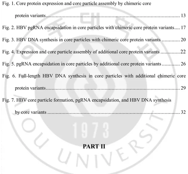
13
Fig. 1. Core protein expression and core particle assembly by chimeric Core protein variants. (A) Schematic diagrams of HBV, DHBV, and chimeric core protein variant
constructs aligned with amino acid sequences of HBV and DHBV core protein carboxyl-terminal domains. Amino acids in bold are identical or homologous. SRPK and PKA phosphorylation sites of HBV are marked with asterisks and arrowheads, respectively. Phosphorylation sites of DHBV (Yu and Summers, 1994; Yeh et al, 1993) are marked with open arrowheads. Amino acid sequences of the HBV and DHBV core proteins are presented as open and closed boxes, respectively. The cytomegalovirus immediate early (CMV IE) promoter is represented as an open arrow. PRE, post-transcriptional regulatory element. (B) Identification of core protein and core particles by chimeric core protein variants. To examine expression of core protein variants, lysates from HuH7 cells transfected with a pHCP, pDCP, pHD192-262, pHD192-220, pHD221-262, pHCP145, pHCP145-R127Q, or
14
C-deficient mutant were electrophoresed on 12% SDS-PAGE gels and protein levels visualized by Western blotting using polyclonal rabbit anti-HBc antibody (top panel). core protein variants (arrowheads) with expected molecular weights are indicated. The C-deficient mutant lacks core protein due to the introduction of a stop codon at Glu 8 in the C ORF. The pHCP and the C-deficient mutant constituted positive and negative controls, respectively. Transfection experiments were repeated four times. To detect core particles formed by core protein variants from native agarose gels, isolated core particles were transferred to PVDF membranes and incubated with polyclonal rabbit anti-HBc antibody (second panel). The Renilla luciferase expression plasmid phRL-CMV was co-transfected into HuH7 cells as a transfection control (third panel). Luciferase and a-tubulin (bottom panel) levels were determined by Western blotting using polyclonal rabbit anti-luciferase and monoclonal mouse anti-tubulin antibodies as transfection and loading controls, respectively. HRP-conjugated secondary antibody and enhanced chemiluminescence were used to visualize C, a-tubulin, and luciferase proteins and core particles. (C) Relative levels of core protein expression and core particle assembly by chimeric core protein variants. Relative levels of core proteins, core particles, and luciferase were measured with the Fujifilm Image Gauge V4.0 program. Relative levels of core protein variant expression and core particle assembly were compared to normalized transfection efficiencies. The data represent the mean ± standard deviation (SD) from four independent experiments.
15
B. HBV RNA encapsidation in core particles with core protein chimeras.
To examine RNA encapsidation by assembly-competent chimeric core variants, various core protein chimeras were co-transfected into HuH7 cells with the C-deficient-RT-YMHA mutant (Fig 2A). To ensure that the nucleic acids within core particles hybridized in situ are encapsidated RNA, not synthesized HBV DNA, the C-deficient-RT-YMHA mutant was used for co-transfection experiments. The conserved YMDD reverse transcriptase motif was modified to YMHA (Kim et al, 2004) in the deficient mutant background in the C-deficient-RT-YMHA mutant; thus, core protein deficient and RT reaction-deficient. In this system, core proteins were supplied in trans from core protein chimeras to trans-complement C-deficient-RT-YMHA mutant, and pgRNA and HBV P protein for pgRNA encapsidation were provided from C-deficient-RT-YMHA mutant to trans-complement core protein chimera. HCP and C-deficient-RT-YMHA co-transfected cells were used as a positive control that complements one another. Core particles from co-transfected cells (Fig 2B, second panel, and 2C) were assembled with efficiency and stability similar to those from singly transfected cells (Fig 1B and C), indicating that core particle stability might not be affected by the existence of pgRNA and P protein. We designated HCP145 as the encapsidation-negative control based on previous reports (Birnbaum and Nassal, 1990; Hatton et al, 1992). Encapsidated RNAs were not detected from 262 and HD192-220 core variant co-transfected cells (Fig 2B, top panel, and 2C). Encapsidated RNAs were detected only from HD221-262 core variant and C-deficient-RT-YMHA co-transfected cells (Fig 2B, top panel, and 2C; see lane 5 in each), indicating that substituting part of HBV is involved in pgRNA encapsidation. To confirm encapsidation efficiency, RNase Protection
16
assay (RPA) was also performed with 5’-end specific probe to show encapsidation of HBV RNA by chimeric core variants. Consistent with encapsidation assays (Fig. 2B, top panel), encapsidation of HBV RNA was only detected in HD221-262 core variant and C-deficient-RT-YMHA co-transfected cells (Fig 2D, lane 5). RPA and encapsidation assay results indicated that 40% identity or 45% homology in the carboxyl-terminus of HBV core protein was sufficient for pgRNA encapsidation.
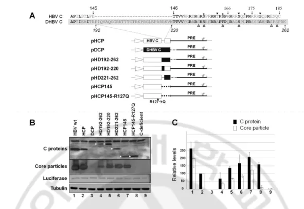
17
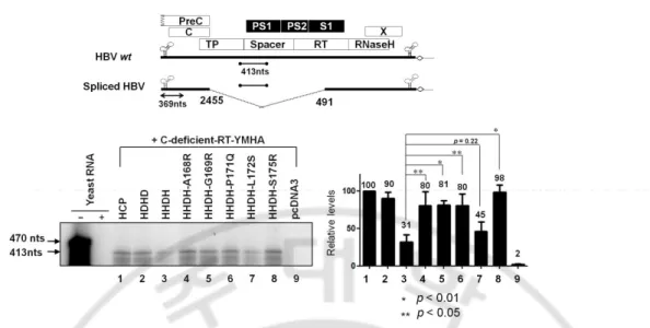
Fig. 2. HBV pgRNA encapsidation in core particles with chimeric core protein variants.
(A) Schematic diagram of HBV wt (Kim et al, 2004), deficient mutant, and C-deficient-RT-YMHA mutant. The C-C-deficient-RT-YMHA mutant is RT- and core protein deficient due to mutation of the YMDD motif to YMHA, in addition to the presence of a stop codon at Glu 8 in the C ORF. The positions of point mutations are indicated as closed arrowheads. Four ORFs of HBV are shown at the top as open boxes. The CMV promoter is
18
denoted by an open arrow. (B) Encapsidation assay to detect HBV nucleic acid in situ from disrupted core particles. To examine encapsidation by chimeric core protein variants, the C-deficient-RT-YMHA mutant was co-transfected into HuH7 cells with the pHCP, pDCP, pHD192-262, pHD192-220, pHD221-262, or pHCP145. HBV wt core protein from pHCP served as a positive control. Isolated core particles were electrophoresed through a 1% native agarose gel and transferred to nylon membrane. A 32P-labeled HBV DNA probe was
hybridized to HBV nucleic acids in core particles after disruption of the particles in situ. Core particles were also detected as described for Figure 1B. (C) Relative levels of RNA encapsidation and core particle assembly by chimeric core protein variants. Relative levels of encapsidated RNA and core particles were measured with the Fujifilm Image Gauge V4.0 program. Relative levels of encapsidated RNA and core particles were compared with normalized transfection efficiencies (n=3). (D) RNase protection assay (RPA) to detect encapsidated pgRNA. In vitro transcribed radiolabeled antisense RNA probe (446 nt) was hybridized overnight at 50°C with pgRNA from isolated core particles. Following RNase digestion, the protected pgRNA (369 nt), nt 1819-2187 of the HBV sequence, was run on a 5% polyacrylamide–8 M urea gel and visualized by autoradiography. Relative levels of encapsidated pgRNA were measured with the Fujifilm Image Gauge V4.0 program. Transfection experiments were repeated three times. The Renilla luciferase expression plasmid phRL-CMV was used as a transfection control and pcDNA3.1 was used to equalize the total amount of DNA transfected. The data represent the mean ± SD from three independent experiments.
19
C. HBV DNA is synthesized in core particles by the HD221-262 core variant.
To further investigate whether this chimeric core protein could support HBV DNA synthesis, Southern blot analysis was performed. As expected, HBV DNA was detected only from HuH7 cells co-transfected with the HD221-262 chimera and C-deficient mutant, and migrated faster than major replicative intermediate (RI) HBV DNAs (Fig 3, lane 6, asterisk), which includes RC, double-stranded linear (DL), and single-stranded (SS) DNA. A shorter exposure to visualize HBV full-length RI DNA from HCP and C-deficient mutant transfected cells clearly revealed production of smaller DNA species from HuH7 cells co-transfected with the HD221-262 chimera and C-deficient mutant (Fig 3, middle panel). A faint band, potentially corresponding to one of major RI DNA, was also detected by longer exposure (Fig 3, lane 6, arrowhead). However, full-length RC HBV DNA was not detected (Fig 3). This result demonstrated that 40% identity or 45% homology at the carboxyl-terminus of the core protein was sufficient to support HBV DNA synthesis, but not that of full-length HBV DNA.
20
Fig. 3. HBV DNA synthesis in core particles with chimeric core protein variants. To
examine HBV DNA synthesis in core particles with chimeric core variants, the C-deficient mutant and the pHCP, pDCP, pHD192-262, pHD192-220, or pHD221-262 were co-transfected into HuH7 cells. HBV DNA was extracted from isolated core particles and Southern blot analysis performed. Briefly, HBV DNA was separated, transferred to nylon membranes, hybridized with a random-primed 32P-labeled HBV specific probe, and
subjected to autoradiography. Transfection experiments were repeated more than three times. The HBV replicative intermediate (RI) DNA is marked with arrows. Core particle formation (bottom panel) was determined as described for Figure 1B. Symbol (*) represents smaller DNA species than full length DNA.
21
D. Core particle formation and RNA encapsidation by chimeric C variants
The detection of fast-migrating, smaller than full-length RI HBV DNA, as a major DNA species from HD221-262 co-transfected cells led us to attempt identification of the motif critical for full-length HBV DNA synthesis. To accomplish this, we constructed and analyzed new chimeric core variants with varying lengths of the DHBV core protein carboxyl-terminus. Since it has been reported that a carboxyl-terminal deleted C164 variant (C166 in adw) can support pgRNA encapsidation and DNA synthesis, even though it is predominantly spliced, we constructed the HD221-241 core variant, with residues 221–241 of DHBV core substituted for residues 146–166 of HBV core, as well as the HD242-262 core variant, with residues 242–262 of DHBV C in place of residues 167–185 of HBV core (Fig 4A). Also, prompted by the suggestion that residues 165–173 (167–175 in adw) of the HBV core protein were important for selective and/or productive viral RNA encapsidation (Pogam et al, 2005), we further constructed the HDHD core chimeric variant with residues 221–241 and 251–262 of DHBV core in the position of residues 146–166 and 176–185 of HBV core, respectively, and the reciprocal HHDH chimeric core variant with residues 242– 250 of DHBV core in the position of residues 167–175 of HBV core (Fig 4A). Expression of chimeric core proteins and assembly of core particles were analyzed from core variants transfected HuH7 cells (Fig 4B, top and second panels, and 4C). All core protein variants were expressed and core particles assembled (Fig 4B and C). Similar to the core particles of the HD192-262 core variant, the core particles of the HD242-262 and HHDH core variants seemed to migrate slightly more slowly than core particles of pHCP core protein (Fig 4B, second panel, lanes 5,7).
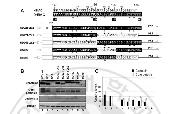
22
Fig. 4. Expression and core particle assembly of additional core protein variants. (A)
Aligned amino acid sequences of carboxyl-terminal domains of HBV and DHBV core proteins and schematic diagrams of additional chimeric core protein variant constructs. Amino acids in bold are identical or homologous. SRPK and PKA phosphorylation sites in the HBV genome are marked with asterisks and arrowheads, respectively. DHBV phosphorylation sites are marked with open arrowheads. The amino acid sequences of HBV and DHBV core protein are presented as open and closed boxes, respectively. The cytomegalovirus immediate early (CMV IE) promoter is represented as an open arrow. PRE, post-transcriptional regulatory element. (B) Expression of chimeric core proteins and core particle assembly by additional chimeric core protein variants. To examine core protein expression by HBV variants with chimeric core sequence, Western blotting was performed on lysates from HuH7 cells and HuH7 cells transfected with pHCP, pDCP, pHD221-262,
23
pHD221-241, pHD242-262, pHDHD, or pHHDH variants, as described for Fig 1B (top panel). Core particle formation by core protein variants was detected as described for Fig 1B (second panel). Transfection experiments were repeated four times. As the respective transfection and loading controls, luciferase (third panel) and a-tubulin (bottom panel) levels were determined as described for Fig 1B. (C) Relative levels of core protein expression and core particle assembly by additional chimeric core protein variants. Relative levels of core proteins, core particles, and luciferase were measured with the Fujifilm Image Gauge V4.0 program. Relative levels of core protein variant expression and core particle assembly were compared with normalized transfection efficiencies. The data represent the mean ± SD from four independent experiments.
24
To examine RNA encapsidation by these core variants, RPAs (Fig 5) was performed in chimeric core variants and C-deficient-RT-YMHA mutant co-transfected HuH7 cells. Core particles from these co-transfections exhibited similar assembly efficiency and migration patterns as those of singly transfected cells (see Figure 4B; Figure 5A, second panel). We could not detect significantly increased RNA encapsidation from cells transfected with these additional core variants compared to that from HD221-262 co-transfected cells (Fig 5A and B). pgRNA from cells co-co-transfected with the HDHD core variant and C-deficient-RT-YMHA mutant displayed slightly increased RNA encapsidation (Fig 5, lane 8). Since RPA with a 5´-end specific probe could not distinguish spliced encapsidated RNA from unspliced full-length pgRNA, encapsidation efficiency determined by RPA represented total encapsidated HBV RNA rather than full-length pgRNA incorporated into core particles. Consistent with RPA, RNAs encapsidated within core particles in situ were also detected from cells co-transfected with HD221-262, HD221-241, HD242-262, HDHD, or HHDH core variant and the C-deficient-RT-YMHA mutant (data not shown).
To our surprise, pgRNA encapsidation from HHDH core variant and C-deficient-RT-YMHA mutant co-transfections was less efficient than from HD221-262 core variant and C-deficient-RT-YMHA mutant co-transfections (Fig 5, and data not shown). The HHDH core variant contains most HBV core protein sequences, with the substitution only of a nine amino-acid motif from residues 242–250 (242RAGSPLPRS250) of DHBV core for residues 167–175 (167RRRSQSPRR175) of HBV core. A closer inspection of the HHDH core
25
variant revealed that only five amino acids (in italics and underlined) differed from the HBV core protein sequence. These results indicated that amino acid residues 167–175 of the HBV core protein were critical for efficient pgRNA encapsidation, and amino acid residues 150– 166 and 176–185 of HBV core protein were not essential, as long as the 40% amino-acid identity or 45% homology or the several critical residues, presumably within the conserved region, were maintained.
26
FIG. 5. pgRNA encapsidation in core particles by additional core protein variants. (A)
RPA to detect encapsidated pgRNA. To detect the pgRNA encapsidated by chimeric core protein variants, the C-deficient-RT-YMHA mutant and the core protein chimeras pHCP, pDCP, pHD192-262, pHD192-220, pHD221-262, pHD221-241, pHD242-262, pHDHD, or pHHDH were co-transfected into HuH7 cells. RPA (top panel) was performed as described for Figure 2D. Core particle formation (second panel) and luciferase levels (bottom panel) were determined as described for Figure 1B. Transfection experiments were repeated three times. (B) Relative levels of encapsidated pgRNA and core particle assembly by additional chimeric core protein variants. Relative levels of encapsidated pgRNA, core particles, and luciferase were measured with the Fujifilm Image Gauge V4.0 program. Relative levels of encapsidated pgRNA and core particle assembly were compared to normalized transfection efficiencies. The data represent the mean ± SD from three independent experiments.
27
E. Full-length HBV DNA is synthesized in core particles by the HDHD core variants
To identify the motif required for full-length HBV DNA synthesis, we analyzed DNA from HuH7 cells co-transfected with these additional chimeric core variants and C-deficient mutant. We clearly detected full-length DL DNA in HD221-241, HD242-262, and HDHD co-transfected HuH7 cells (Fig 6A and 6B, lanes 6–8). RC DNA was detected only from HDHD core variant co-transfected HuH7 cells (Fig 6A, lane 8). Full-length DNA was barely detectable from HHDH co-transfected cells. Collectively, these results further suggest that the amino acid residues 167–175 of the HBV core protein (167RRRSQSPRR175) are important for full-length DNA synthesis, while residues 150–166 and 176–185 are not. Since the HHDH core variant encapsidated pgRNA less efficiently (Fig 5 and 7C), the low level of DNA produced by cells co-transfected with the HHDH core variant and C-deficient mutant likely reflects this low encapsidation efficiency (Figs 6A, 6B, and 7D). Similar to transfection with HD221-262 (Fig 3), we also detected small-sized DNA from all cells co-transfected with core variants (Figure 6A, asterisks). We speculate that these small-sized DNAs were synthesized from spliced RNA, since HBV DNAs produced by the carboxyl-terminally deleted C164 variant are predominantly from spliced RNA (Kock et al, 2004; Pogam et al, 2005). When we used a probe corresponding to the region most frequently removed during splicing (Park et al, 2008; Gunther et al, 1997), the intensities of these small DNA forms were significantly decreased in core variant co-transfected cells (data not shown) and were barely detectable in core particles from HD221-262 and HHDH co-transfected cells (data not shown); this is consistent with the forms synthesized by HD221-241, HD242-262, and HDHD variant core proteins primarily comprising DNA from spliced RNA. We
28
also used a minus-strand RNA probe to detect plus-stranded HBV DNA (data not shown) and a plus-strand RNA probe to detect minus-stranded HBV DNA (data not shown). Both probes detected small-sized DNA, indicating that small DNA was double-stranded DNA synthesized from spliced RNA (data not shown). To further analyze small-sized DNA in detail, (Fig 6A, asterisks), polymerase chain reaction (PCR) was performed from core particles isolated from C-deficient mutant and various core variants co-transfected HuH7 cells. Consistent with the results from Southern blotting using strand-specific probes (data not shown) and spliced-out region specific probe (data not shown), small-sized DNA was amplified from HCP, HD221-262, HD221-241, HD242-262, HDHD, HHDH core variants co-transfected cells (Fig 6C, lanes 1, 5–9), further indicating that small DNA was from spliced RNA. Consistent with Southern blotting (Figure 6A), full-length DNA was also amplified from HCP, HD221-241, HD242-262, and HDHD core variants co-transfected cells (Fig 6C, lanes 1, 6–8), but not or hardly detectable from DCP, HD192-262, HD192-220, HD221-262, HHDH core variant co-transfected cells (Fig 6C, lanes 2–4, and 9). Sequence analysis revealed that the PCR-amplified small DNA was generated from encapsidated RNA which is spliced from nucleotides (nt) 2455–491, one of the most spliced sites, deleting 1257 nt (Figure 6C). Since the intensity of the small-sized DNA from cells co-transfected with HHDH was even weaker than those from cells co-transfected with HD221-241 and HD242-262 (Figure 6A and C, lanes 6 and 7 vs 9), we further speculate that the HHDH may be less competent to encapsidate spliced RNA also (Fig 7F)
29
FIG. 6. Full-length HBV DNA synthesis in core particles with additional chimeric core protein variants. (A) To examine synthesis of HBV DNA in core particles with chimeric
core variants, the C-deficient mutant and the core protein chimeras pHCP, pDCP, pHD192-262, pHD192-220, pHD221-pHD192-262, pHD221-241, pHD242-pHD192-262, pHDHD, or pHHDH were co-transfected into HuH7 cells. HBV DNA was extracted from isolated core particles and Southern blot analysis performed as described for Figure 3. Transfection experiments were repeated five times. The HBV replicative intermediate (RI) DNA is marked. Core particle formation (bottom panel) was determined as described for Figure 1B. (B) Relative levels of
30
HBV double-stranded linear (DL) DNA from isolated core particles were measured with the Fujifilm Image Gauge V4.0 program and compared after normalization to transfection efficiencies (top right panel). The data represent the mean ± SD from five independent experiments. * p < 0.001, ** p < 0.01, *** p < 0.05 (n=5). (C) PCR and sequence alignment of the spliced junction of DNAs from isolated core particles. HBV DNA was extracted from isolated core particles and PCR was performed. Primers were shown as dotted arrows. The 814 base-pair (bp) DNA that was 1257 nt smaller than 2,071 bp of full-length HBV DNA and full-length HBV DNA were amplified (arrowheads).
31
F. Residues R169 and R175 are important for HBV replication
To identify the amino acid residues from 167–175 of HBV core protein (167RRRSQSPRR175) that are important for rescue of full-length HBV DNA synthesis from HHDH core variant co-transfected cells, we singly altered amino acids in the HHDH background motif, comprising 167RAGSPLPRS175, to the corresponding residues in HBV, resulting in A168R, G169R, P171Q, L172S, and HHDH-S175R core variants (Fig 7A). Core particle formation was examined by particle Western blotting from HuH7 cells co-transfected with A168R, G169R, HHDH-P171Q, HHDH-L172S, or HHDH-S175R core variants and the C-deficient mutant (Fig 7B). Core particle assembly efficiency of the HHDH-A168R, HHDH-G169R, and HHDH-171Q was not restored to the level of HBV wt core protein and, although not significant, was slightly less efficient than that of the HDHD core variant (Fig 7B, n=5). However, core particle assembly efficiency of the HHDH-L172S (p<0.001, n=5) and HHDH-S175R (p<0.0001, n=5) was restored and was more efficient than that of the HDHD core variant and HBV wt core protein (Fig 7B).
RNA encapsidation was examined by RPA in HuH7 cells co-transfected with additional core variants and the C-deficient-RT-YMHA mutant (Fig7C, n=4). Consistent with Figure 5, RNA encapsidation by the HHDH C variant was markedly reduced compared to HBV wt core protein or HDHD core variants (Fig 7C, lanes 1 and 2 vs lane 3). HHDH-A168R, HHDH-G169R, and HHDH-P171Q C variants could rescue pgRNA encapsidation, although less efficiently than HBV wt core protein or the HDHD C variant (Fig 7C).
33
Fig 7. HBV core particle formation, pgRNA encapsidation, and HBV DNA synthesis by core variants. (A) Aligned amino acid sequences of HDHD and HHDH and amino acid
substitutions in the derived core variants A168R, G169R, HHDH-P170Q, HHDH-L172S, and HHDH-S175R. Amino acids in bold are identical or homologous. SRPK and PKA phosphorylation sites of HBV are marked with asterisks and arrowheads, respectively. Phosphorylation sites of DHBV are marked with open arrowheads. Amino acid sequences of HBV and DHBV core proteins are presented as black and white letters, respectively, on contrasting background. (B-D) HBV core particle formation, pgRNA encapsidation, and HBV DNA synthesis by core variants. To examine HBV core particle formation (B), pgRNA encapsidation (C), and HBV DNA synthesis (D) in core particles with the C-deficient or C-deficient-RT-YMHA mutants and pHCP or the core protein chimeras, pHDHD, pHHDH, A168R, G169R, P170Q, HHDH-L172S, or HHDH-S175R, were co-transfected into HuH7 cells. pcDNA3.1 was used to
34
equalize the amount of DNA transfected. (B) Core particle formation and luciferase levels (data not shown) were determined as described for Figure 1B. The data represent the mean ± SD (n=5). * p < 0.001, ** p < 0.01, and * p < 0.05 (n=5). (C) To examine pgRNA encapsidation, RPA was performed as described for Figure 2D. The data represent the mean ± SD from four independent experiments. (D) HBV DNA was extracted from isolated core particles and Southern blot analysis performed as described for Figure 3. The HBV replicative intermediate (RI) DNA is marked. DNAs from spliced RNAs are indicated by asterisks. Relative levels of core particles and encapsidated pgRNA and HBV DL DNA from isolated core particles were measured with the Fujifilm Image Gauge V4.0 program and compared after normalization to transfection efficiencies. The data represent the mean ± SD from five independent experiments. * p < 0.001 HHDH vs HHDH-G169R, ** p < 0.01 HHDH vs S175R, p=0.21 HDHD vs G169R, or p=0.24 HDHD vs HHDH-S175R (n=5). (E) PCR and sequence alignment of the spliced junction. HBV DNA was extracted from isolated core particles and PCR was performed as described for Figure 6C. The 814 base-pair (bp) DNA that was 1257 nt smaller than 2,071 bp of full-length HBV DNA and full-length HBV DNA were amplified (arrowheads). (F) RPA to discriminate encapsidated full-length pgRNA and spliced RNA. To detect the pgRNA encapsidated by chimeric core protein variants, the C-deficient-RT-YMHA mutant and the core protein chimeras were co-transfected into HuH7 cells as described for Fig7. RPA was performed as described for Fig 2D using spliced-region probe. The 470 nts of the HBV sequence was synthesized in vitro and the protected sequence, nt 2680-3092 of HBV sequence, was 413 nts long.
35
The HHDH-S175R could encapsidate pgRNA more efficiently than HBV wt core protein (Fig 7C, lanes 1 vs 8). However, the HHDH-L172S encapsidated pgRNA very poorly (Fig 7C, lane 7), even though core particles by HHDH-L172S were assembled more efficiently than HBV wt core protein or the HDHD core variant (Fig 7B and C, lanes 7).
HBV DNA synthesis was also examined by Southern blotting, using a probe specific for full-length HBV, of cells co-transfected with HHDH-A168R, HHDH-G169R, HHDH-P171Q, HHDH-L172S, or HHDH-S175R core variants and the C-deficient mutant (Fig 7D). Full-length RC HBV DNA synthesis was observed from cells co-transfected with HCP, HDHD, HHDH-G169R, or HHDH-S175R core variants and the C-deficient mutant, indicating the importance of the R169 and R175 residues for full-length RC HBV DNA synthesis (Fig 7D, lanes 1, 2, 5, and 8). However, HBV DNA synthesis was not rescued by HHDH-A168R, HHDH-P171Q, or HHDH-L172S core variants (Fig 7D, lanes 4, 6, and 7). The low level of HBV DNA synthesis by the HHDH-L172S C variant was due to its very inefficient pgRNA encapsidation (Fig 7C and D, lanes 7). Since residues R167, S170, P173, and R174 are identical to the corresponding regions from DHBV (242RAGSPLPRS250 in DHBV vs 167RRRSQSPRR175 in HBV), their importance remains to be determined.
Polymerase chain reaction (PCR) was also performed as in Figure 6C to further examine the small-sized DNA (see Fig7D, asterisks). Consistent with Figure 6C and the result from spliced-out region specific probe (data not shown), small-sized DNA was amplified from core variants co-transfected cells (Fig7E, lanes 1–8), further indicating that small-sized DNA was from spliced RNA. In consistent with Southern blotting (Fig 7D), full-length DNA was also amplified from HCP, HDHD, HHDH-G169R, and HHDH-S175R
co-36
transfected cells (Fig 7E, lanes 1, 2, 5, and 8), but not from HHDH, A168R, HHDH-P171Q, or HHDH-L172S co-transfected cells (Fig 7E, lanes 3, 4, 6, and 7). Consistent with the result from Figure 6C, sequence analysis revealed that one of the most spliced sites, nucleotides (nt) 2455–491, was deleted by splicing (Fig 7E).
To further confirm this result, RPA was also performed to discriminate encapsidated full-length pgRNA from spliced RNA using a spliced-out region probe which encompasses nt 2689-3092 of HBV sequence (Fig 7F). If chimeric core variants encapsidated spliced RNA more efficiently than full-length pgRNA, the encapsidation level by spliced-out region probe would be low compare to that by 5-end specific probe. As shown in Figure 7F, the encapsidation efficiencies did not differ significantly between spliced-out region and 5`-end specific probes (Figure 7C vs F), we could conclude that the low level of HBV DNA synthesis by the HHDH and HHDH-L172S core variant was due to very inefficient pgRNA encapsidation (Fig 7C-D and Fig 7F, lanes 3 and 7).
37
IV. DISCUSSION
In this study, chimeric core variants by substituting the carboxyl-terminal regions of HBV core protein with the corresponding regions of DHBV core protein were generated and core particle assembly, pgRNA encapsidation, and HBV DNA synthesis were examined. Unlike DHBV core protein, which fails to trans-complement HBV core protein (Okamoto et al, 1990), various chimeric core variants could trans-complement HBV replication, including the HD221-262 with carboxyl-terminal 42 amino acids of DHBV core protein for those in HBV. We therefore hypothesize that the amino-terminus of HBV core protein may interact with viral or host components to form a tertiary structure and/or to support HBV replication. Core particle assembly for HBV replication. All chimeric core variants can assemble into core particles as long as the N-terminal 145 residues of HBV core proteins are intact (Fig 1B). However, core particles formed by chimeric core variants migrated slowly and demonstrated differing assembly competencies (Fig 1B, 2B, 4B, 5A, and 6A), suggesting that the carboxyl-terminal nucleic acid binding domain may affect core particle formation to some extent or interact with the amino-terminal assembly domain for particle stability. The presence of slowly migrating core particles (HD192-262, HD242-262, and HHDH core chimeras) suggests that these core particles might be less stable or differ in net charge, thus affecting HBV replication (Pogam et al, 2005; Newman et al, 2003; Kim et al, 2004). HHDH core particles migrated more slowly than HDHD, providing evidence of inefficient pgRNA encapsidation and DNA synthesis (Fig 5–7). However, this speculation did not apply to all C
38
chimeras, since pgRNA encapsidation and HBV DNA synthesis by HD221-242 and HD242-262 were similar, even though core particle migration patterns were different (Fig 5 and 6). Core particles assembled from truncated core proteins or those with insertions are unstable (Kock et al, 2004; Pogam et al, 2005; Kock et al, 1998); destabilization through insertional mutagenesis may explain the failure of trans-complementation by HD192-262 and 220. It is still possible to speculate that the insertion of 29 residues in HD192-220 destabilizes core particles, thus preventing protection of encapsidated pgRNA from nuclease treatment during core particle isolation. Since HD192-262 has more extensive alterations, these alterations may cause the destabilizations of chimeric core protein and/or core particles or reduce the efficiency of core particle assembly. Sequence conservation in the carboxyl-terminal domain of hepadnavirus core protein. Using a series of core protein carboxyl-terminal deletion mutants, Le Pogam et al. (Pogam et al, 2005) suggested that residues 167–175 (165–173 of ayw) of HBV core protein are important for selective and/or productive viral RNA encapsidation by charge balance and core particle stability through the arginine-rich domain. The carboxyl-terminal 10 amino acids of HBV core protein are dispensable for HBV DNA replication (Kock et al, 2004; Pogam et al, 2005). The present study extends these results by showing that residues 167–175 (165–173 of ayw), as well as the 62% homologous residues from 146–166, of HBV core protein are sufficient for full-length HBV DNA synthesis using the HDHD core variant (Fig 6A and 6B). From the 27% homology between HBV and DHBV core proteins, the carboxyl-terminus is 45% homologous or 40% identical, and residues 146–166 are 62% homologous, suggesting that several critical residues from 146–166 may be conserved or have coevolved to encapsidate
39
pgRNA and subsequently synthesize DNA. Even though our results also demonstrated that residues 167–175 (165–173 of ayw) of HBV core protein are important for HBV replication, several questions are still unanswered. First, if these residues are solely essential for HBV replication as long as residues 146–166 are at least 62% homologous, the replication efficiencies of HD221-241 and HDHD should be similar, and the former should engage in full-length RC DNA synthesis, as does the HDHD. Second, replication of the HD242-262 should be inefficient, similar to that of the HD221-262 and/or HHDH. However, HD221-241 and HD242-262 exhibited similar replication efficiencies; replication efficiency was improved relative to HD221-262. Although not significant (p=0.065, n=5), HBV DNA synthesis was little more efficient by HD242-262 than HD221-241 (Fig 6B).
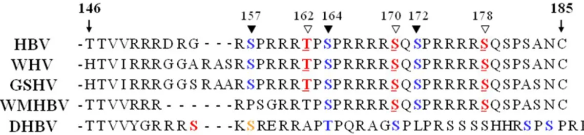
Putative phosphorylation sites in the carboxyl-terminal domain of HBV core protein. Hepadnavirus core proteins are heavily phosphorylated (Gazina et al, 2000; Lan et al, 1999; Machida et al, 1991; Liao et al, 1995; Yeh et al, 1991). The core protein of DHBV is phosphorylated at six sites (S230, S232, T239, S245, S257, and S259) on an S/TP motif within the carboxyl-terminal domain (Perlman et al, 2005; Yu and Summers, 1994). Three phosphorylation sites (S155, S162, and S170 in subtype ayw and S157, S164, and S172 in subtype adw) in the carboxyl-terminal domain of HBV core protein have been identified as having an SPRRR motif (Liao and Ou, 1995). Several intracellular protein kinases such as protein kinase C (Kann and Gerlich, 1994), the cyclin-dependent kinase cdc2 (Yeh et al, 1993), the 46 kDa serine protein kinase (Kau and Ting, 1998), and serine/arginine protein-specific kinases 1 (95 kDa SRPK1) and 2 (105 kDa SRPK2) (Daub et al, 2002) have been shown to phosphorylate these serine residues in vitro. The synthesis of smaller than
full-40
length DNA was also demonstrated for major phosphorylation-site mutants (S155E, S162E, and S170E) (Kock et al, 2004). Phosphorylation at these sites is important for pgRNA encapsidation and HBV replication (Kock et al, 2004; Gazina et al, 2000; Lan et al, 1999; Melegari et al, 2005) and these serine phospho-acceptor sites contribute pleiotropically toward modulating HBV replication (Lewellyn and Loeb, 2011b). Three additional putative cAMP-dependent protein kinase A (PKA) phosphorylation sites (RRXS/T: T162, S170, S178) have been identified, and two α-type CK2-activated PKAs (PKAIα and PKAIIα) phosphorylate both S170 and S178 in vitro in the absence of cAMP (Enomoto et al, 2006).
Phosphorylation and dephosphorylation states may not be drastically altered in the core protein chimeras, since both HBV and DHBV core proteins have six phospho-acceptor sites (Fig 1A and 4A). However, for the HHDH, the S172L and R175S substitutions may have reduced the number of putative phosphorylation sites to four (Fig 4A). Also, even though S178 was retained in the HHDH, the 175SRRS178 could have disrupted 175RRRS178 motif, the putative PKA phosphorylation site at S178, contributing to inefficient pgRNA encapsidation and HBV replication (Fig 5–7). HDHD, however, have seven putative phosphorylation sites, ensuing efficient pgRNA encapsidation and HBV replication.
Arginine-rich domains in the carboxyl-terminus of HBV core protein. The carboxyl-terminal domain of HBV core protein has 16 (ayw) or 17 (adw) arginine residues with four clusters (150RRR152, 159RRR161, 166RRRR169, and 174RRRR177) comprising arginine-rich domains (ARD) I–IV and conferring a net positive charge (Fig 4A). The carboxyl-terminal domain of DHBV core protein, in contrast, has 12 positively charged
41
amino acids (arginine or lysine) but does not conserve the four ARDs, although the 227RRR229 and 233RERR236 motifs may be equivalent to ARD-I and -II (Fig 4A). Recently, mutagenesis of the ARDs of HBV core protein demonstrated their pleiotropic contribution to HBV replication (Lewellyn and Loeb, 2011a). R to A (RRRR→AAAA) mutation in ARD-III impaired in pgRNA encapsidation and minus-strand DNA template switching most strikingly (Lewellyn and Loeb 2011a).
Since ARD-I and -II remained intact in HHDH, inefficient pgRNA encapsidation and subsequent poor replication might be attributed to the 166RRAG169 (ARD-III) and 174RSRR177 (ARD-IV) changes. In HD242-262, ARD-III and ARD-IV were disrupted to 166RRAG169 and 174RSSS177, respectively, but HBV replication was more efficient than HHDH. HDHD has 158RERR161 (ARD-II), 166QRRR169 (ARD-III), and 174RRSS177 (ARD-IV), indicating that these changes may be tolerated to maintain full-length DNA synthesis. Also, HD221-241, HD242-262, HHDH, and HDHD have 15, 14, 15, and 14 positively charged amino acids (arginine or lysine) respectively, in their carboxyl-terminal domains (Fig 4A).
Important amino acids in the carboxyl-terminal domain of HBV core protein for HBV replication. The HHDH-A168R rescued pgRNA encapsidation, even though HBV DNA synthesis was not fully rescued (Fig 7C and 7D, lane 4), suggesting that R168 itself, partial restoration of ARD-III (166RRR168), and/or S170 in 167RRGS170 motif (a putative PKA phosphorylation site) may be important for encapsidation, but are not sufficient to support HBV DNA synthesis. HHDH-G169R restored pgRNA encapsidation and HBV DNA synthesis, suggesting that the 167RARSPLPRS175 motif might be sufficient to form a