Introduction
Several anatomy textbooks describe that the ulnar nerve is composed of the ventral rami of the C8, T1 and often C7 [1-4]. Those of the C7 components may supply the flexor carpi ulnaris muscle in the anterior compartment of the forearm. However, there are few literatures regarding contributing frequency and quantities of the C7 to the ulnar nerve. Fuss (1989) [5] reported the lateral cord con- tribution to the ulnar nerve was in 56% of the 158 brachial
plexus. Pyun et al. (2010) [6] reported that the connection between lateral cord and ulnar nerve was observed at 13.1%
(5/38) in their anatomic study and the flexor carpi ulnaris muscle was abnormal electromyographic findings at the 46.2% (36/78) in the C7 radiculopathy. Therefore differ- encies between anatomic study and electromyographic diagnosis exist. This study was aimed to determine frequ- ency and the number of the myelinated axons of the C7 in the ulnar nerve.
Materials and Methods
Fifty cadavers of brachial plexus were obtained from cadavers of Korean adults (Male: 58, Female: 42, average age: 69.7 years). The brachial plexus containing the ulnar nerve were extracted from the axilla and the extracted sam- ples were immersed in Guanidine-HCl (0.2 M) for 2 weeks
Frequency and Quantity of the C7 Contribution to the Ulnar Nerve
Mi-Sun Hur, Jung-Su Woo, Ho-Jeong Kim, Kyu-Seok Lee
Department of Anatomy, Kwandong University College of Medicine
(Received 16 August 2013, revised 16 September 2013, accepted 17 September 2013, Published Online 30 September 2013)
Abstract : Many anatomists and clinicians who investigate the peripheral nerve concern about the composition of the spinal roots of each terminal nerve of the brachial plexus. From this viewpoint, the spinal root composition of the ulnar nerve is still unclear. Several anatomy textbooks describe that the ulnar nerve is composed of the ventral rami of the C8, T1 and often C7. There is no literature regarding the frequency and contribution quantity of C7 to the ulnar nerve. The purpose of present study was to determine frequency and contribution quantity of the C7 to the ulnar nerve.
Fifty cadavers of brachial plexus were obtained from cadavers of Korean adults. The brachial plexus containing the ulnar nerve were extracted from the axilla and the extracted samples were immersed in Guanidine-HCl (0.2 M) for 2 weeks to soften the connective tissue around the nerve bundles. C7 was contributed to the ulnar nerve in all sides (100%). The numbers of the myelinated axons of C7 participating to the ulnar nerve was 1,452±429 (mean
±S.D.). Thus the C7 can be considered as always participating component of the ulnar nerve, not often partici- pation, although numbers of the myelinated axons of C7 was lesser than those of the C8, but similar to those of the T1.
The results of the study provide a reference for accurate diagnosis and treatment regarding ulnar nerve injury due to various accidents.
Keywords:C7, Frequency, Quantity, Ulnar nerve
*This work was supported by Basic Science Research Program through the National Research Foundation of Korea (NRF) funded by the Ministry of Education and Technology (2009-0071338).
The author (s) agree to abide by the good publication practice guideline for medical journals.
The author (s) declare that there are no conflicts of interest.
Correspondence to : Kyu-Seok Lee (Department of Anatomy, Kwan-dong University College of Medicine)
E-mail : [email protected] 대한체질인류학회지 제26권 제3호
Korean J Phys Anthropol Vol. 26, No. 3 (2013) pp. 101~104
http://dx.doi.org/10.11637/kjpa.2013.26.3.101 Original Article
to soften the connective tissue around the nerve bundles.
Each processed brachial plexus was dissected under a sur- gical microscope at a magnification of 7.5x (OPMI-FC, Carl Zeiss, German). After determining contribution of the C7, 20 samples were processed with a routine histolo- gical staining procedure (H&E) to count the number of myelinated axons. Imbroglio Modometer version 2.0 soft- ware was used to count the number of myelinated axons.
Results
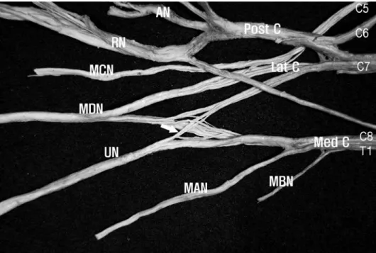
The C7 was found to contribute to the ulnar nerve in all fifty cadavers (100/100, 100%). In all cases, the C7 fasci- cles branched out at the distal portion of the fascicles com- posing the lateral root of the median nerve, decussating with the medial root of the median nerve (Fig. 1). The
quantity of C7 contribution to the ulnar nerve was 1,452
±429 myelinated axons, C8 was 11,448±1,473 and T1 was 1,720±382 in 20 sides (Table 1).
Discussion
Before counting the number of myelinated axons of C7, we compared the total number of myelinated axons of the ulnar nerve with that of another study. The total number of the myelinated axons of the ulnar nerve was 16,400 [7].
In the present study, total number of the myelinated axons of ulnar nerve was 14,620. It showed that it is similar with that of another study. The C7 of the ulnar nerve innervated the flexor carpi ulnaris muscle, along with the C8 compo- nents [3-4,8-10]. In the cases that it has a large number of nerve fibers, it can be considered that the C7 innervates other muscles and/or the C7 is responsible for sensory innervation to the skin.
In this study, the C7 was found to consistently contri- bute to the ulnar nerve, which was different to the result of Fuss (1989) [5] who reported 56% contribution of C7 to the ulnar nerve. And the results of Pyun et al. (2010) [6]
reported 13.1% in their anatomic study. However, electro- 102 Mi-Sun Hur, Jung-Su Woo, Ho-Jeong Kim, Kyu-Seok Lee
Table 1. Numbers of myelinated axons of C7, C8 and T1 roots participating to the ulnar nerve.
C7 C8 T1
Number of 1,452±429 11,448±1,473 1,720±382
myelinated axons (9.9%) (78.3%) (11.8%)
Fig. 1. A photograph showing the C7 fascicles (arrow) constituting the ulnar nerve in the posterior aspect of the brachial plexus. C7 fasci- cles constituting the ulnar nerve branched out at the distal portion of the lateral cord. AN, axillary nerve, Lat C, lateral cord; MAN, medial antebrachial cutaneous nerve; MBN, medial brachial cutaneous nerve; MCN, musculocutaneous nerve; MDN, median nerve; Med C, medial cord; Post C, posterior cord; RN, radial nerve; UN, ulnar nerve.
diagnostic findings were different from the anatomic study.
Dumitru and Zwarts (2002) [11] described the presence of lateral root of ulnar nerve at 15.0-92.0% in their electro- diagnostic study.
Hur and Lee (2011) [12] reported that the C7 was con- stant components of ulnar nerve and the myelinated fiber number of spinal root (C7, C8, T1) of ulnar nerve was 1,339±680, 10,075±1,473, 1,032±582, respectively.
Therefore, it can be considered that the C7 nerve root is a constant component of the ulnar nerve, not often, although the contribution quantity of C7 was lesser than those of the C8 and was similar to those of the T1.
The results of the study provide a reference for accurate diagnosis and treatment regarding ulnar nerve injury due to various accidents.
References
1. Romanes GJ. Cunningham’s Textbook of Anatomy. 12th ed.
New York: Oxford University Press; 1981. p. 328-9.
2. Ellis H. Clinical Anatomy - A revision and applied anatomy for clinical students. 8th ed. London: Blackwell Scientific Publications; 1992. p. 209.
3. Moore KL, Dalley AF. Clinically Oriented Anatomy. 5th ed. Baltimore: Lippincott Willams & Wilkins; 2006. p. 777.
4. Standring S. Gray’s Anatomy. 39th ed. Edinburgh: Elsevier Churchill Livingston; 2005. p. 847, 877.
5. Fuss FK. The lateral root of the ulnar nerve. Acta Anat (Basel). 1989; 134:199-205.
6. Pyun SB, Kang S, Kwon HK. Anatomical and electrophy- siological myotome corresponding to the flexor carpi ulnaris muscle. J Korean Med Sci. 2010; 25:454-57.
7. Norkus T, Norkus M, Ramanauskas T. Donor, recipient and nerve grafts in brachial reconstruction: anatomical and tech- nical features of facilitating the exposure. Surg Radiol Anat.
2005; 27:524-30.
8. Lindner HH. Clinical Anatomy. International ed. London:
Appleton & Lange; 1989. p. 554.
9. MacKinon PCB, Morris JF. Oxford Textbook of Functional Anatomy. Musculo-Skeletal System (Vol. 1). 2nd ed. Oxford:
Oxford University Press; 2005. p. 85.
10. Russell SM. Examination of Peripheral Nerve Injuries. 1st ed. New York: Thieme; 2006. p. 22-7.
11. Dumitru D, Zwarts M. Brachial plexopathies and proximal mononeuropathies. In: Dumitru M, Amato A, Zwarts M, editors. Electrodiagnostic Medicine. 2nd ed. Philadelphia:
Hanley & Belfus; 2002. p. 777-836. Cited from Pyun et al.
(2010).
12. Hur MS, Lee KS. Quantification of the nerve fiber of the terminal branches of the typical brachial plexus. Korean J Phys Anthropol. 2011; 24:135-40.
Frequency and Quantity of the C7 Contribution to the Ulnar Nerve 103
자신경에 분포하는 C7의 빈도와 참여량
허미선, 우정수, 김호정, 이규석
관동대학교 의과대학 해부학교실
간추림 : 말초신경을 연구하는 많은 해부학자와 임상가들은 팔신경얼기 끝신경의 척수신경 뿌리구성에 대해 관 심을 가지고 있다. 이러한 관점에서, 자신경의 척수신경 뿌리구성은 명확하지 않다. 여러 해부학 교과서에는 자 신경이 C8, T1의 앞가지로 구성되어 있고, 종종 C7도 포함되는 것으로 기술되어 있다. 그러나 자신경에 참여하 는 C7의 빈도와 참여량에 대한 문헌은 있지 않다. 이 연구의 목적은 자신경에 참여하는 C7의 빈도와 참여량을 관찰하는 데 있다.
한국성인시신의 팔신경얼기 100쪽을 사용하였다. 자신경을 포함하는 팔신경얼기를 겨드랑에서 떼어낸 후, Guanidine-HCl (0.2 M)에 2주 동안 담구어 신경다발을 둘러싸는 결합조직을 부드럽게 하였다.
C7은 모든 경우에서 자신경에 분포하였다(100%). 자신경에 분포하는 C7의 말이집축삭의 수는 1,452±429 (mean±S.D.)였다. C7의 말이집축삭의 수가 C8의 말이집축삭의 수보다 적었고, T1의 말이집축삭의 수와 비슷 하였지만, C7은 자신경에 자주 참여하는 것이 참여하는 것이 아니라, 항상 참여하는 성분으로 여겨질 수 있다.
이 연구의 결과는 다양한 사고로 인한 자신경 손상시 정확한 진단과 치료에 대한 자료를 제공할 것이다.
찾아보기 낱말 : C7, 빈도, 참여량, 자신경
104 Mi-Sun Hur, Jung-Su Woo, Ho-Jeong Kim, Kyu-Seok Lee
교신저자 : 이규석(관동대학교 의과대학 해부학교실) 전자우편 : [email protected]