INTRODUCTION
“One airway, one disease” is a well-established concept that links the upper and lower airways. Anatomically, these 2 air- ways have common respiratory epithelium and similar muco- sal susceptibility to disease. Epidemiologic studies have consis- tently shown that asthma and rhinitis often coexist in the same patients. Between 19% and 38% of patients with allergic rhinitis have coexisting asthma, and the prevalence rate of asthma is much higher in patients with allergic rhinitis than in the gener- al population.1,2 In Korea, the prevalence of bronchial hyper-re- sponsiveness was 55.7% and 25.5% in 6- to 15-year-old patients with allergic and non-allergic rhinitis, respectively.3
Currently, this concept is applied to the middle ear space, giv-
en that the middle ear space is an anatomical extension of the airway through the Eustachian tube and capable of mounting an allergic inflammatory response. A previous study showed the incidence of atopy was 24% in the study population com- prising patients who had otitis media with effusion (OME) per- sisting for more than 3 months and unresponsiveness to antibi-
Synergistic Effect of Dermatophagoides farinae and
Lipopolysaccharides in Human Middle ear Epithelial Cells
Ji-Eun Lee,
1,2Yeon Hoo Kim,
1Chae-Seo Rhee,
3,4,5,6Dong-Young Kim
7*
1Department of Otorhinolaryngology-Head and Neck Surgery, Chosun University College of Medicine, Gwangju, Korea
2Department of Otorhinolaryngology-Head and Neck Surgery, Graduate school of Medicine, Seoul National University, Seoul, Korea
3Sensory Organ Research Center, Seoul National University Biomedical Research Institute, Seoul, Korea
4Institute of Allergy and Clinical Immunology, Seoul National University Biomedical Research Institute, Seoul, Korea
5Graduate School of Immunology, Seoul National University, Seoul, Korea
6 Department of Otorhinolaryngology-Head and Neck Surgery, Seoul National University Bundang Hospital, Seoul National University College of Medicine, Seongnam, Korea
7Department of Otorhinolaryngology-Head and Neck Surgery, Seoul National University College of Medicine, Seoul, Korea
This is an Open Access article distributed under the terms of the Creative Commons Attribution Non-Commercial License (http://creativecommons.org/licenses/by-nc/3.0/) which permits unrestricted non-commercial use, distribution, and reproduction in any medium, provided the original work is properly cited.
Purpose: Although the concept of “one airway, one disease,” which includes the middle ear space as part of the united airway is well recognized, the role of allergens in otitis media with effusion (OME) is not clearly understood. We aimed to investigate the effect of the interaction between Dermatophagoides farinae (Der f) and lipopolysaccharide (LPS) on the induction of epithelial inflammatory response in vitro. Methods: Primary hu- man middle ear epithelial cells were exposed to Der f, LPS, or both in different sequences, and the magnitude of the immunologic responses was compared. The mRNA expressiona of mucin (MUC) 4, 5AC, 5B, 8, GM-CSF, TNF-α, TLR4, and MD-2 were evaluated using real-time PCR. MUC levels before and after siRNA-mediated knockout of TLR4 and MD-2 were assessed. Lastly, the involved cell signaling pathway was evaluated. Results:
The expressiona of cytokines, and the MUC 4, 5AC, 5B, and 8 genes were augmented by pretreatment with Der f followed by LPS; however, reverse treatment or combined treatment did not induce the same magnitude of response. Increased MUC expression was decreased by TLR4 knockdown, but not by MD-2 knockdown. The signal intensity of MUC 8 was higher in MD-2 over-expressed cells than in those exposed to LPS only. The translo- cation of nuclear factor-κB was observed in cells pretreated with Der f followed by LPS. Conclusions: When Der f treatment preceded LPS expo- sure, Der f and LPS acted synergistically in the induction of pro-inflammatory cytokines and the MUC gene, suggesting an important role in the de- velopment of OME in patients with concealed allergy airway sensitization.
Key Words: Dermatophagoides farinae; innate immunity; lipopolysaccharides; mucins; toll-like receptors
Correspondence to: Dong-Young Kim, MD, PhD, Department of Otorhinolaryngology, Seoul National University College of Medicine, Seoul National University Hospital, 101 Daehak-ro, Jougno-gu, Seoul 03080, Korea.
Tel: +82-2-2072-2440; Fax: +82-2-745-2387; E-mail: dongkim@snu.ac.kr Received: December 7, 2015; Revised: January 3, 2016;
Accepted: January 6, 2016
•There are no financial or other issues that might lead to conflict of interest.
Allergy Asthma Immunol Res. 2016 September;8(5):445-456.
http://dx.doi.org/10.4168/aair.2016.8.5.445 pISSN 2092-7355 • eISSN 2092-7363
otics.4 Another study suggested that 35% of patients with recur- rent OME have allergic rhinitis.5 Significantly higher numbers of eosinophils and T lymphocytes, as well as significantly high- er levels of IL-4 and IL-5 mRNA+ cells, have been reported in patients with atopic OME than in non-atopic controls.6 Further- more, other studies have shown increased expression of IL-5 and major basic proteins in the middle ear mucosa of patients with OME than that in normal controls, as well as higher levels of eosinophilic cationic proteins in the supernatant of middle ear effusion of atopic patients with OME than in that of non- atopic controls.7,8 These findings support the concept that the middle ear may be part of the united airway and may behave similarly to the nose or lung under allergic inflammatory con- ditions. Although allergy has been considered one of the most important factors of OME based on clinical observations, the role of allergy has not been investigated extensively and is easi- ly ignored. In general, the possible integration of the middle ear as part of the united airway concept will have major clinical im- plications for the diagnosis and management of allergic airway disease.
Dermatophagoides pteronyssinus (Der p) and Dermatopha- goides farinae (Der f) are the most common house dust mite (HDM) species, which produce allergens that are widely dis- tributed in Korea. Recently, Der p and Der f have been shown to activate toll-like receptors (TLRs) 2 or 4 and thereby stimu- late the innate immune response. TLRs recognize repetitive patterns in diverse microbes, including gram-positive and gram-negative bacteria and viruses, and they are key compo- nents of innate immunity.9,10 In the innate immune response, airway epithelial cells aid the immune system by inducing re- cruitment of immune-competent cells to local tissues and thereby modulating their activity.11 In addition, lipopolysaccha- ride (LPS), a major component of the outer membrane of gram- negative bacteria, is a virulence factor that can induce inflam- matory responses. LPS may induce the release of multiple pro- inflammatory cytokines and chemokines from airway epitheli- al cells, and this initial inflammatory response plays a key role in the containment of infection. The inflammatory response in- duced by LPS in epithelial cells has been linked to TLR 4.12
The airway mucosa is lined with ciliated columnar epithelium interspersed with goblet cells. Accumulation of secretions and mucostasis in the airway mucosa due to allergic airway disease can alter the viscoelasticity of the mucus, ciliary motility, and epithelial defense mechanisms, thereby increasing the suscep- tibility to various stimuli, such as bacteria and viruses.13 Viruses are clinically linked to allergy: viral infection can induce asth- ma/allergic exacerbations, and viral clearance and persistence of symptoms are prolonged in allergic individuals.14-16 Con- versely, chronic allergic inflammation may impair anti-viral re- action to acute rhinovirus infection, as indicated by the sup- pression of IFN-α, IFN-γ, and IL-12 induction.17 Meanwhile, air- borne LPS might adversely affect asthmatic individuals by en-
hancing an established airway inflammation and airway ob- struction.18,19
Considering the above findings, it is natural to think that epi- thelial cells of the middle ear might be involved in recognizing and formulating an innate immune response to Der f and LPS, and a kind of reciprocal effect may be developed by subsequent exposure to Der f and LPS. Although allergy or allergen sensiti- zation could be a concealed, potent stimulant of inflammation in sinus and middle ear disease, the effect of allergen sensitiza- tion on infection and its mechanism in airway epithelium is not clearly understood. Moreover, although allergy has been impli- cated in OME, the role of middle ear epithelial cells in the in- nate immune response has not been characterized. Thus, this study was design to investigate whether allergen pre-sensitiza- tion within airway mucosa can affect subsequent viral or bacte- rial infection. For this, we performed in vitro experiments using primary human middle ear epithelial cells (HMEECs) and as- sessed the signaling pathways activated by Der f and LPS as well as the production of pro-inflammatory cytokines and the expression of the mucin (MUC) gene.
MATERIALS AND METHODS Reagents
LPS, SB203580, and Bay were obtained from Sigma-Aldrich (Munich, Germany). Der f crude body extract was purchased from Arthropods of Medical Importance Resource Bank (Seoul, Korea). Other chemicals used were of the purest grade avail- able from Sigma (St. Louis, MO, USA).
Cell culture and stimulation
HMEECs (kindly provided by Dr. David J. Lim, House Ear In- stitutes, Los Angeles, CA, USA) were maintained in a mixture of Dulbecco’s modified Eagle’s medium (DMEM, Invitrogen, Carlsbad, CA, USA) and bronchial epithelial basal medium (Lonza, Walkersville, MD, USA) (1:1).20-22 Cells were grown to 60% confluence in 100-mm culture plates and kept at 37°C in a carbon dioxide-enriched (95% air, 5% CO2) humidified atmo- sphere.
HMEECs were seeded onto 60-mm culture plates, with 2.2×
105 cells per well for the experimental condition. Predeter- mined non-cytotoxic doses of Der f or LPS by MTT (3-(4,5-di- methylthiazol-2-yl)-2,5-diphenyltetrazolium bromide) assay (data not shown) were applied to HMEECs. By performing the pilot study using real-time PCR, treatment with 1.0 μg/mL LPS for 24 hours was identified to be the optimal and effective in that it not only elicits a moderate level of inflammatory re- sponse but also has an augmentative effect following the ad- ministration of 2.0 μg/mL Der f. Cells were then stimulated in several ways: 1) Der f 2.0 μg/mL only for 48 hours (Df48h); 2) LPS 1.0 μg/mL only for 24 hours (LPS24h); 3) Der f 2.0 μg/mL for 48 hours and add LPS 1.0 μg/mL for 24 hours (Df48h/
LPS24h); 4) LPS 1.0 μg/mL for 48 hours and add Der f 2.0 μg/
mL for 24 hours (LPS48h/Df24h); or 5) Der f 2.0 μg/mL and LPS 1.0 μg/mL both simultaneously for 48 hours (Df48h/LPS48h) (Fig. 1).
Real-time polymerase chain reaction (qRT-PCR)
Total RNA was isolated from the epithelial cells using TRIzol (Invitrogen) as per the manufacturer’s instructions and reverse transcribed into cDNA using the Quantitect Reverse Transcrip- tion kit (Qiagen, Venlo, Netherlands). Then qRT-PCR analyses were performed using a 7,500 FAST qRT- PCR System (Applied Biosystems, Foster City, CA, USA). Each reaction mixture con- tained 10 μL of SYBR® Green PCR Master Mix (Applied Biosys- tems), 4 pmol each of the forward and reverse primers, and 1 μL of cDNA in a final volume of 20 μL. Reaction mixtures were incubated at 95°C for 5 minutes to activate FastStartTaq DNA Polymerase, followed by 40 cycles of amplification. Data were analyzed using Sequence Detection Software version 1.9.1 (Ap- plied Biosystems). Target mRNA expression was normalized to glyceraldehyde-3-phosphate dehydrogenase (GAPDH) expres- sion and calculated using the comparative Ct method. Primers used in this study are shown in Table.
RNA interference and plasmid DNA transfection
Cells were transfected with control small interfering (si) RNA, TLR 4 siRNA or myeloid differentiation (MD)-2 siRNA, and 10 μL of Lipofectamine RNAiMAX (Invitrogen) in 60- mm plates as per the manufacturer’s protocol. The plasmid pFlag-CMV1- hMD2 was a kind gift from Doug Golenbock (Addgene plasmid
#13028). After cells were washed with OPTI-MEM (Gibco, Carlsbad, CA, USA), DNA was transfected to cells using X- treme GENE HP-DNA transfection reagent (Roche Diagnostics, Indianapolis, IN, USA) as per the supplier’s protocol. After 4 hours of incubation, the medium was exchanged for a com- plete medium containing 10% serum and antibiotics. Cells were incubated for an additional 24 hours and treated with Df or LPS. Cell viability was measured by light microscopy, and the gene-silencing or expression efficacy was evaluated by evaluat- ing the mRNA levels. The siRNAs were NM_138554.2 for TLR4 and NM_014364.2 for MD-2 (Bioneer, Daejeon, Korea).
Western blot analysis
At specific time points after the Der f or LPS treatment, the medium was removed, and cells were washed with phosphate- buffered saline (PBS; 10 mM, pH 7.4). Cells were then lysed with lysis buffer (50 mM Tris pH 7.7, 150 mM NaCl, 1% NP-40, 5 mM EGTA, 50 mM -glycerophosphate, 20 mM NaF, 1 mM Na3VO4, 2 mM phenylmethylsulfonyl fluoride, 10 mg/mL leupeptin, and 10 mg/mL aprotinin) and incubated for 20 minutes at 4°C.
Briefly after sonication, cells were centrifuged at 13,000×g for 10 minutes at 4°C. The supernatant containing the total cell ly- sate was collected. Protein concentration of the lysates was measured using the Bio-Rad Protein Assay (Bio-Rad, Hercules, CA, USA). Equal amounts of protein were mixed with sample buffer (62.5 mM Tris–HCl [pH 6.8], 1% sodium dodecyl sulfate, 2.5% glycerol, 0.5% 2-β-mercaptoethanol, and bromophenol blue), boiled for 5 minutes, and separated by electrophoresis on 10%-12% Tris-HCl gels. Protein content of the gels was trans- ferred to a PVDF membrane (Amersham, Buckinghamshire, UK), and the membranes were blocked with TBS-T (20 mM Tris, 500 nM NaCl, with 0.1% Tween-20) containing 5% (w/v) skim milk for 1 hour at room temperature. Membranes were probed with antibodies against MUC 8 (Sigma–Aldrich, St. Lou- Table. Primers for real-time polymerase chain reaction
Primers
MUC 4 F:5’-ATG GTCATCTCG GAG TTC CAG-3’
R:5’-GTAGACCAGGTCGTAGCCCTT-3’
MUC 5AC F: 5’-GGGACTTCTCCTACCAAT-3’
R:5’-TATATGGTGGATCCTGCA GGGTAG-3’
MUC 5B F:5’-CAC ATC CAC CCT TCC AAC-3’
R:5’-GGC TCA TTG TCG TCT CTG-3’
MUC 8 F:5’-GAC AGG GTT TCT CCT CAT TG-3’
R:5’-CGT TTA TTC CAG CAC TGT TC-3’
IL-1b F: 5’-ACAGATGAAGTGCTCCTTCCA -3’
R: 5’-GTCGGAGATTCGTAGCTGGAT-3’
IL-33 F: 5’-CAAAGAAGTTTGCCCCATGT-3’
R: 5’-AAGGCAAAGCACTCCACAGT-3’
GM-CSF F: 5’-CCTTGACCATGATGGCCAG-3’
R: 5’- TGGAGGGCAGTGCTGTTTG-3’
TNF-α F:5’-AGACGCCACATCCCCTGACAA-3’
R:5’-AGACGGCGATGCGGCTGATG-3’
MD2 F: 5’-CCG AGG ATC TGA TGA CGA TT -3’
R: 5’- TGG GCT CCC AGA AAT AGC TT -3’
CD14 F: 5’-AGC CAC AGG ACT TGC ACT TT -3’
R: 5’-TGG GCA ATG CTC AGT ACC TT- 3’
TLR4 F: 5’-TCC CTG AAC CCT ATG AAC -3’
R: 5’-CRA AAC CAG CCA GAC CTT -3’
GAPDH F: 5’-ATCATCCCTGCCTCTACTGG-3’
R: 5’-GTCAGGTCCACCACTGACAC-3’
F, forward; R, reverse.
Fig. 1. Study design. Cells were treated with medium only, a pre-determined, non-cytotoxic dose of Der f for 24/48 h or LPS for 24/48 h, or both in different sequences or simultaneously as indicated. UT, untreated; Df, Dermatophagoi- des farina; LPS, lipopolysaccharide; HMEEC, human middle ear epithelial cell.
is, MO, USA), TLR-4, p-p38, p38, p-CREB (Santa Cruz Biotech- nology, Dallas, TX, USA) and GAPDH (Santa Cruz Biotechnolo- gy) followed by peroxidase-conjugated anti-mouse IgG or anti- rabbit IgG (Jackson Immuno Research, West Grove, PA, USA).
Membranes were developed using the enhanced chemilumi- nescent analysis system (SuperSignal® West Pico Chemilumi- nescent Substrate, Pierce, Waltham, MA, USA) and the signal was captured using an image reader (LAS4000; Fuji Photo Film, Tokyo, Japan). Results were obtained from three independent experiments.
Immunofluorescence
HMEECs were transfected with siRNA-TLR4, MD-2, and con- trol, and treated with Der f 24h and then with LPS24h on a cover slide in 12-well plates. Cells were rinsed 3 times with PBS, fixed with 4% paraformaldehyde for 10 minutes at room temperature,
and rinsed again. Cells were then blocked with 1% bovine se- rum albumin, followed by the addition of the primary antibod- ies anti- Der f and anti-TLR4. After extensive washing with PBS, fluorescein isothiocyanate-conjugated IgG was added. Follow- ing incubation, the slides were rinsed, mounted, and viewed at 488 nm on a confocal microscope (FV1000, Olympus, Japan).
Statistical analysis
Statistical analyses were performed using SPSS for Windows (Ver. 12.0, SPSS Inc., Chicago, IL, USA). All data are expressed as mean ± standard deviation (SD). A one-way analysis of variance (ANOVA) was used to determine statistical significance differenc- es between control and groups at each time or dose point. Schef- fe’s F test was used to correct for multiple comparisons when sta- tistically significant differences were identified in the ANOVA. A P value of <0.05 was considered statistically significant.
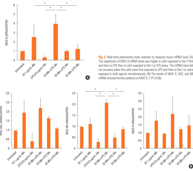
Fig. 2. Real-time polymerase chain reaction to measure mucin mRNA level. (A) The expression of MUC 8 mRNA level was higher in cells exposed to Der f first and then to LPS than in cells exposed to Der f or LPS alone. The mRNA level did not increase when the cells were first exposed to LPS and then to Der f or were exposed to both agents simultaneously. (B) The levels of MUC 4, 5AC, and 5B mRNA showed similar patterns to MUC 8. (*P<0.05).
MUC 8 mRNA/GAPDH MUC 4 mRNA/GAPDH
Untreated
Untreated Df 1 μg/mL 48h
Df 1 μg/mL 48h LPS 0.5 μg/mL 24h
LPS 0.5 μg/mL 24h Df 48h+LPS 24h
Df 48h+LPS 24h Df 48h+LPS 24h
Df 48h+LPS 24h Df 48h+LPS 48h
Df 48h+LPS 48h 6
5
4
3
2
1
0
3.5 3.0 2.5 2.0 1.5 1.0 0.5 0 A
B
MUC 5AC mRNA/GAPDH
Untreated Df 1 μg/mL 48h
LPS 0.5 μg/mL 24hDf 48h+LPS 24hDf 48h+LPS 24hDf 48h+LPS 48h 3.0
2.5
2.0
1.5
1.0
0.5
0
*
*
*
*
MUC 5B mRNA/GAPDH
Untreated Df 1 μg/mL 48h
LPS 0.5 μg/mL 24hDf 48h+LPS 24hDf 48h+LPS 24hDf 48h+LPS 48h 2.5
2.0
1.5
1.0
0.5
0
*
*
*
*
Fig. 3. Immunoblotting to determine MUC 8 and Toll-like receptor 4 signal intensity. (A) Treatment with Der f followed by LPS had a synergistic effect and increased MUC 8 production. (B) The signal intensity of MUC 8 and Toll-like receptor 4 induced by combined treatment with Der f and LPS was higher than that induced by treatment with LPS alone.
UntreatedDf 2 μg/mL 48h+ LPS 1 μg/mL 24hLPS 1 μg/mL 24hLPS 2 μg/mL 24hLPS 4 μg/mL 24hLPS 8 μg/mL 24hLPS 10 μg/mL 24h
Df 48h
24h
48h 24h LPS
MUC 8 GAPDH
MUC 8 TLR4 GAPDH
A B
RESULTS
Induction of MUC and pro-inflammatory cytokine gene mRNAs expression in HMEECs stimulated by Der f and LPS
Treatment with Df48h increased the level of MUC 8 mRNA expression. The expression level was significantly augmented when cells were exposed to Df48h/LPS24h. We further ana- lyzed the interaction of Der f and LPS under different treatment conditions. When we treated the cells in either with 1) LPS48h/
Df24h or 2) Df48h/LPS48h, MUC8 mRNA expression did not reach the levels induced by Df48h/LPS24h (Fig. 2A). With re- gard to the MUC 4, 5AC and 5B mRNA expression levels in- creased in a similar fashion, showing the greatest increase in the cells treated Df48h/LPS24h without statistical significance (Fig. 2B). This pattern was confirmed at the protein level. Im- munoblot assay showed that Df48h/LPS24h synergistically ele- vated the MUC 8 protein production; however, this was not ob- served in Df48h or LPS24h (Fig. 3A). Next, we analyzed the magnitude of the synergistic effect of both Der f and LPS by evaluating the MUC 8 and TLR 4 protein expression levels, be- cause both Der f and LPS are known to bind to TLR 4 to trigger the inflammatory signaling pathway. To further demonstrate the synergistic effect of Der f and LPS, we treated HMEECs with LPS 2, 4, 6, 8, and 10 μg/mL each for 24 hours, and then com- pared the intensity of MUC8 and TLR 4 proteins produced by Df48LPS24 treatment. Df48h/LPS24h showed a higher intensi- ty than that produced by LPS24h only (>10 μg/mL) (Fig. 3B). In terms of mRNA expression of pro-inflammatory cytokines, such as IL-1ß, GM-CSF, IL-33, and TNF-α, the augmented pat- tern was also similar to that of MUC, although they did not reach statistical significance (Fig. 4). These results suggest a possibility that Der f and LPS can act synergistically.
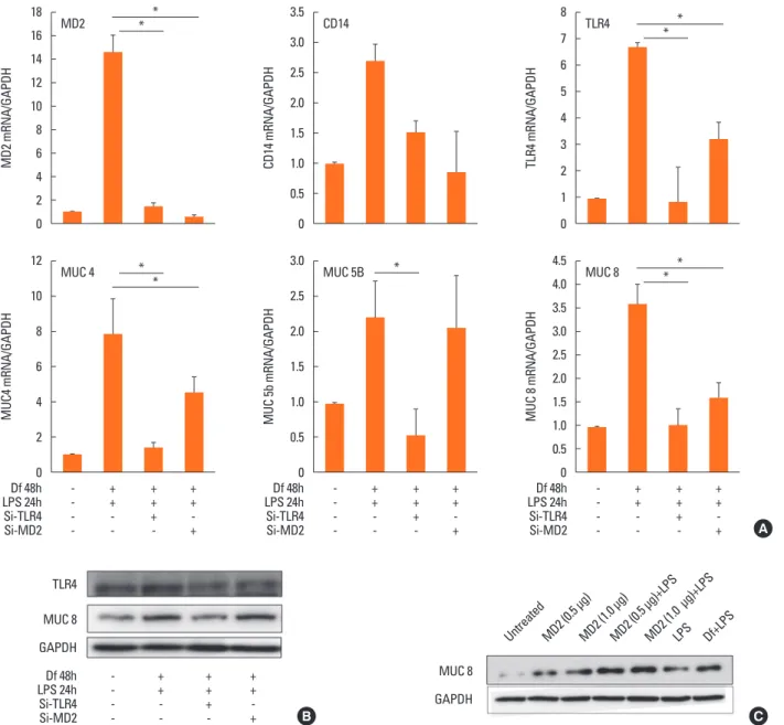
Role of TLR4 in the induction of the MUC gene in Der f- and LPS-stimulated HMEECs
To elucidate the mechanism underlying the difference in mu- cin gene expression in cells treated with LPS48h/Df24h or Df24h/LPS48h, we further determined the roles of TLR 4 and MD-2. We therefore transfected HMEECs with siRNAs to knock
down the expression of TLR4 or MD-2. Suppression of TLR4 and MD-2 by each siRNA were confirmed by real-time PCR.
CD14 expression was also suppressed by both siRNA without statistical significance (Fig. 5A, upper panel). Expressions of MUC 4, 5B and 8 mRNAs by the treatment with Df48h/LPS24h decreased significantly upon siRNA-TLR4 transfection. Howev- er, a significant amount of mRNAs were still expressed in siR- NA-MD-2 transfected HMEECs (Fig. 5A, lower panel). Immu- noblot analysis further confirmed decreased or sustained in- crease in MUC 8 protein signals in siRNA-TLR4- and siRNA- MD-2-transfected cells, respectively (Fig. 5B). These results in- dicated that TLR 4 could be partially activated in the absence of MD-2 protein.
Alternatives to MD-2 for MUC gene induction in Der f- and LPS-stimulated HMEECs
Our above mentioned results suggest the possibility of Der f being a functional substitute for the MD-2 protein; thus, the TLR 4 signal could be activated without the help of MD-2 pro- tein. Therefore, to determine the exact role of MD-2 in Der f and LPS synergism, a mammalian expression vector contain- ing MD-2 (pCMV1-hMD-2) was transfected into HMEECs for transient expression studies. MD-2 was over-expressed in HMEECs subsequently exposed to LPS for 24 hours, which showed a higher MUC 8 protein signal density than those ex- posed to LPS only and similar MUC 8 protein signals compared to those exposed to Df48h/LPS24h (Fig. 5C). Moreover, we con- ducted the co-immunofluorescence staining of TLR 4 and Der f in siRNA-treated cells. Der f and TLR 4 were co-localized not only in the siRNA-negative control transfected cells but also in the siRNA-MD-2 transfected cells (Fig. 6A and B). These results suggest that the MD-2 protein may play an important role in the pro-inflammatory Der f and LPS synergism.
p38 MAPK and CREB phosphorylation inhibition and NF-κB translocation in Der f-and LPS-stimulated HMEECs
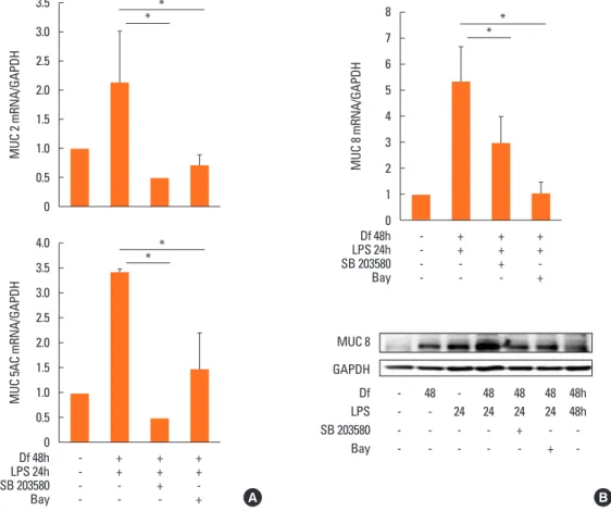
To further investigate the mechanism underlying the syner- gistic functions of Der f and LPS, we treated cells with various inhibitors, such as SB203580 (a bicyclic inhibitor of p38),
PD98059 (a specific inhibitor of MAPK/MEK-1), Bay (a NF-κB inhibitor), LY294002 (PI3K inhibitor), and a JNK inhibitor (data not shown). Cells were treated with each inhibitor 2 hours prior to LPS treatment. Of these, the mRNA expression levels of MUC 2, 5AC, and 8 decreased significantly under SB203580 or Bay treatment (Fig. 7A). MUC 8 protein signal intensity was lower in SB203580-or Bay-treated cells than in Df48h/LPS24h cells and most lowest in Df48h/LPS48h cells (Fig. 7B). This suggests that p38 MAPK and NF-κB transcriptional factors may be engaged in the synergism of Der f and LPS. The p-p38 signal post LPS treatment peaked at 5 minutes, and the expression of p-p38 and p-CREB was inhibited by SB203580 (Fig. 8A). GAPDH was constitutively expressed and not affected by SB203580. More- over, NF-κB translocation was seen in Df48h/LPS24h by confo- cal microscopy (Fig. 8B).
DISCUSSION
We investigated the synergistic effect of Der f and LPS on
MUC gene expression and pro-inflammatory cytokine produc- tion in HMEECs. LPS is known to induce and potentiate MUC gene expression;3 however, the interaction between LPS and Der f has not been studied so far. Before starting the experi- ment, to rule out possible endotoxin contamination of Der f crude body extract, we first measured the endotoxin level via the LAL method (Toxin Sensor chromogenic LAL Endotoxin assay kit, Gen Script, Piscataway, NJ, USA). The endotoxin level in Der f crude body extract was found to be 0.16 EU/mL, which was considered negligible. In this study, Der f and LPS synergis- tically induced MUC 8 gene expression and pro-inflammatory cytokines, and this effect depends on the sequence of exposure, i.e., it only occurs when Der f stimuli precedes LPS stimuli. Ad- ditionally, pre-treated Der f is likely to function as an MD-2 pro- tein and induce p-p38 and p-CREB expressions. These findings provide evidence for the interaction between 2 environmental stimuli associated with allergic airway disease and infectious diseases in the airway epithelium.
We measured mucin gene production as the final product be- Fig. 4. Real-time PCR to measure the levels of various cytokine mRNAs. The mRNA levels of pro-inflammatory cytokines IL-1b, IL-33, GM-CSF, and TNF-α tended to increase when the cells were exposed to the Der f initially and then to LPS.
IL-1
β mRNA/GAPDH
GM-CSF mRNA/GAPDH IL-33 mRNA/GAPDHTNF-
α mRNA/GAPDH Untreated
Untreated
Untreated
Untreated Df 1 μg/mL 48h
Df 1 μg/mL 48h
Df 1 μg/mL 48h
Df 1 μg/mL 48h LPS 0.5 μg/mL 24h
LPS 0.5 μg/mL 24h
LPS 0.5 μg/mL 24h
LPS 0.5 μg/mL 24h Df 48h+LPS 24h
Df 48h+LPS 24h
Df 48h+LPS 24h
Df 48h+LPS 24h Df 48h+LPS 24h
Df 48h+LPS 24h
Df 48h+LPS 24h
Df 48h+LPS 24h Df 48h+LPS 48h
Df 48h+LPS 48h
Df 48h+LPS 48h
Df 48h+LPS 48h 2.0
1.8 1.6 1.4 1.2 1.0 0.8 0.6 0.4 0.2 0
2.5
2.0
1.5
1.0
0.5
0
3.0 2.5 2.0 1.5 1.0 0.5 0
4.0 3.5 3.0 2.5 2.0 1.5 1.0 0.5 0
cause it is one of the major molecules that the airway epitheli- um produces in large amounts in response to several stimuli, such as TNF-α, IL-1β, LPS, oxidative stress, and neutrophil elas- tase, and because it has been implicated in numerous airway diseases.24 Mucins are broadly classified as either cell mem- brane-bound or secreted; notably, MUC2, MUC5AC, MUC5B, MUC6, MUC7, MUC8, MUC9, and MUC19 are categorized as secreted mucins.25,26 MUC5AC and MUC5B are predominant in
the sinus and nasal cavity, respectively, and considered primar- ily responsible for the gel-like characteristics of mucoid middle ear fluids.27-30 The MUC 8 gene is over-expressed in chronic rhi- nosinusitis and nasal polyp epithelium, and its expression lev- els are also increased by inflammatory mediators in nasal epi- thelial cells.31,32 Airway mucus obstruction is a shared feature among lower airway diseases, such as cystic fibrosis, asthma, and chronic obstructive lung disease.33 Hence, investigating the
MD2 mRNA/GAPDHMUC4 mRNA/GAPDH CD14 mRNA/GAPDHMUC 5b mRNA/GAPDH TLR4 mRNA/GAPDHMUC 8 mRNA/GAPDH
18 16 14 12 10 8 6 4 2 0 12 10 8 6 4 2 0
3.5 3.0 2.5 2.0 1.5 1.0 0.5 0 3.0 2.5 2.0 1.5 1.0 0.5 0
8 7 6 5 4 3 2 1 0 4.5 4.0 3.5 3.0 2.5 2.0 1.5 1.0 0.5 0
*
*
*
* *
*
*
*
* MD2
MUC 4
CD14
MUC 5B
TLR4
MUC 8
A
- + + + - + + + - + + +
Df 48h Df 48h Df 48h
- + + + - + + + - + + +
LPS 24h LPS 24h LPS 24h
- - + - - - + - - - + -
Si-TLR4 Si-TLR4 Si-TLR4
- - - + - - - + - - - +
Si-MD2 Si-MD2 Si-MD2
Fig. 5. mRNA and protein levels in cells transfected with small interfering RNA or in those overexpressing MD-2. (A, upper panel) The level of MD-2 or TLR4 mRNA was decreased in cells transfected with small interfering (si) RNA-MD-2 and siRNA-TLR4, respectively. (A, lower panel) The expressions of MUC 4, 5B, and 8 mRNA were also decreased in si-RNA-transfected cells. However, unlike in the cells transfected with si-RNA-TLR4, the expression of the target genes was not completely abolished in cells transfected with si-RNA-MD-2. (B) The decreased expression level of TLR4 and MUC 8 was confirmed at the protein level. The signal of MUC 8 protein was observed not only in the negative control, but also in the si-RNA-MD-2–transfected cells. (C) Overexpression of MD-2 enhanced the LPS-driven produc- tion of MUC 8. The signal intensity of MUC 8 was higher when LPS was added to the cell transfected with the Flag-pCMV-hMD-2 vector than with vector only. The pattern of increase when the cells were treated with LPS only for 24 hours was similar to that observed when those were treated first with Der f and then with LPS (Df48h/LPS24h).
Untreated MD2 (0.5 μg)MD2 (1.0 μg)MD2 (0.5 μg)+LPSMD2 (1.0 μg)+LPSLPS Df+LPS MUC 8
TLR4
GAPDH
MUC 8 GAPDH
B C
- + + +
Df 48h
- + + +
LPS 24h
- - + -
Si-TLR4
- - - + Si-MD2
Fig. 6. Confocal laser scanning image. (A) Red fluorescence indicates Der f expression; green, TLR4 expression; and blue, nuclear location. The lower right panel is a merged image of the other 3 panels. TLR4 was mainly expressed in the cell membrane, and it can be seen as yellow fluorescence after the overlap, suggesting that Der f and TLR4 co-localized in the cell membrane. Scale bar, 40 μm. (B) In high magnification, yellow fluorescence is seen not only in the negative control, but also in the membrane of cells transfected with si-RNA MD-2. Scale bar, 10 μm.
A B
Untreated Untreated
SiRNA-TLR4 SiRNA-TLR4
SiRNA-Negative control SiRNA-Negative control
SiRNA-MD2 SiRNA-MD2
MUC 2 mRNA/GAPDH MUC 8 mRNA/GAPDH
MUC 5AC mRNA/GAPDH
3.5 3.0 2.5 2.0 1.5 1.0 0.5 0
8 7 6 5 4 3 2 1 0 4.0
3.5 3.0 2.5 2.0 1.5 1.0 0.5 0
* *
*
* *
* - + + +
- + + + Df 48h
Df 48h
- + + +
- + + + LPS 24h
LPS 24h
- - + -
- - + - SB 203580
SB 203580
- - - +
- - - +
Bay
Bay
Fig. 7. Inhibition of MUC gene mRNA expression by SB203580 and Bay. (A) Expression of MUC 2, 5AC, and 8 mRNA was down-regulated by treatment with SB 203580 and Bay. (B) Down-regulation of MUC8 protein expression was confirmed by immunoblotting. Results are representative of those obtained from 3 indepen- dent experiments.
MUC 8 GAPDH
A B
- 48 - 48 48 48 48h Df
- - 24 24 24 24 48h LPS
- - - - + - - SB 203580
- - - + - Bay
TLR4DF DAPI
mucin gene expression in HMEECs is considered an appropri- ate approach to evaluating the “extended one airway concept.”
Interactions between allergens, viruses, bacteria and/or other pro-inflammatory stimuli have been previously reported.3 The synergistic effects of human rhinovirus and nitric oxide 2 on IL-8 release and an additive effect on ICAM-1 expression have been demonstrated.34 The interaction between the human rhi- novirus or respiratory syncytial virus and Der p I has been stud- ied, and it has been found that they synergistically induce IL-8 expression in bronchial epithelial cells.35 With regard to the in- teraction between bacteria and allergens, a previous study has shown that systemic LPS administration suppresses early and late allergic reactions in vivo via the TLR4-dependent pathway that triggers nitric oxide synthase 2 activity in a murine model of asthma;36 the generation of Th1 or Th2 responses is found to be dependent on the dose of LPS. At a low LPS level, inhaled LPS induces a Th2 response to inhaled antigens, whereas at a high level, inhaled LPS results in a Th1 response to the anti- gen.37 In an HDM allergen-evoked asthma murine model, LPS dose-dependently inhibits HDM-induced eosinophilic recruit- ment into the lungs, mucus production in the airways, and pro- duction of Th2 cytokines (IL-4, IL-5, IL-10, and IL-13).38 How- ever, the interaction between Der f and LPS at a cellular level has not been addressed previously, especially in the middle ear epithelium in vivo or in vitro, despite being frequently implicat- ed in middle ear disease pathogenesis.
It is well known that co-molecules, such as MD-2, CD14, myD88, and LPS-binding protein are needed to activate the TLR 4 signaling pathway; in particular, MD-2, a glycoprotein
co-expressed with TLR4 at the surface of various cell types, has received recent attention. Theoretically, to transmit the LPS- driven inflammatory signals, LPS bound to LBP is shuttled to CD14, which transfers the LPS to an MD-2 molecule. Activation of MD-2 occurs in 2 ways: it combines with endogenously ex- pressed TLR-4 to increase the surface expression of TLR-4 and MD-2,39 and participates directly in endotoxin recognition by extracting the endotoxin from monomeric endotoxin:CD14 complex.40
Previous studies demonstrate that MD-2 is essential for LPS- driven inflammatory responses, and that lack of MD-2 protein expression in human corneal epithelial cells account for the lack of response to LPS exposure in the cornea.41 MD-2 binds to TLR 4 and greatly enhances the response of TLR4-transfected cells to LPS exposure.42 Patients with the single nucleotide poly- morphisms of MD-2 promoter increase mRNA expressions of MD-2 and tended to have high levels of IL-1β, IL-6, IL-8, IL-10, and tumor necrosis factor (TNF)-α after Der p 2 and LPS stimu- lation.43 These facts prompted us to investigate whether the MD-2 protein might have a specific role in allergen and LPS synergism in HMEECs. In our study, to evaluate the role of MD- 2, we knocked-down the MD-2 gene by transfecting siRNA- MD-2. Interestingly, the production of MUC genes was not completely decreased in the absence of MD-2. Moreover, pre- treated Der f did not significantly alter the MD-2 mRNA expres- sion level (data not shown). Overexpression of the MD-2 gene using the pCMV-hMD-2 vector had a similar induction effect of MUC 8 protein when followed by LPS exposure for 24 hours.
This implies that Der f may combine with LPS, TLR4, and Fig. 8. Inhibition of phosphorylation of p38 MAPK and CREB, and translocation of nuclear factor κB (NF-κB) by SB203580 and Bay in HMEECs stimulated with Der f + LPS. (A) HMEECs were treated with Der f for 24 hours and then with SB203580 2 hours prior to LPS treatment. Subsequently, the cells were treated with LPS for 5, 15, and 30 minutes in the presence of Der f. The phosphorylation of p38 MAPK and CREB was analyzed using Western blotting. (B) Localization of NF-κB p65 was vi- sualized under a fluorescence microscope after immunofluorescence staining with NF-κB p65 antibody (green). Cells were stained with DAPI to visualize nuclei (blue). NF-κB translocation in HMEECs stimulated with Der f + LPS was inhibited by treatment with SB203580 and Bay. Results are representative of those obtained from 2 independent experiments.
P-P38
P38 P-CREB GAPDH
A B
- + + + + + + + Df
- - - + + + SB 203580
- - 5 15 30 5 15 30
LPS min
Untreated
SB 203580
Df + LPS
Bay
DAPI NF-KB
CD14, and have a role in endotoxin recognition in cases of defi- ciency of the MD-2 protein. Notably, the crystal structures of MD-2 and Der p 2 exhibit structural homology with 2 anti-par- allel β-pleated sheets stabilized by disulfide bonds.44-46 In an in vivo study, airway sensitization and Der p 2 challenge resulted in allergic asthma in wild-type and MD-2-deficient mice, but not in TLR4-deficient mice.47 A previous study stated that hu- man airway epithelium internalizes Der p2, which in turn, in- creases the endogenous cellular expression of MD-2.48 Der p 2 has not only structural homology but also functional homology with MD-2, facilitating signaling through direct interactions with the TLR4 complex and reconstituting LPS-driven TLR4 signaling in the absence of MD-2.47 Taken together, these facts suggest that Der f may have functional homology with MD-2.
Meanwhile, there are several additional factors that may influ- ence augmented mucin gene expression induced by Der f and LPS. Notably, at least 3 different microbial pathogen-associated molecular patterns can be detected in mite feces and/or in the mite environment: LPS, β-glucan, and chitin. Both LPS and β-glucan mediate innate immunity through reactive oxygen species production and can subsequently affect the mucin gene production.49 The endotoxin level of Der f crude body ex- tract used in this study was 0.16 EU/mL, and thus the contribu- tion might not be significant. Chitin can polarize Th1, Th2, and Th17 immune responses, and recruit IL-4 positive innate im- mune cells, including eosinophils and basophils;50,51 however, its precise contribution in the HDM-LPS synergism remains to be elucidated.
HDM allergenic proteins can be categorized into 4 main fami- lies: proteases, proteins displaying affinity for lipids, non-pro- teolytic enzymes, and non-enzymatic components.52 In the context of innate immunity activation, all these protein and non-protein compounds could putatively participate in stimu- lation. Among them, group 2 HDM allergen is described as an MD2-like lipid binding protein based on its structural/se- quence homology,53 supporting the hypothesis of our study.
Finally, the lack of synergism when the HMEECs were treated with Der f and LPS simultaneously (Df48h/LPS48h) or LPS48h/
Df24h may be attributed to the following aspects. First, it has been noted that Der f binds to LPS with a molar ratio of 1:1.54 Thus, when treated simultaneously, a fraction of these mole- cules may bind before reaching and binding to TLR4 and thus cannot activate the signaling pathway as expected. Second, an insufficient number of TLR 4 on the cell surface or endogenous MD-2 protein may interact with Der f and LPS when they are added at the same time. Third, the “hygiene hypothesis” states that the high levels of exposure to bacterial products, such as LPS, early in life are inversely correlated with the development of atopy and allergic disease.55-57 Thus, it is thought that LPS ex- posure leads to the exhaustion of TLR 4 and the development of a counter-regulatory response.58 Finally, unlike in LPS48h/
Df24-treated cells, Der f would act like MD-2 protein in Df48h/
LPS24h-treated cells, more signal activity could be triggered.
In summary, these experiments demonstrate that Der f and LPS can act synergistically when Der f exposure precedes LPS exposure in HMEECs. Based on our findings, the likely under- lying mechanism is that Der f has functions similar to those of MD-2, combining with TLR-4 and participating in endotoxin recognition. Such synergy suggests its important role in the de- velopment of OME in patients with concealed allergen-sensi- tized airway epithelium. Thus, targeted inactivation of innate immune signals to allergen exposure may be useful for the de- velopment of specific therapeutics for otitis media in concealed allergic sensitization.
ACKNOWLEDGMENTS
This study was supported by a research fund from Chosun University 2015.
REFERENCES
1. Blair H. Natural history of childhood asthma. 20-year follow-up.
Arch Dis Child 1977;52:613-9.
2. Pedersen PA, Weeke ER. Asthma and allergic rhinitis in the same patients. Allergy 1983;38:25-9.
3. Kim SW, Han DH, Lee SJ, Lee CH, Rhee CS. Bronchial hyperre- sponsiveness in pediatric rhinitis patients: the difference between allergic and nonallergic rhinitis. Am J Rhinol Allergy 2013;27:e63-8.
4. Döner F, Yariktas M, Demirci M. The role of allergy in recurrent oti- tis media with effusion. J Investig Allergol Clin Immunol 2004;14:
154-8.
5. Bernstein JM, Lee J, Conboy K, Ellis E, Li P. Further observations on the role of IgE-mediated hypersensitivity in recurrent otitis media with effusion. Otolaryngol Head Neck Surg 1985;93:611-5.
6. Sobol SE, Taha R, Schloss MD, Mazer BD, Manoukian JJ, Tewfik TL, et al. T(H)2 cytokine expression in atopic children with otitis me- dia with effusion. J Allergy Clin Immunol 2002;110:125-30.
7. Wright ED, Hurst D, Miotto D, Giguere C, Hamid Q. Increased ex- pression of major basic protein (MBP) and interleukin-5(IL-5) in middle ear biopsy specimens from atopic patients with persistent otitis media with effusion. Otolaryngol Head Neck Surg 2000;123:
533-8.
8. Hurst DS, Venge P. Evidence of eosinophil, neutrophil, and mast- cell mediators in the effusion of OME patients with and without at- opy. Allergy 2000;55:435-41.
9. Ozinsky A, Underhill DM, Fontenot JD, Hajjar AM, Smith KD, Wil- son CB, et al. The repertoire for pattern recognition of pathogens by the innate immune system is defined by cooperation between toll-like receptors. Proc Natl Acad Sci U S A 2000;97:13766-71.
10. Akira S, Takeda K, Kaisho T. Toll-like receptors: critical proteins linking innate and acquired immunity. Nat Immunol 2001;2:675- 80.
11. Kitajima T, Muroi M, Yamashita N, Tanamoto K. Toll-like receptors required for dermatophagoides farinae to activate NF-κB. Biol Pharm Bull 2014;37:74-80.
12. Chow JC, Young DW, Golenbock DT, Christ WJ, Gusovsky F. Toll- like receptor-4 mediates lipopolysaccharide-induced signal trans-
duction. J Biol Chem 1999;274:10689-92.
13. Vlastos I, Athanasopoulos I, Mastronikolis NS, Panogeorgou T, Margaritis V, Naxakis S, et al. Impaired mucociliary clearance in al- lergic rhinitis patients is related to a predisposition to rhinosinus- itis. Ear Nose Throat J 2009;88:E17-9.
14. Papadopoulos NG, Xepapadaki P, Mallia P, Brusselle G, Watelet JB, Xatzipsalti M, et al. Mechanisms of virus-induced asthma exacer- bations: state-of-the-art. A GA2LEN and InterAirways document.
Allergy 2007;62:457-70.
15. Papadopoulos NG, Christodoulou I, Rohde G, Agache I, Almqvist C, Bruno A, et al. Viruses and bacteria in acute asthma exacerbations- -a GA² LEN-DARE systematic review. Allergy 2011;66:458-68.
16. Copenhaver CC, Gern JE, Li Z, Shult PA, Rosenthal LA, Mikus LD, et al. Cytokine response patterns, exposure to viruses, and respira- tory infections in the first year of life. Am J Respir Crit Care Med 2004;170:175-80.
17. Rochlitzer S, Hoymann HG, Müller M, Braun A; U-BIOPRED con- sortium. No exacerbation but impaired anti-viral mechanisms in a rhinovirus-chronic allergic asthma mouse model. Clin Sci (Lond) 2014;126:55-65.
18. Rizzo MC, Naspitz CK, Fernández-Caldas E, Lockey RF, Mimiça I, Solé D. Endotoxin exposure and symptoms in asthmatic children.
Pediatr Allergy Immunol 1997;8:121-6.
19. Michel O, Kips J, Duchateau J, Vertongen F, Robert L, Collet H, et al.
Severity of asthma is related to endotoxin in house dust. Am J Respir Crit Care Med 1996;154:1641-6.
20. Song JJ, Lee JD, Lee BD, Chae SW, Park MK. Effect of diesel exhaust particles on human middle ear epithelial cells. Int J Pediatr Otorhi- nolaryngol 2012;76:334-8.
21. Preciado D, Kuo E, Ashktorab S, Manes P, Rose M. Cigarette smoke activates NFκB-mediated Tnf-α release from mouse middle ear cells. Laryngoscope 2010;120:2508-15.
22. Jun HJ, Lim HW, Choi J, Jung HH, Chae SW. Ciglitazone inhibits cigarette smoke solution-induced inflammatory responses in hu- man middle ear epithelial cells. Int J Pediatr Otorhinolaryngol 2012;76:1136-9.
23. Smirnova MG, Guo L, Birchall JP, Pearson JP. LPS up-regulates mu- cin and cytokine mRNA expression and stimulates mucin and cy- tokine secretion in goblet cells. Cell Immunol 2003;221:42-9.
24. Voynow JA, Gendler SJ, Rose MC. Regulation of mucin genes in chronic inflammatory airway diseases. Am J Respir Cell Mol Biol 2006;34:661-5.
25. Rose MC. Mucins: structure, function, and role in pulmonary dis- eases. Am J Physiol 1992.263:L413-29.
26. Rose MC, Voynow JA. Respiratory tract mucin genes and mucin glycoproteins in health and disease. Physiol Rev 2006;86:245-78.
27. Kerschner JE. Mucin gene expression in human middle ear epithe- lium. Laryngoscope 2007;117:1666-76.
28. Giebink GS, Le CT, Paparella MM. Epidemiology of otitis media with effusion in children. Arch Otolaryngol 1982;108:563-6.
29. Thai P, Loukoianov A, Wachi S, Wu R. Regulation of airway mucin gene expression. Annu Rev Physiol 2008;70:405-29.
30. Alenmyr L, Herrmann A, Högestätt ED, Greiff L, Zygmunt PM.
TRPV1 and TRPA1 stimulation induces MUC5B secretion in the human nasal airway in vivo. Clin Physiol Funct Imaging 2011;31:
435-44.
31. Cho KN, Choi JY, Kim CH, Baek SJ, Chung KC, Moon UY, et al.
Prostaglandin E2 induces MUC8 gene expression via a mechanism involving ERK MAPK/RSK1/cAMP response element binding pro-
tein activation in human airway epithelial cells. J Biol Chem 2005;
280:6676-81.
32. Lee HM, Kim DH, Kim JM, Lee SH, Hwang SJ. MUC8 mucin gene up-regulation in chronic rhinosinusitis. Ann Otol Rhinol Laryngol 2004;113:662-6.
33. Yoshida T, Tuder RM. Pathobiology of cigarette smoke-induced chronic obstructive pulmonary disease. Physiol Rev 2007;87:1047- 82.
34. Spannhake EW, Reddy SP, Jacoby DB, Yu XY, Saatian B, Tian J. Syn- ergism between rhinovirus infection and oxidant pollutant expo- sure enhances airway epithelial cell cytokine production. Environ Health Perspect 2002;110:665-70.
35. Foster S, Bedford KJ, Gould ME, Coward WR, Hewitt CR. Respira- tory syncytial virus infection and virus-induced inflammation are modified by contaminants of indoor air. Immunology 2003;108:
109-15.
36. Rodríguez D, Keller AC, Faquim-Mauro EL, de Macedo MS, Cunha FQ, Lefort J, et al. Bacterial LPS signaling through TLR4 suppresses asthma -like response via NP synthase 2 activity. J Immunol 2003;
171:1001-8.
37. Eisenbarth SC, Piggott DA, Huleatt JW, Visintin I, Herrick CA, Bot- tomly K. Lipopolysaccharide-enhanced, toll-like receptor 4-de- pendent T helper cell type 2 responses to inhaled antigen. J Exp Med 2002;196:1645-51.
38. Daan de Boer J, Roelofs JJ, de Vos AF, de Beer R, Schouten M, Hom- mes TJ, et al. Lipopolysaccharide inhibits Th2 lung inflammation induced by house dust mite allergens in mice. Am J Respir Cell Mol Biol 2013;48:382-9.
39. O’Neil DA, Porter EM, Elewaut D, Anderson GM, Eckmann L, Ganz T, et al. Expression and regulation of the human beta-defen- sins hBD-1 and hBD-2 in intestinal epithelium. J Immunol 1999;
163:6718-24.
40. Gioannini TL, Teghanemt A, Zhang D, Coussens NP, Dockstader W, Ramaswamy S, et al. Isolation of an endotoxin-MD-2 complex that produces Toll-like receptor 4-dependent cell activation at picomo- lar concentrations. Proc Natl Acad Sci U S A 2004;101:4186-91.
41. Zhang J, Kumar A, Wheater M, Yu FS. Lack of MD-2 expression in human corneal epithelial cells is an underlying mechanism of li- popolysaccharide (LPS) unresponsiveness. Immunol Cell Biol 2009;87:141-8.
42. Shimazu R, Akashi S, Ogata H, Nagai Y, Fukudome K, Miyake K, et al. MD-2, a molecule that confers lipopolysaccharide responsive- ness on Toll-like receptor 4. J Exp Med 1999;189:1777-82.
43. Liao EC, Hsieh CW, Chang CY, Yu SJ, Sheu ML, Wu SM, et al. En- hanced Allergic Inflammation of Der p 2 Affected by Polymor- phisms of MD-2 Promoter. Allergy Asthma Immunol Res 2015;7:
497-506.
44. Ohto U, Fukase K, Miyake K, Satow Y. Crystal structures of human MD-2 and its complex with antiendotoxic lipid IVa. Science 2007;
316:1632-4.
45. Derewenda U, Li J, Derewenda Z, Dauter Z, Mueller GA, Rule GS, et al. The crystal structure of a major dust mite allergen Der p 2, and its biological implications. J Mol Biol 2002;318:189-97.
46. Kim HM, Park BS, Kim JI, Kim SE, Lee J, Oh SC, et al. Crystal struc- ture of the TLR4-MD-2 complex with bound endotoxin antagonist Eritoran. Cell 2007;130:906-17.
47. Trompette A, Divanovic S, Visintin A, Blanchard C, Hegde RS, Madan R, et al. Allergenicity resulting from functional mimicry of a Toll-like receptor complex protein. Nature 2009;457:585-8.
48. Yin SC, Liao EC, Chiu CL, Chang CY, Tsai JJ. Der p2 internalization by epithelium synergistically augments toll-like receptor-mediated proinflammatory signaling. Allergy Asthma Immunol Res 2015;7:
393-403.
49. Ryu JH, Yoo JY, Kim MJ, Hwang SG, Ahn KC, Ryu JC, et al. Distinct TLR-mediated pathways regulate house dust mite-induced allergic disease in the upper and lower airways. J Allergy Clin Immunol 2013;131:549-61.
50. Lee CG, Da Silva CA, Dela Cruz CS, Ahangari F, Ma B, Kang MJ, et al. Role of chitin and chitinase/chitinase-like proteins in inflam- mation, tissue remodeling, and injury. Annu Rev Physiol 2011;73:
479-501.
51. Reese TA, Liang HE, Tager AM, Luster AD, Van Rooijen N, Voeh- ringer D, et al. Chitin induces accumulation in tissue of innate im- mune cells associated with allergy. Nature 2007;447:92-6.
52. Thomas WR, Smith WA, Hales BJ, Mills KL, O’Brien RM. Charac- terization and immunobiology of house dust mite allergens. Int Arch Allergy Immunol 2002;129:1-18.
53. Thomas WR, Hales BJ, Smith WA. House dust mite allergens in asthma and allergy. Trends Mol Med 2010;16:321-8.
54. Ichikawa S, Takai T, Yashiki T, Takahashi S, Okumura K, Ogawa H, et al. Lipopolysaccharide binding of the mite allergen Der f 2. Genes Cells 2009;14:1055-65.
55. Braun-Fahrländer C, Riedler J, Herz U, Eder W, Waser M, Grize L, et al. Environmental exposure to endotoxin and its relation to asth- ma in school-age children. N Engl J Med 2002;347:869-77.
56. Gehring U, Bischof W, Fahlbusch B, Wichmann HE, Heinrich J.
House dust endotoxin and allergic sensitization in children. Am J Respir Crit Care Med 2002;166:939-44.
57. Riedler J, Braun-Fahrländer C, Eder W, Schreuer M, Waser M, Maisch S, et al. Exposure to farming in early life and development of asthma and allergy: a cross-sectional survey. Lancet 2001;358:1129- 33.
58. Wills-Karp M, Santeliz J, Karp CL. The germless theory of allergic disease: revisiting the hygiene hypothesis. Nat Rev Immunol 2001;
1:69-75.