INTRODUCTION
The ear, a member of the special sensory organs, consists of three parts: the external, middle and internal ear. At the embryonic stage, the internal ear develops first. The first evidence of development of the human internal ear is the formation of the otic placode after thickening of the surface ectoderm early in the 4th week. Next, the otic placode inva- ginates and sinks into the underlying mesenchyme and forms an otic pit. Towards the end of the 4th week, around the 26th day of development, the superficial epithelial opening of the otic pit is closed and forms the otic vesicle. By the 5th week, the endolymphatic appendage is formed within the posterolateral part of the otic vesicle and begins to grow.
At the same time, other parts of the otic vesicle are differen- tiated as primordia of the utricle and saccule. Around the 6th week of development, the cochlear duct begins to be formed from the saccule, and flat disk-like diverticula grow out of the utricular portion. The terminal portions of each semicircular canal are enlarged and form the ampulla (1-4).
The process by which the internal ear is formed in mouse is known to be almost identical to that in human. The time when the otic vesicle is shaped in mouse is the 9.5th day, which corresponds to the stage 15 in human, namely about the 33rd postovulatory day according to the Carnegie stage
(5). From the otic vesicle, the endolymphatic appendage is formed around the 10th day.
On the 11.5th day, it is possible to distinguish the por- tions where the utricle and saccule are formed. The flat diver- ticula, which will form the semicircular canals and primordi- um of the cochlear duct, are formed from the utricle and saccule, respectively, at around day 12 (6, 7). In rat, compared with in mouse, the formation of the internal ear progresses more slowly, but the order of the developmental stages is similar.
The transforming growth factor (TGF ) is a polypeptide growth factors family composed of many isomers. This pro- tein group is known as the TGF family (8). Based on the structural differences of the specific region, TGF is gener- ally classified as four isomers: TGF 1, 2, 3 and 5.
TGF stimulates or inhibits the growth and differentia- tion of cells, and also controls the production and secretion of the extracellular matrix (9). The time when these functions of TGF are well displayed in individuals is the developmen- tal process of embryos (10-12).
Recently, there have been many studies focused on the functions of these growth factors during the development of experimental animals. Therefore, TGF is thought to play important roles in the parts where various interactions occur between ectodermal cells and mesodermal cells during the
Ho-Jeong Kim, Ki-Young Kang*, Jin-Ghi Baek*, Hyoung-Chul Jo�, Hyun Kim�
Department of Anatomy, College of Medicine, Kwandong University, Gangneung; Department of Anatomy*, College of Medicine, Seonam University, Namwon; Department of Otorhinolaryngology�, College of Medicine, Seonam University, Namwon; Department of Anatomy�, College of Medicine, Kosin University, Busan, Korea
Address for correspondence Hyun Kim, M.D.
Department of Anatomy, College of Medicine, Kosin University, 34 Amnam-dong, Seo-gu, Busan 602-703, Korea
Tel : +82.51-990-6410, Fax : +82.51-990-3081 E-mail : drhkim@kosin.ac.kr
136
Expression of TGF Family in the Developing Internal Ear of Rat Embryos
In order to investigate the expression patterns of the transforming growth factor (TGF) isoforms in the internal ear, an immunohistochemical study of rat embryos was performed. Rat embryos were taken on the 13th, 15th, 17th, and 19th day after conception and their internal ears were immunohistochemically stained against TGF 1, 2, and 3. As a result, the 13-day-old embryo showed a very weak posi- tivity to TGF 1. After the 15th day of pregnancy, no reactivity to TGF 1 was defect- ed. Immunoreactivity to TGF 2 was observed from the 15th day of pregnancy throughout the rest of the period. The ampulla of the semicircular canal and the cochlear duct showed a notably strong immunohistochemical reaction. A strong reaction to TGF 3 was observed on the 15th day of pregnancy. However, no posi- tive reactions were observed thereafter. A strong immunoreactivity was observed especially on the apical cytoplasms, the surfaces of the epithelial cells, and base- ment membranes of the cochlear duct, as well as the semicircular canals of the developing internal ear of rat embryo.
Key Words : Labyrinth; Ear, Inner; Growth and Development; Transforming Growth Factor Beta
Received : 1 October 2004 Accepted : 5 September 2005
development, especially in the internal ear. It is also known that the hair cells of the cochlea of developing mouse express TGF 2 mRNA (12-14). By using the immunohistochemi- cal technique, it was shown that TGF 2 is also expressed in the internal ear, but unlike the TGF 2 mRNA, it is limited on the basement membrane of the cochlear epithelia. The expression of TGF 2 continues from the 12th day to the 17th day of development. TGF 1 is not expressed in the cochlear duct of mouse embryo. TGF 3 is expressed on all epithelial cells of the embryonic cochlear duct on the 13th day after fertilization and this positive signal continues until the 17th day. This protein is detected mostly on cell surface.
Therefore, during the development of the internal ear of mouse embryo, the expression of TGF isoforms undergoes the most dramatic change (15).
The effects of TGF s on cells are confirmed by numerous studies that demonstrated the expression and disappearance of these isoforms in developing tissues or organs. Since most studies on the development of the internal ear has so far been focused on the expression of TGF isoform, without con- sideration of the developmental stages, we performed this study to confirm the process of the development of the internal ear and to observe the expression patterns and various influ- ences upon the neighboring cells of TGF s in rat embryos during the period from the 13th day to the 19th day of ges- tation at intervals of two days.
MATERIALS AND METHODS Experimental animals
In order to observe the expressions of TGF s during the development of the internal ear, we used female Sprague- Dawley (SD) rats. We raised these animals in isolated condi- tions with free feeding until their weight reached 230-250 g.
Then, we mated them with males in the evening and con- firmed the fertilization by observing the vaginal plug or sperm on the vaginal smear next morning. We designated this day as day 0 of gestation. On the 13th, 15th, 17th, and 19th day of gestation, the embryos were extracted. On each selected day of pregnancy, five female rats were sacrificed and two embryos from each were selected and used for this study.
Tissue processing and basic stain
The embryos were fixed overnight in 10% neutrally buffered formalin immediately after extraction from the uterus. The fixed embryos were washed, dehydrated, and embedded in paraffin. Serial sections were made by 5- m thickness, attached onto the aminopropyl-triethoxysilane coated slide glass, and air-dried. The tissue sections were deparaffinized, hydrated, stained with hematoxylin-eosin (HE), dehydrated, cleared and sealed with cover glass. Histologic characteristics were
examined under a light microscope (BX50, Olympus, Japan).
Immunohistochemical stain
The sections were deparaffinized, hydrated, and then treat- ed with 3% H2O2in methanol to block the activity of the endogenous peroxidase. After washing with phosphate buffered saline (PBS 0.1M, pH 7.4) for 5 min, the sections were treated with a 10% protein blocking agent (Shandon, Waltham, MA, U.S.A.) for 1 hr at room temperature to block nonspecific antigen-antibody reactions. These sections were incubated with 1:400 anti-TGF 1, 2 and 3 antibody (Santa Cruz Biotechnology, Santa Cruz, CA, U.S.A.) diluents for 12 hr at 4℃and washed with PBS. Thereafter, they were incu- bated with biotinylated secondary antibodies (Shandon) for 60 min at room temperature. After being washed with PBS, these were treated with streptavidin peroxidase reagent (Shan- don) for 30 min at room temperature. After administering the peroxidase treatment, the tissue sections were washed with PBS and colorized with a 0.05% diaminobenzidine (DAB, Vector Laboratories Inc., Burlingame, CA, U.S.A.) solution for less than 1 min.
Photographs and reorganization
We took pictures of the HE stained and immunohisochem- ically stained sections using a light microscope and compared the degrees of staining intensities from each slide. On the 17th day of gestation, the embryos were prepared and sec- tioned serially at 25- m thickness. These sections were stained with HE. Then, we took photographs and reconstructed the structure of the developing labyrinth using a desktop per- sonal computer.
RESULTS Embryos on the 13th day after fertilization
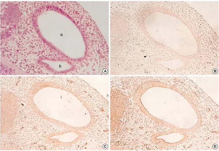
From the 13th day of gestation, the embryos showed the otic vesicle clearly and the endolymphatic appendage was sunken posteromedially. The endolymphatic appendage was observed as a large elliptical structure with a small projec- tion in the anterior portion (Fig. 1). At this stage, all TGF beta isoforms showed very weak signals on the epithelial cells and also on the subepithelial tissues of the otic vesicle and endolymphatic appendage, while TGF 3 showed rather intense signals. Positive immunoreactivity was observed on the posterior part of the otic vesicle, which was composed of columnar epithelial cells. Indeed, it appeared clearly on the surface of those columnar cells. However, there were no sig- nificant differences in the expression patterns of TGF fam- ily on the epithelia of the otic vesicle and endolymphatic appendage at this stage (Fig. 1).
Embryos on the 15th day after fertilization
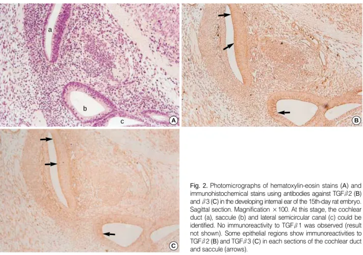
On the 15th day, the internal ear was observed to develop as a form of labyrinth composed of tubes. It was also possi- ble to observe parts of the saccule, utricle, cochlear duct, and lateral semicircular canal (Fig. 2). The immunoreactivity against TGF 1 was not detected in the above developing internal ear structures, but TGF 2 and 3 showed appar- ently positive immunoreactivity.
TGF 2 showed strong immunoreactivity in the thick epithelial layer within the developing cochlear duct, espe- cially on the surfaces of these sensory epithelial cells. The epithelial cells of the saccule from which the cochlear duct grows showed positive immunoreactivity with anti-TGF 2 antibody. Furthermore, we found positive reactions in the basement membrane of the saccular epithelia. The epithelia of the lateral semicircular canal showed DAB coloring which was more distinct on the apical domain of these cells (Fig. 2).
TGF 3 showed a similar immunoreactivity to that of TGF 2 except for some different results on the epithelial surface of the developing saccule and lateral semicircular canal.
In the basement membrane of the saccule, TGF 3 showed less intense DAB coloring than TGF 2. On the other hand, it is evident that some columnar cells of the developing sac- cule adjacent to the cochlear duct and the plate of lining cells of the developing cochlear duct expressed both TGF 2 and TGF 3 (Fig. 2).
Embryos on the 17th day after fertilization
The ampullar portions, where the posterior and anterior semicircular canals grow, were observed clearly in the embryo.
From the results of three dimensional reconstruction of serial sections, the structures of the saccule, utricle, cochlea, lateral and posterior semicircular canal were partially observed. The cochlear duct further developed and it made 1.5 turns (Fig.
3, 4).
In the internal ear, immunoreactivity to TGF 3 was greatly reduced, and positive signals were not observed as in the case of TGF 1. Very weak immunoreactivity of TGF 1 and 3 was detected on the surface of epithelia of the ampulla of the semicircular canal and the cochlear duct. In contrast to TGF 1
Fig. 1. Photomicrographs of hematoxylin-eosin stains (A) and immunohistochemical stains using antibodies against TGF 1 (B), 2 (C), and 3 (D) in the developing internal ear of the 13th-day rat embryo. Horizontal section. The otic vesicle (a) is observed and endolymphatic appendage (b) forms another cavity medial to the otic vesicle. At this stage, the immunoreactivities to all the TGF isoforms are very weak (×200).
A B
C D
a
b
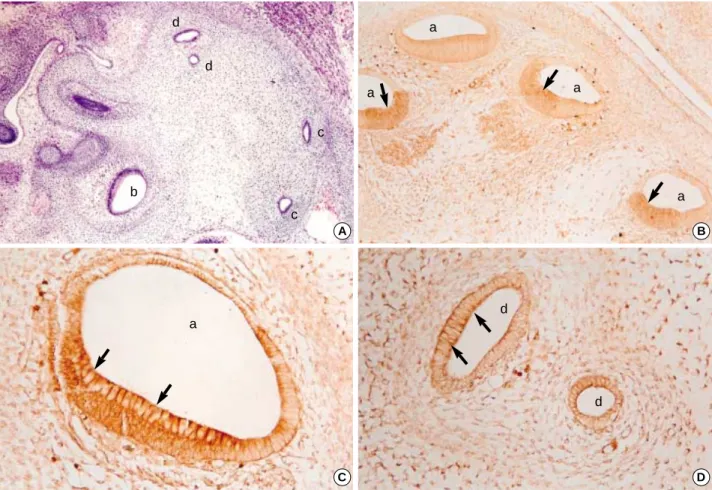
and 3, TGF 2 showed strong immunoreactivity in all parts of the internal ear, especially in the cellular mass lining along the portion toward the site of future spiral ganglion of the cochlea, the surface of epithelial cells, and the basement mem- brane of the ampullae of the semicircular canals (Fig. 3, 4).
Embryos on the 19th day after fertilization
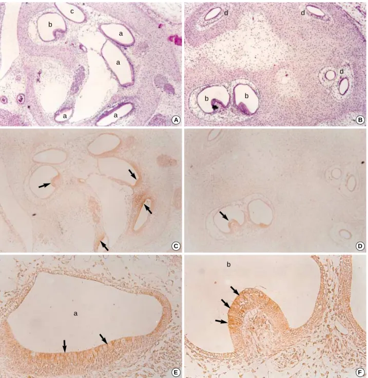
On the 19th day of pregnancy, TGF 1 and 3 showed no DAB coloring in the internal ear, similar to what we observed on the 17th day. However, the positive immunoreactivity to TGF 2 was observed in all of the semicircular canals, ampul- lae and cochlear ducts, but was relatively weaker than those of the 17th-day embryos.
The epithelia of the semicircular canal showed DAB col- oring on the apical surfaces. In the ampullae of the semicir- cular canal, the development of the ampullary crest was observed and some of the ciliated epithelia of the ampullary crest, especially on the apical surfaces, showed more intense signal with the TGF 2 antibody. In the cochlear duct, a thick epithelial layer settled on the lateral wall of the duct, and these cells showed strong immunoreactivity on the api- cal cytoplasms and on their surfaces (Fig. 5).
DISCUSSION
In the developing internal ear of mouse, especially in the hair cells of the cochlear duct, it is known that the expression of TGF 2 continues from the 12th day to the 17th day of pregnancy (11-14). In contrast to TGF 2, TGF 1 is not expressed in the cochlear duct of mouse embryo, and TGF 3 is expressed somewhat later than 2 in the cochlear duct from the 13th day to the 17th day after fertilization (15, 16).
In this study, SD rats were used as experimental animals.
Compared with previous studies, we found that the develop- ment of the internal ear in SD rats followed the same course as that in mouse. Thus, we speculate that these growth fac- tors have similar functions in different species such as mouse and rat. In addition, cells from a specific location did not show the same immunoreactivity concordantly, and positive reac- tivity was restricted within the specific group of cells, while other cells show expression of two or more phenotypes simul- taneously. This suggests the development and differentiation of cells are not uniform and their role in the developmental process may differ greatly.
According to the results from the above experimental data, it might be suggested that TGF 2 plays important roles during the whole developmental process of the internal ear.
Indeed, the cochlear duct, spiral limbus, and interdental cells
Fig. 2. Photomicrographs of hematoxylin-eosin stains (A) and immunohistochemical stains using antibodies against TGF 2 (B) and 3 (C) in the developing internal ear of the 15th-day rat embryo.
Sagittal section. Magnification ×100. At this stage, the cochlear duct (a), saccule (b) and lateral semicircular canal (c) could be identified. No immunoreactivity to TGF 1 was observed (result not shown). Some epithelial regions show immunoreactivities to TGF 2 (B) and TGF 3 (C) in each sections of the cochlear duct and saccule (arrows).
A B
C c
b a
showed a morphological abnormality when the TGF 2 gene had intentionally been removed (17).
Recently, bone morphogenetic protein 4 (BMP-4), a mem- ber of the TGF family, has also been reported to play impor- tant roles in the development of the internal ear of frog and chicken (18-21).
During the internal ear development, mesodermal tissues are differentiated and form the otic capsule. In addition, it was asserted that many interactions occur between the epithe- lial cells and the mesodermal tissues and that growth factors participate in these interactions during the otic capsule for- mation. In particular, it is known that TGF 1 is expressed in both epithelial cells and mesodermal tissues. Thus, TGF 1 may play certain roles in the process of formation of the otic capsule (22, 23). Another line of evidence that supports the above statement is that the retinoic acid, a potent teratogen, produces abnormalities also in the internal ear and it was suggested that TGF 1 might play an important role in this phenomenon (24). In this case, abnormalities were mainly due to the hypoplasia of the cartilaginous tissue. Moreover, they were associated with an apparent decrease of immunore- activity in adjacent mesodermal tissues with the treatment
Fig. 3. Photomicrographs of hematoxylin-eosin stains (A, ×100) and immunohistochemical stains using antibodies against TGF 2 (B,
×100; C, D, ×400) in the developing internal ear of the 17th-day rat embryo. Sagittal sections. At this stage, cochlear duct (a), ampulla (b), and semicircular canals (c, d) can be identified. In the epithelium of cochlear duct and semicircular canals, strong immunohistochemi- cal reactions of TGF 2 antibodies are observed, especially at the apices of lining columnar cells (arrows).
A B
C D
d
d
a
a
a
a
a
d
d c
c b
Fig. 4.A 3-dimensional reconstruction of the sagittal sections of the developing internal ear of 17th-day rat embryo. In the middle, the utricle (Utr) and the saccule (Sac) are interconnected and the ampulla (Amp) made a growth upward from them. Two semicir- cular canals, namely anterior (ASC) and posterior (PSC), formed loops lateralward, and the cochlear duct (Coc) roughly had a spi- ral form located medially.
ASC
Amp
Utr Sac
PSC Coc
of anti-TGF 1 antibody. It was also reported that the TGF 1 expression of the periotic tissue disappears right after finish- ing its role around the embryonic day 14 (23), and the obser- vation of very weak immunoreactivity to TGF 1 in a 13-day embryo logically suggests that the chondrogenesis occurring around the otic capsule had already been completed. Another recent study showed that the expression of TGF receptor II and Smad2, a downstream component of the TGF sig-
nal transduction pathway, were downregulated after the treat- ment with a high dose of retinoic acid (25).
In this study, we could not observe immunoreactivity with the anti-TGF 1 antibody except at early stages of develop- ment. To explain these differences, further studies involving a wider time-window during the development, with shorter intervals. When we consider the differences between species, it is necessary to search other growth factors that participate
Fig. 5.Photomicrographs of hematoxylin eosin stains (A, B, ×100) and immunohistochemical stains using antibodies against TGF 2 (C, D, ×100; E, F, ×400) in the developing internal ear of the 19th-day rat embryo. Sagittal sectons. At this stage, the cochlear duct (a), ampulla (b), saccule (c) and semicircular canals (d) are clearly identified. The immunoreactivity to TGF 2 was relatively weaker than those of 17th- day embryo is observed at the apices of columnar cells of cochlear duct and crista ampullaris (arrows).
A B
C
E
D
F c
b
b a
a a
a
b b
d d
d
a
in the differentiation of cartilaginous tissues.
The internal ear is composed of many tubular tissues, and the epithelial cells of these ducts are not a single cellular group.
Thus, in order to confirm how these different cells under dif- ferentiation, we need elaborate reorganizations of the serial sections and special staining tools, such as the immunohis- tochemical or in situ hybridization techniques.
REFERENCES
1. Larsen WJ. Human Embryology. 3rd Edition, Oxford: Churchill Livingstone, 2001; 392-412.
2. Moore KL, Persaud TV. The Developing Human: Clinically orient- ed embryology. 7th Edition, Philadelphia: Saunders, 2003; 476-9.
3. Park HW. Human embryology. 3rd Edition, Seoul: Koonja Publish- ing Inc., 2005; 503-7.
4. Streit A. Origin of the vertebrate inner ear: evolution and induction of the otic placode. J Anat 2001; 199: 99-103.
5. O’Rahilly R, Muller F. Developmental Stages in Human Embryos.
Washington DC: Carnegie Institution of Washington, 1987; 186.
6. Theiler K. The House Mouse. New York: Springer-Verlag, 1972;
44-108.
7. Rugh R. The mouse. Its reproduction and development. Oxford:
Oxford University Press, 1991; 249-51.
8. Miller DA, Pelton RW, Derynck R, Moses HL. Transforming growth factor-beta. A family of growth regulatory peptides. Ann N Y Acad Sci 1990; 593: 208-17.
9. McCartney-Francis NL, Frazier-Jessen M, Wahl SM. TGF-beta: a balancing act. Int Rev Immunol 1998; 16: 553-80.
10. Akhurst RJ, Lehnert SA, Gatherer D, Duffie E. The role of TGF beta in mouse development. Ann N Y Acad Sci 1990; 593: 259-71.
11. Nilsen-Hamilton M. Transforming growth factor-beta and its actions on cellular growth and differentiation. Curr Top Dev Biol 1990; 24:
95-136.
12. Millan FA, Denhez F, Konaiah P, Akhurst RJ. Embryonic gene expres- sion patterns of TGF 1, 2 and 3 suggest different development functions in vivo. Development (Camb.) 1991; 111: 131-43.
13. Pelton RW, Dickinson ME, Moses HL, Hogan BL. In situ hybridiza- tion analysis of TGF 3 RNA expression during mouse development:
comparative studies with TGF 1 and 2. Development (Camb.)
1990; 110: 609-20.
14. Schmid P, Cox D, Bilbe G, Maier R, McMaster GK. Differential expression of TGF 1, 2 and 3 genes during mouse embryo- genesis. Development (Camb.) 1991; 111: 117-30.
15. Pelton RW, Saxena B, Jones M, Moses HL, Gold LI. Immunohisto- chemical localization of TGF 1, TGF 2 and TGF 3 in the mouse embryo: Expression patterns suggest multiple roles during embryonic development. J Cell Biol 1991; 4: 1091-105.
16. Takemura T, Sakagami M, Takebayashi K, Umemoto M, Nakase T, Takaoka K, Kubo T, Kitamura Y, Nomura S. Localization of bone morphogenetic protein-4 messenger RNA in developing mouse cochlea. Hear Res 1996; 95: 26-32.
17. Paradies NE, Sanford LP, Doetschman T, Friedman RA. Develop- mental expression of the TGF in the mouse cochlea. Mech Dev 1998; 79: 165-8.
18. Wu DK, Oh SH. Sensory organ generation in the chick inner ear. J Neurosci 1996; 16: 6454-62.
19. Morsli H, Choo D, Ryan A, Johnson R, Wu DK. Development of the mouse inner ear and origin of its sensory organs. J Neurosci 1998; 18: 3327-35.
20. Cole LK, Le Roux I, Nunes F, Laufer E, Lewis J, Wu DK. Sensory organ generation in the chicken inner ear: contributions of bone morphogenetic protein 4, serrate1, and lunatic fringe. J Comp Neu- rol 2000; 424: 509-20.
21. Kil SH, Collazo A. Origin of inner ear sensory organs revealed by fate map and time lapse analyses. Dev Biol 2001; 233: 365-79.
22. Frenz DA, Van de Water TR, Galinovic-Schwartz V. Transforming growth factor beta: does it direct otic capsule formation? Ann Otol Rhinol Laryngol 1991; 100: 301-7.
23. Frenz DA, Galinovic-Schwartz V, Liu W, Flanders KC, Van de Water TR. Transforming growth factor beta 1 is an epithelial- derived signal peptide that influence otic capsule formation. Dev Biol 1992; 153: 324-36.
24. Frenz DA, Liu W. Treatment with all-trans-retinoic acid decreases levels of endogenous TGF-beta (1) in the mesenchyme of the devel- oping mouse inner ear. Teratology 2000; 61: 297-304.
25. Butts SC, Liu W, Li G, Frenz DA. Transforming growth factor- 1 signaling participates in the physiological and pathological regula- tion of mouse inner ear development by all-trans retinoic acid. Birth Defects Res A Clin Mol Teratol 2005; 73: 218-28.