INTRODUCTION
Electrical stimulation of acupoint (ESA; electroacupunc- ture) has been used to treat a wide range of musculoskeletal disorders and reported to relieve pain and inflammation, strengthen muscle, and reduce abnormal muscle tone (1-5).
Its mechanisms of action are not fully elucidated, but some of its action on the endogenous opioids system through mul- tiple neuronal pathways have been identified in experimen- tal studies.
Previous studies have revealed that ESA with low (2-10 Hz) and high (≥100 Hz) frequencies has different mecha- nisms with selective release of -endorphins, enkephalins or dynorphins (6-8). ESA at different frequencies activates the distinct different regions in the spinal cord and the central nervous system (9, 10), and evokes the different responses of autonomic nervous system (11, 12). Based on these findings, ESA with low and high frequencies seems to produce the dif- ferent therapeutic effect.
Ankle sprain is a very common condition of acute muscu- loskeletal injuries and is induced by accidental overextension of ligaments in ankle joint, resulting in pain, reduction of weight bearing during walking, and edema around ankle. Early weight bearing, bracing, and functional rehabilitation are the generally accepted management of mild and moderate ankle
sprain (13, 14). In addition, cyclo-oxygenase inhibitors and opioids are known to be useful in reducing pain and edema and to achieve earlier recovery of normal function (15, 16).
The ankle sprain model in rats was demonstrated to gen- erate similar clinical symptoms of mild degree of ankle sprain (3). There have been few studies on the effect of ESA for ankle sprain (3), and moreover, discrete effects of ESA at low and high frequencies on pain and edema induced by ankle sprain have not been reported.
Therefore, we aimed to investigate whether ESA with two frequencies, 2 Hz and 100 Hz, can reduce pain and edema induced by ankle sprain and whether there is a difference in therapeutic effects between 2 Hz and 100 Hz ESA in a recent- ly developed ankle sprain model in rats.
MATERIALS AND METHODS Animal preparation
Male Sprague-Dawley rats were housed in separate cages and allowed to acclimate for 7 days by using a 12/12 hr day/
night cycle. The experiments were performed on rats weigh- ing 210-270 g. The experimental protocol was approved by our Institutional Animal Care and Committee.
Tae Soo Hahm
Department of Anesthesiology and Pain Medicine, Samsung Medical Center, Sungkyunkwan University School of Medicine, Seoul, Korea
Address for correspondence Tae Soo Hahm, M.D.
Department of Anesthesiology and Pain Medicine, Samsung Medical Center, 50 Ilwon-dong, Gangnam-gu, Seoul 135-710, Korea Tel : +82.2-3410-0364, Fax : +82.2-3410-0361 E-mail : [email protected]
*This study was partly supported by Samsung Biomedical Research Institute Grant (# SBRI C-A5- 341-1).
347
The Effect of 2 Hz and 100 Hz Electrical Stimulation of Acupoint on Ankle Sprain in Rats
The electrical stimulation of acupoint (ESA) releases several endogenous neuropep- tides, which play important roles in management of pain and inflammation. ESA with low and high frequencies has been shown to release different neuropepides, suggesting its various therapeutic effects. Pain and edema are major problems for ankle sprain. However, there have been few reports on the effects of ESA for ankle sprain. We aimed to investigate that ESA can reduce pain and edema resulting from ankle sprain, and whether there is a difference in therapeutic effects between low and high frequency ESA. To induce ankle sprain in Sprague-Dawley rats, the ankle of right hindpaw was overextended in direction of simultaneous inversion and plan- tar flexion. Stepping force and edema in the paw of the sprained ankle were mea- sured by electronic balance and plethysmometer, respectively. In both 2 and 100 Hz ESA groups, stepping force was increased significantly in similar degrees (p<
0.05). Only 2 Hz ESA produced the significant rapid decrease in ankle edema. This study demonstrates that ESA of 2 Hz and 100 Hz shows comparable analgesic effects, but only 2 Hz ESA can facilitate the reduction of edema caused by ankle sprain.
Key Words : Electrical Stimulation; Acupoint ; Frequency, Sprains and Strains; Ankle
Received : 20 June 2006 Accepted : 22 August 2006
Procedure for ankle sprain
Rats were anesthetized with 2% enflurane in O2via a mask.
The ankle sprain model was made according to the method described by Koo et al. (3). Ankle sprain was produced by manually overextending the lateral ligament.
Electrical stimulation
Under general anesthesia, electrical stimulation was deliv- ered to contralateral SI6 point (Yangno) through a pair of bipolar needles at 24 hr after ankle sprain. It was controlled by Grass S88 electrical stimulator (Grass Telefactor, West Warwick, RI, U.S.A.) equipped with SIU5 isolation unit (Grass Telefactor, West Warwick, RI, U.S.A.). The SI6 acu- point is known as an effective analgesic point for pain induced by ankle sprain in rats (3) and located at the posterior distal end of the forearm between the radius and ulna. Electric cur- rent (1 ms pulse duration, 5 times intensity as muscle twitch) was delivered for 30 min. Rats were allocated randomly into three groups (2 Hz ESA, 100 Hz ESA or control groups: n=
9/group). In the control group, needles were not inserted and electrical current was not delivered.
Measurement of pain
The pain was determined by the stepping force in the paw of the sprained ankle during walking (3). Rats were allowed to acclimate within plastic walking channel (10 cm width, 10 cm high, 60 cm long) for 10 min and then allowed to walk spontaneously. The stepping force in the paw of the sprained ankle was recorded by Pocket Pro 250-B electronic balance (Acculab, Newton, PA, U.S.A.), which is placed on the floor at the midway area of the right walking path. The signal of the balance was converted to digital signal by an AD converter (Physiolab, Seoul, Korea), which was fed into a computer monitor. The waves of stepping force were ob-
served, and the peak amplitude was measured during walk- ing. To estimate the level of pain produced by ankle sprain, stepping forces were measured before ankle sprain and at 24 hr after ankle sprain. To evaluate the analgesic effect of ESA, stepping forces were measured at 1, 2, 4 hr after the termi- nation of ESA. The rats with a weight bearing ratio less than 20% or more than 40% were excluded from the study. The data were converted by the following formula: % recovery=
(improvement of stepping force by EA in gram/reduction of stepping force by sprain in gram)×100.
Measurement of paw volume
The volume of the right hindpaw was measured before and 24 hr after ankle sprain. To evaluate the anti-edematous effect of ESA, the volume of the right hindpaw was measured at pre-ESA and at 2, 4, 6, 12 and 24 hr after the termination of ESA. The volume of paw was measured by a water replace- ment plethysmometer (Ugo Basile, Comerio, Italy). The posi- tion at about 5 mm proximal to the ankle is clearly divided by dense long hairs and sparse thin hairs. This clearly demar- cated boundary has been used as a convenient landmark (3).
The paw was immersed into water up to the marked line. The volume of paw edema was calculated from experimental paw volume minus pre-sprained paw volume. The data were con- verted by the following formula: decreased paw edema (%)=
(volume of paw edema after ESA in mL/volume of paw edema at 24 hr after sprain in mL)×100.
Statistical analysis
Data are presented as mean±SEM. Statistical tests were done using one-way repeated measured ANOVA or one-way ANOVA followed by least significant difference posthoc test.
A probability level <0.05 was considered to be statistically significant.
B Stepping forces of right front and hindpaws (g)
120 100 80 60 40 20 0
0 200 400 600 800 1,000 1,200
Time (ms) A
Stepping forces of right front and hindpaws (g) 120 100 80 60 40 20 0
0 200 400 600 800 1,000 1,200
Time (ms)
RF RH RF RH
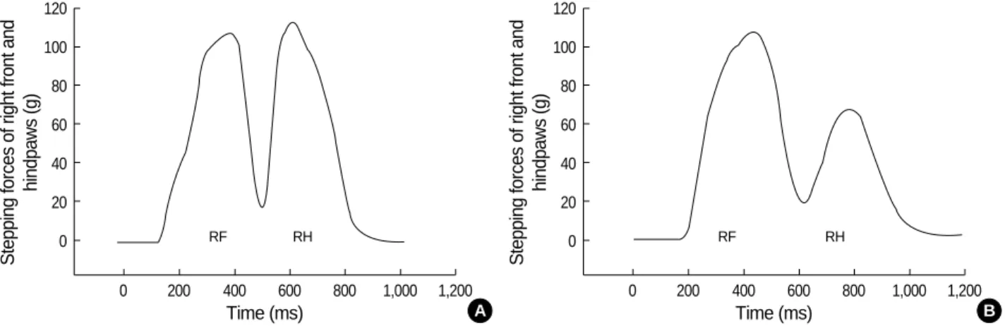
Fig. 1.Changes in stepping forces of right paws in a rat. (A) shows the normal pattern of stepping forces of right frontpaw (RF) and hind- paw (RH) during walking before ankle sprain. (B) shows the pattern of stepping forces at 24 hr after ankle sprain of right hindpaw. The ankle sprain induces the marked decrease in stepping force of right hindpaw.
RESULTS
The sprain injury of right hindpaw produced a marked decrease (p<0.001) in stepping force during walking (Fig. 1) and a significant increase (p<0.001) in foot volume (Fig. 2).
The weight-bearing ratios were decreased significantly (p<
0.001) from 53.1±0.5% (2 Hz ESA), 53.5±0.6% (100 Hz ESA) and 52.9±0.3% (control) to 33.4±0.9%, 34.2±1.0%
and 32.3±1.5%, respectively. The foot volumes at 24 hr after sprain were increased (p<0.001) to 170.4±5.33% (2 Hz ESA), 174±5.03% (100 Hz ESA), 174±5.03% (con- trol), compared to the values at pre-sprain.
In 2 and 100 Hz ESA groups, the % recovery of stepping force was significantly (p<0.05) increased at 1 and 2 hr after post-ESA compared to the values at pre-ESA. In the control group, no differences between pre-ESA and post-ESA were observed (Fig. 3).
The maximal % recovery at 2 Hz ESA and 100 Hz ESA
were 32% and 33%, respectively, showing no differences bet- ween two groups, but showed a significant (p<0.05) increase compared to the values in the control group (Fig. 4).
In three groups, the edema volumes were reduced as time elapsed. The edema volumes in the 2 Hz ESA group were reduced more significantly (p<0.05) than the values in 100 Hz ESA and control groups at 6 and 12 hr after ESA (Fig. 5).
DISCUSSION
In the present study, ESA at 2 and 100 Hz showed impro- vement in stepping force and produced a similar degree of anal- gesic effects, but only ESA at 2 Hz facilitated the reduction of edema in ankle sprain.
The analgesic effect of ESA observed in the current study is consistent with several studies in which ESA shown to be effective in several pain models (8, 17, 18). It has been also
Foot volume of right hindpaw (mL)
3.5 3.0 2.5 2.0 1.5 1.0 0.5
0.0 Control 2 Hz 100 Hz
Before ankle sprain
Fig. 2.Changes in paw volumes of right hindpaw before ankle sprain and at 24 hr after sprain. Data are expressed as mean± SEM. *p<0.001 compared to the value before sprain.
* *
*
24 hr after ankle sprain
% Recovery of stepping force
40
30
20
10
0
-10
-1 0 1 2 4
Post-ESA (hr)
ESA
Control
Fig. 3.Effects of 2 Hz and 100 Hz ESA on recovery of stepping forces in the paw of the sprained ankle at 24 hr after ankle sprain.
Data are expressed as mean±SEM. *p<0.05 compared to the value at pre-ESA.
2 Hz 100 Hz
*
*
*
*
*
*
*
*
Maximal % recovery
40
30
20
10
0 Control 2 Hz 100 Hz
Fig. 4.Comparison of maximal % recovery in 2 Hz and 100 Hz ESA.
There is no difference between two groups. Data are expressed as mean±SEM. *p<0.05 compared to the value the control group.
Decreased paw edema (%)
70 60 50 40 30 20 10 0
-1 0 2 4 6 12 24
Post-ESA (hr)
ESA Control
Fig. 5.Effects of 2 Hz and 100 Hz ESA on reduction of edema vol- umes in the paw of the sprained ankle at 24 hr after ankle sprain.
Data are expressed as mean±SEM. *p<0.05 compared to the value at 6 and 12 hr in 100 Hz ESA and control groups.
2 Hz 100 Hz
reported that ESA effectively alleviated ankle sprain pain in rats (3). However, they used the electrical current with train of four pulses (100 Hz of intra-train frequency) repeated at 2 Hz, which was different from continuous electrical stimu- lation used in the present study and did not specifically exam- ine the analgesic and anti-edema effects of low and high fre- quency ESA.
In our study, analgesic effectiveness of ESA at 2 Hz was similar to that at 100 Hz. In the previous studies, ESA at low and high frequencies produced different analgesic effects in various pain conditions. Some studies have reported that high frequency was more effective against inflammatory hy- peralgesia (19) and post-operative pain (20). Another study has reported that low frequency ESA produced superior anal- gesic effects in response to noxious radiant heat stimuli (10).
Moreover, it has been reported that ESA at 100 Hz reduced mechanical, but did not thermal inflammatory hyperalgesia (21). These inconsistent results reflect that ESA of specific frequencies may produce different effects to the several types of pain and hyperalgesia.
The reasons of similar analgesic effect of both frequencies on ankle sprain in the present study might be due to the spe- cific nature of ankle sprain itself or the measurement method of pain. Ankle sprain has a combined property of mechani- cal and inflammatory pain. We measured the stepping force during walking as an index of ankle pain. This index has been also widely used in several arthritic pain models (3, 22, 23).
However, the changes in stepping forces during walking may be affected by ankle sprain pain as well as muscle activity and joint movement. Also, ESA can strengthen muscle power and improve the range of motion of joint (4, 5). Therefore, improvement of motor activity induced by ESA at 2 and 100 Hz may affect positively the changes in the amount of stepping forces.
In this study, ESA at 2 Hz produced more rapid reduction of edema compared to spontaneous progression of edema reduction, but ESA at 100 Hz failed to show any effects on edema reduction. This is consistent with a previous report that showed only low frequency ESA inhibited paw edema in the complete Freund’s adjuvant-induced inflammation model (2). This finding, together with the previous data, sug- gests that ESA at 2 and 100 Hz have different therapeutic effects on edema resulting from ankle sprain.
The anti-inflammatory effect of low frequency ESA has been observed in several studies. ESA at low frequency pro- duced significant inhibition of paw edema in capsaicin or carrageenan-induced inflammation (24-26) and suppressed the activity of macrophages induced by lipopolysaccharide (27). The anti-inflammatory effect of low frequency ESA is considered to be related to opioid receptor agonist. Its effect was reduced or suppressed by low doses of naloxone (25, 27).
These findings are in line with the observation that morphine suppressed carrageenan-induced edema and anti-edema effect of morphine was reversed by low doses of naloxone (28). On
the other hand, some studies showed that low frequency ESA induced the release of ACTH from the pituitary gland and elevated the levels of peripheral cortisols, which can suppress inflammation by shutting off the production of inflamma- tory mediator at the sites of injury (2, 29, 30). In addition, some investigators showed that anti-inflammatory effects of low frequency ESA were not affected by naloxone (31), sug- gesting that a non-opioid mechanism may be involved in anti-inflammatory effect by low frequency ESA. This is dif- ferent from previous findings that naloxone inhibited anti- inflammatory effect of low frequency ESA (25, 27). They assumed that the discrepancy might be due to different inflam- mation-induced agents (carrageenan vs. capsaicin).
Although the mechanism underlying ESA is not clear, it has been demonstrated that ESA is mediated by the endoge- nous opioids system and ESA at 2 Hz facilitates the release of -endorphin, enkephalin, or endomorphin, whereas ESA at 100 Hz releases dynorphin. The effect produced by 2 Hz ESA was naloxone-reversible, while high frequency stimula- tion was not (6). In a cross-tolerance study, high frequency ESA tolerant rats showed cross-tolerance to opioid recep- tor agonist (dynorphin) but did not show cross-tolerance to a specific agonist for opioid receptor (7). Also, endomor- phin-1 was involved in 2 Hz, but not 100 Hz, ESA-induced analgesia (8). These results suggest that ESA with low and high frequencies have different mechanisms, and optimiza- tion of stimulation frequency is important in determining the therapeutic effect in various pathological conditions.
Interestingly, 2 Hz ESA reduced ankle sprain pain at 1 and 2 hr after the electrical stimulation, but inhibited edema at 6 and 12 hr after the electrical stimulation. This time disso- ciation between analgesic effect and anti-edema effect of ESA is in agreement with the previous observation that low fre- quency ESA attenuated inflammatory hyperalgesia at 2.5 hr, but inhibited edema at 24 hr (24). These results, together with present data, suggest that the analgesic effect of ESA is independent of an effect on peripheral inflammation, and the anti-inflammatory mechanisms of ESA may be different from analgesic mechanism. Although the endogenous opioids sys- tems are closely involved in mechanisms of action of ESA, further studies are required to clarify the mechanisms under- lying ESA-induced anti-inflammatory effects.
In conclusion, the present study shows that 2 and 100 Hz ESA are similarly effective against ankle sprain pain, but only 2 Hz facilitates the reduction of ankle sprain edema. This result suggests that there is different therapeutic effects bet- ween ESA at 2 Hz and 100 Hz, and 2 Hz ESA may be con- sidered as a preferred therapeutic method for ankle sprain.
ACKNOWLEDGMENTS
We thank Dr. Koo SungTae and Kwon HyukNam for their technical assistances, and also thank Dr. Ko Justin and
Ahn HyunJoo for their helpful comments on the manuscript.
REFERENCES
1. Deluze C, Bosia L, Zirbs A, Chantraine A, Vischer TL. Electroa- cupuncture in fibromyalgia: results of a controlled trial. Brit Med J 1992; 305: 1249-52.
2. Zhang RX, Lao L, Wang X, Fan A, Wang L, Ren K, Berman BM.
Electroacupuncture attenuates inflammation in a rat model. J Altern Complement Med 2005; 11: 135-42.
3. Koo ST, Park YI, Lim KS, Chung K, Chung JM. Acupuncture anal- gesia in a new rat model of ankle sprain pain. Pain 2002; 99: 423-31.
4. Naeser MA, Alexander MP, Stiassny-Eder D, Lannin LN, Bachman D. Acupuncture in the treatment of hand paresis in chronic and acute stroke patients-improvement observed in all cases. Clin Rehabil 1994;
8: 127-41.
5. Moon SK, Whang YK, Park SU, Ko CN, Kim YS, Bae HS, Cho KH.
Antispastic effect of electroacupuncture and moxibustion in stroke patients. Am J Chin Med 2003; 31: 467-74.
6. Wang JQ, Mao L, Han JS. Comparison of the antinociceptive effects induced by electroacupuncture and transcutaneous electrical nerve stimulation in the rat. Int J Neurosci 1992; 65: 117-29.
7. Chen XH, Han JS. Analgesia induced by electroacupuncture of dif- ferent frequencies is mediated by different types of opioid receptors:
another cross-tolerance study. Behav Brain Res 1992; 47: 143-9.
8. Han Z, Jiang YH, Wan Y, Wang Y, Chang JK, Han JS. Endomor- phine-1 mediates 2 Hz but not 100 Hz electroacupuncture analgesia in the rat. Neurosci Lett 1999; 274: 75-8.
9. Lee JH, Beitz AJ. The distribution of brain-stem and spinal cord nuclei associated with different frequencies of electroacupuncture analgesia. Pain 1993; 52: 11-28.
10. Zang WT, Jin Z, Cui GH, Zang KL, Zang L, Zeng YW, Luo F, Chen ACN, Han JS. Relations between brain network activation and anal- gesic effect induced by low vs. high frequency electrical acupoint stim- ulation in different subjects: functional magnetic resonance imag- ing study. Brain Res 2003; 982: 168-78.
11. Liao JM, Lin CF, Ting H, Chang CC, Lin YJ, Lin TB. Electroacu- puncture at Hoku elicits dual effect on autonomic nervous system in anesthetized rats. Neurosci Res 2002; 42: 15-20.
12. Stener-Victorin E, Kobayashi R, Kurosawa M. Ovarian blood flow responses to electro-acupuncture stimulation at different frequencies and intensities in anaesthetized rats. Auton Neurosci 2003; 108: 50-6.
13. Liu SH, Jason WJ. Lateral ankle sprains and instability problems.
Clin Sports Med 1994; 13: 793-809.
14. Renstrom PA, Konradsen L. Ankle ligament injuries. Br J Sports Med 1997; 31: 11-20.
15. Ekman EF, Fiechtner JJ, Levy S, Fort JG. Efficacy of celecoxib ver- sus ibuprofen in treatment of acute pain: a multicenter, double-blind, randomized controlled trial in acute ankle sprain. Am J Orthop 2002;
31: 445-51.
16. Aghababian RV. Comparison of diflunisal and acetaminophen with codeine in the management of grade 2 ankle sprain. Clin Ther 1986;
8: 520-6.
17. Zhang YQ, Ji GC, Wu GC, Zhao ZQ. Excitatory amino acid recep- tor antagonists and electroacupuncture synergetically inhibit car- rageenan-induced behavioral hyperalgesia and spinal fos expres- sion in rats. Pain 2002; 99: 525-35.
18. Hwang BG, Min BI, Kim JH, Na HS, Park DS. Effects of electroa- cupuncture on the mechanical allodynia in the rat model of neuro- pathic pain. Neurosci Lett 2002; 320: 49-52.
19. Lao L, Zhang RX, Zhang G, Wang X, Berman BM, Ren K. A para- metric study of electroacupuncture on persistent hyperalgesia and Fos protein expression in rats. Brain Res 2004; 10: 18-29.
20. Lin JG, Lo MW, Wen YR, Hsieh CL, Tsai SK, Sun WZ. The effect of high and low frequency electroacupuncture in pain after lower abdominal surgery. Pain 2002; 99: 509-14.
21. Huang C, Hu ZP, Long H, Shi YS, Han JS, Wan Y. Attenuation of mechanical but not thermal hyperalgesia by electroacupuncture with the involvement of opioids in rat model of chronic inflammatory pain.
Brain Res Bull 2004; 63: 99-103.
22. Otsuki T, Agatsuma Y, Jokura H, Sakurada S, Kisara K, Yoshimoto T. Monosodium urate test: a new analgesic test by crystal-induced monoarthritis in rats. J Neurosci Methods 1990; 33: 229-31.
23. Min SS, Han JS, Kim YI, Na HS, Yoon YW, Hong SK, Han HC. A novel method for convenient assessment of arthritic pain in volun- tarily walking rats. Neurosci Lett 2001; 308: 95-8.
24. Lao L, Zhang G, Wei F, Berman BM, Ren K. Electro-acupuncture attenuates behavioral hyperalgesia and selectively reduces spinal fos protein expression in rats with persistent inflammation. J Pain 2001; 2: 111-7.
25. Ceccherelli F, Gagliardi G, Ruzzante L, Giron G. Acupuncture modu- lation of capsaicin-induced inflammation: effect of intraperitoneal and local administration of naloxone in rats. A blind controlled study.
J Altern Complement Med 2002; 8: 341-9.
26. Lee JH, Choi YH, Choi BT. The anti-inflammatory effects of 2 Hz electroacupuncture with different intensities on acute carrageenan- induced inflammation in the rat paw. Int J Mol Med 2005; 16: 99-102 27. Aoki E, Kasahara T, Hagiwara H, Sunaga M, Hisamitsu N, Hisamit- su T. Electroacupuncture and moxibustion influence lipopolysaccha- ride-induced TNF-alpha production macrophages. In Vivo 2005;
19: 495-500.
28. Joris A, Costello A, Dubner R, Hargreaves KM. Opiates suppress carrageenan-induced edema and hyperthermia at doses that inhibit hyperalgesia. Pain 1990; 43: 95-103.
29. Cheng R, Mckibbin L, Roy B, Pomeranz B. Electroacupuncture ele- vates blood cortisol levels in naive horses: sham treatment has no effect. Int J Neurosci 1980; 10: 95-7.
30. Pan B, Castro-Lopes JM, Coimbra A. Activation of anterior lobe corticotrophs by electroacupuncture or noxious stimulation in the anaesthetized rats, as shown by colocalization of Fos protein with ACTH and -endorphin and increased hormone release. Brain Res Bull 1996; 40: 175-82.
31. Zhang SP, Zhang JS, Yung KK, Zhang HQ. Non-opioid-dependent anti-inflammatory effects of low frequency electroacupuncture. Brain Res Bull 2004; 62: 327-34.
. .