인삼 사포닌과 다당류 혼합물의 활성화된 RAW264.7 세포주에 대한 염증조절 효과
이 도 익#
중앙대학교 약학대학 면역학교실
(Received September 16, 2009; Revised February 26, 2010; Accepted February 26, 2010)
The Inflammation-modulatory Effects of Ginseng Saponin and Polysaccharide on Activated RAW264.7 Cell-line
Lee Do Ik#
Department of Immunology, College of Pharmacy, Chung-Ang University, Seoul 000-000, Korea
Abstract
— It is well known that the numbers and functions of immune-associated cells are increased by saponins and polysaccharides in ginseng. In this study, the mixture of polysaccharide and saponin (MPS) from Panax ginseng is applied to LPS- activated RAW 264.7 cells. The production of NO and the gene expression of IL-6 and TNF-
αare decreased in LPS- activated RAW 264.7 cells and the expression of arginase II and PD-1L genes is decreased in LPS-untreated macrophages.
Therefore, the mixture of saponin and polysaccharide from Panax ginseng could be used in order to regulate immune responses.
Keywords □
saponin, Polysaccharide, mixture, inflammation, immunosuppression
인삼은세계적으로널리이용되는천연약으로많은효과가있 다고알려져왔다
.
그중가장큰비중을차지하는성분은사포 닌과다당체(
진산)
로이들에대한연구는현재꾸준히진행되고있다
.
인삼에서정제된다당체(polysaccharides)
는생쥐를이용한생체내실험결과인삼다당체투여생쥐군에서정상생쥐군에 비해골수및비장세포가증가하는것을관찰할수있었으며
,
인삼 다당체는
TNF-
α, IL-1, IL-2, IL-6, IL-12, IFN-
γ 및GM- CSF
와같은다양한cytokines
의생산과림프계세포가증식하도 록자극하는강력한면역조절기능도가지고있었다.
1)그러나인삼다당체에의하여유도되는
cytokine mRNA
를관찰한결과, Thl
세포에서분비되는것으로알려진IL-2
와IFN-
γmRNA, macrophage
로부터분비되는것으로알려진IL-l
α와GM-CSF mRNA
의발현을용량의존적으로증가시켰으나, Th2
세포에서 분비되는것으로알려진IL-4
와IL-5 mRNA
의발현은차이가없 었다.
2)또한,
인삼다당체에의해증가된임파구의subset
을flow
cytometry
로분석한결과,
배양3
일째에IgM+
세포의수가증 가하고, NK
활성은2.4
배, T
세포에대한반응은2.1
배가증가됐 고방사선방어효과도7.46 Gy
에서10.96 Gy
로크게늘어나는효과가있었다
.
1)인삼다당체는암세포의전이를44%
억제하였고골수모세포의경우
4.7
배,
백혈구수치는2
배증가하는등 의효능을보였으며암세포살해면역세포생성작용3.5
배,
방사선민감작용에서일반대조군에비해
45%
의방사선감소수치 를나타났다.
배양5
일째에Thy 1.2
+세포와CD4
+세포, CD8
+세포의수가증가하였다
.
하지만,
감마방사선에의해오히려면역반응 억제가상쇄될 수 있다는연구 또한 있다
.
3) 사포닌(ginsenoside)
은인삼에서가장많이들어있는구성물로4
개의steroid
유사체(4
개의고리구조)
로되어있고이는타식물에서발견되는사포닌과다른구조를지니고있다
.
화학구조의특성에 따라protopanaxadiol(PD)
계, protopanaxatrol(PT)
계, oleanane
계사포닌으로구분되며4)항산화효과
,
암세포억제효과,
항염작용이뛰어나다고알려져있다
.
5)염화탄소로간손상을일으킨 모델동물에서인삼의간보호효과는우수한것으로밝혀졌고,
인삼의대표적인성분인
ginsenoside Rb1, Rb2, Re, Rg1
에대해간보호효과를실험한결과역시모두간보호효과가있었다
.
#본논문에관한문의는저자에게로
(
전화) 02-820-5608 (
팩스) 02-820-5608 (E-mail) [email protected]
종설
또한
, Macrophage
세포의cyclooxygenase(COX)-2
의발현을억제하고
prostaglandin E2
의생합성을억제하는효과와함께유도형
NO
합성효소(iNOS)
의활성을억제하여NO
의생합성을억 제하는효과가있었다.
이러한효과는인삼사포닌들이이효소들을직접저해하기보다는
NF-
κB
등의전사인자를조절하는 양식으로prostaglandin E2
와NO
의생합성을억제하였다.
한편활성화된
macrophage
는외부항원에대한방어에중요한역할을하며
IL-1
β, TNF-
α, IL-6
등의cytokine
을생성하여 비정상세포를파괴한다.
특히IL-6
는활성화된macrophage
가 항암효과를나타내는데중요한인자로알려져있다.
6-8)NO
또한활성화된
macrophage
에의해생성되어,
바이러스제거등면역 반응에중요한역할을하지만염증반응이나조직손상등을일으 킬수있다.
본실험에서는다당체와사포닌의혼합물(mixture of Polysaccharides and saponin, MPS)
을비활성및활성화된RAW264.7
에 처리하여면역 증강 및활성 억제 효과를pro-
inflammatory cytokine
과NO
생성변화및arinase, PD-L1
발현변화를통해확인하였고
polysaccharides(PS)
와비교평가하 였다.
실험방법
Mixture of Saponin and Polysaccharide(MPS)와 Poly- saccharide(PS)제조
인삼분말
100 g
에1%(W/W)
염기성용액1500 m
l를가하고잘흔든뒤
80
oC
에서12
시간동안추출하고여과하여여액을4
oC
에서10,000 rpm
으로10
분간원심분리한다음,
상등액200 m
l을취하여시료를만들고순수알콜1000 m
l를가하여혼합한다
.
생성된침전물은필터처리한후Ultra filteration
을이용하여염과저분자물질은제거하고다당체를얻었으며상등액은농축
한후
HP20
컬럼에통과시켜사포닌을흡착한후알콜로탈착시켜사포닌을얻어두액을동결건조하여인삼추출분말
12.5 g (
다당체10 g,
사포닌2.5 g)
을만들었다.
인삼추출물은솔빛P&F
에서제공하였다
.
세포배양
RAW264.7
은macrophage like cell line
으로American Type Culture Collection(ATCC)
에서구입하였다,
실험시에는동결되었 던세포를해동하여10% FBS(Cellgro, U.S.A) DMEM
배지에서5
일동안배양하여5×10
5개의세포를well
에부착시켜안정화시킨후에사용하였다
. 10% FBS DMEM
배지에는100 U/m
l의penicillin, streptomycin(Cellgro, U.S.A), 2 mM L-glutamine
이첨가되었으며
,
세포는37
oC, 5% CO
2환경에서배양되었다. FBS
는55
oC
에서30
분간비활성시킴으로써세포활성화에영향을 주지 않게 하였다
. RAW264.7
활성화 시키기 위해LPS
(lipololysaccharide, sigma, U.S.A)
을사용하였으며LPS
원액은마이크로필터
(0.2
µm)
를통과한배지에녹여준비했다.
농도가1 mg/m
l로원액을준비하여사용을위해4
oC
에서보관하거나세 포배양실험을위해배지에직접희석했다.
MTT assay
RAW264.7
세포는24 well plate
에5×10
5cells/well
의분포가되도록하였으며각각의농도별로
MPS, PS
또는LPS
를처리하 였다. 24
시간 배양후10
µl의MTT
용액을(5 mg/m
l, Sigma- Aldrich)
각각의well
에첨가하였고, 4
시간동안배양되었다.
배양후
100
µl의solubilization solution(0.04 N HCl in isopropanol)
을첨가하여생성된결정을용해시킨후
570 nm
에서흡광도를측정하였다
.
측정값은대조군에대한백분율로표현하여독성평가에이용되었다
.
NO측정NO
는Griess reagent(1% sulfanilamide in 5% phosphoric acid and 0.1% naphthylethylenediamine dihydrochloride in distilled water, sigma)
을이용하여측정하였다.
즉NO
측정을위해각군의상징액
100
µl을취하여96 well plate
로옮긴후Griess reagent 100
µl을첨가하고차광하여10
분동안실온에서반응시켰다
.
이후540 nm
의파장에서흡광도를측정하여표준검량곡선을통해농도를계산하였다
.
RT-PCR(Reverse Transcription Polymerase Chain Reaction)
각군의
5×10
5세포로부터Trizol Reagent(Invitrogen, U.S.A.)
를이용하여
total RNA
를추출하였다. Trizol reagent 1 m
l을첨가하여세포를용해시키고실온에서
5
분동안방치후chloroform 200
µl를첨가하여13500 rpm
에서15
분동안원심분리하였다.
투명한상층액을취하여새로운
tube
로옮기고동량의isopropyl alcohol
을첨가한 후13500 rpm
에서10
분동안원심분리하여RNA
를침강시켰다. RNA
침전을75% EtOH in DEPC 1 m
l로세척한후공기중에서건조시켰다
. RT-PCR
반응용량은20
µl 가되도록하였고ethidium bromide
로염색된1.0% agarose gel
에서전기영동하였다
.
ELISA(Enzyme-linked immunosorbant assay)
AW264.7
세포는24 well plate
에5×10
5cells/well
의분포가되도록하고
,
각농도별로MPS, PS
또는LPS
를처리하였다.
각 군에서cytokine
의생성량을측정하기위하여세포배양24
시간후 상층액을 취하였고
,
다음의ELISA
방법을 사용하였다.
Microplate
를IL-1
β(eBioscience, San Diego, CA, USA), IL-6
(BD Biosciences, San Jose, CA, USA), TNF-
α(BD Biosciences)
capture antibody
로4
oC
에서밤새코팅한후0.05% Tween 20
이포함된
PBS(PBS-T)
로 세척하고, 3% bovine serum albumin (BSA)
가포함된PBS
로blocking
하였다. 1
시간후세척하고상 층액과표준액을well
에넣어4
oC
에서밤새배양하였고,
세척후biotinylated antibody
를넣어실온에서45
분동안반응시켰다.
이 어서세척하고streptavidin-alkaline phosphatase
를넣은후실 온에서30
분 동안 배양하였고,
세척 후 각well
에 기질p- nitrophenyl phosphate(Sigma)
용액을넣어발색시켰다.
흡광도 는microplate reader
의405 nm
파장에서측정했으며, cytokine
의농도는표준검량곡선으로계산하였다
.
Fig. 1 −
Effect of MPS and PS from Ginseng on the viability of RAW274 cells. RAW 274 cells were incubated with or without LPS and MPS or PS. Cytotoxicity of these mixture was determined by MTT assay as described in Materials and Methods.
실험결과 및 고찰
RAW264.7세포에대한MPS와PS의세포독성평가
RAW264.7
에대한세포독성은MTT assay
를통해측정되었으며
,
이는대조군에대한백분율로값을평가하였다. MPS
와PS
모두세포에대한독성이적으며이는앞으로비교실험에크게 영향을주지않을것으로보인다
(Fig. 1A). LPS
에의해활성화된
RAW264.7
세포또한크게영향을받지않지만LPS
의영향에의해활성화되지않은세포보다는생존성이떨어지는것으 로보인다
(Fig. 1B).
활성화된RAW264.7
세포에비해비활성화Fig. 2 −
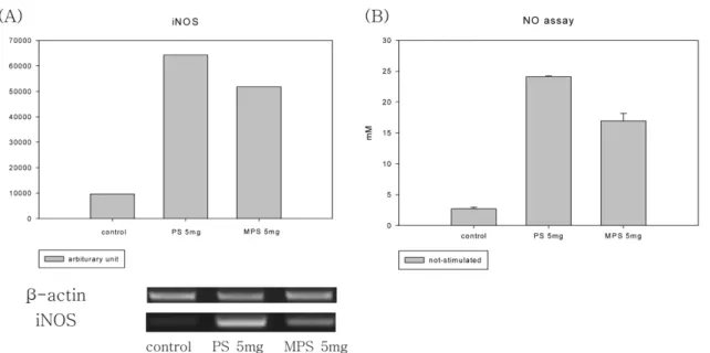
Effect of NO production by RAW264.7 in the presence of MPS and PS 5×10
5RAW264.7 cells were incubated with MPS or PS for
24 hr and then NO assay was performed and expression level of iNOS was measured by RT-PCR. RT-PCR products were
electrophorased in 1% agarose gel and the bands were analyzed by UVIDoc Mw program.
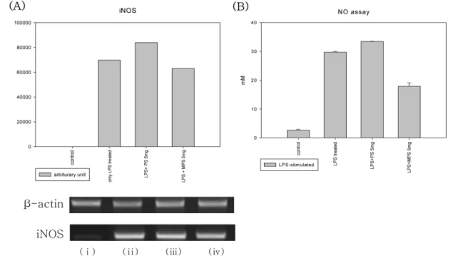
Fig. 3 −
Effect of NO production by LPS-activated RAW264.7 in the presence of MPS and PS. 5×10
5RAW264.7 cells were incubated with MPS or PS for 24 hr after activation of cells with 10 LPS for hr RT-PCR products were electrophorased in 1% agarose gel and the bands were analyzed by UVIDoc Mw program. (i); control, (ii); only LPS treated, (iii); LPS+PS 5 mg, (iv); LPS+MPS 5 mg.
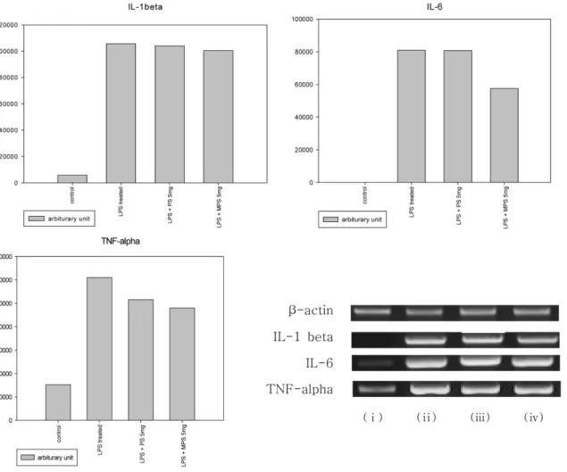
Fig. 4 −
Comparison for genes expression ofpro-inflammatory cytokines by MPS and PS. 5×10
5RAW264.7 cells were incubated with MPS or
PS for 24 hr and then the gene expression of pro-inflammatory cytokines were measured by RT-PCR. RT-PCR products were
electrophorased in 1% agarose gel and the bands were analyzed by UVIDoc Mw program.
된
RAW264.7
세포에서농도의존적으로세포에대한독성이증가함을알수있었다
.
MPS와PS가NO생성및iNOS발현에미치는영향
NO
는pro-inflammatory cytokines
와더불어macrophage
가활성화되었을때생성되는인자이다
. iNOS
에의해양적증가가일어나며이들의증가는
macrophage
가항원을제거하는데도움을주어면역력을증강시켜준다고볼수있다
. iNOS
의발현변화는
PCR
을통해확인하였고,
결과물을arbiturary unit
으 로환산하여그값의변화를관찰하였다. Fig. 2A
와같이MPS
처리군은
iNOS
의생성이증가되며,
이는PS
에의한것보다는 적게일어남을알수있다. iNOS
의증가는Fig. 2B
와같이NO
의증가로바로이어지며
, NO
또한PS
보다MPS
에서약간적은양으로증가함을보였다
.
이와같이MPS
는PS
와마찬가지 로iNOS gene expression
및NO
생성을모두증가시킨다는 것을통해macrophage
의활성화에영향을미친다는것을알수있었다
.
Macrophage
는항원에의해활성화되어NO
등의인자를생성하여면역력을증강시키는반면
,
지나친활성화에의해세포 손상을일으키는원인이되기도한다.
고농도의NO
가존재하게되면만성염증및세포사멸과관련된질환을야기한다
. LPS
에 의해활성화된RAW264.7
세포는control
군에비해현저하게높 은NO
생성증가를보이며, PS
와는달리MPS
처리군에서LPS
에의해증가된
NO
생성이감소함을보였다(Fig. 3B). NO
를생 성하는iNOS
의gene expression
또한LPS
에의해증가되었다가
MPS
처리에 의해 약간의 감소가 있는것이확인되었다(Fig. 3A).
MPS와 PS가 RAW264.7 세포주의 pro-inflammatory cytokines관련gene expression에미치는영향
Pro-inflammatory cytokine
은염증을매개하는cytokine
으로NO
와함께macrophage
가활성화되었을때생성이증가되는대Fig. 5 −
Comparison for genes expression ofpro-inflammatory cytokines by MPS and PS. 5×10
5RAW264.7 cells were incubated with MPS or PS for 24 hr and then the gene expression of pro-inflammatory cytokines were measured by RT-PCR. RT-PCR products were electrophorased in 1% agarose gel and the bands were analyzed by UVIDoc Mw program. (i); control, (ii); only LPS treated, (iii);
LPS+PS 5 mg, (iv); LPS+MPS 5 mg.
표적물질이다
.
대표적인pro-inflammatory cytokine
으로는IL- 1
β, TNF-
α, IL-6
등이 있으며PCR
을 통해 이들의gene expression
변화를확인하였다. PCR
생성물은arbiturary unit
으 로환산하여그값을비교하였다. MPS
와PS
는control
군에비해
IL-1
β, TNF-
α, IL-6
의생성이증가시킴을알수있었으며,
MPS
보다PS
를통해증가가더크게일어난다는것이확인되었다
(Fig. 4).
LPS
에의해활성화된RAW264.7
에PS
를처리시IL-1
β, TNF-
α는크게변화가일어나지않았고
, IL-6
의gene expression
만약간의감소를보였다
.
이와달리MPS
를처리하였을때IL-1
β에서는약간의감소가보였고
, TNF-
α, IL-6
은PS
를처리하였을때보다발현이감소됨을관찰하였다
(Fig. 5). Gene expression
은세 포에LPS
와동시에PS, MPS
를처리하여24
시간배양후RT- PCR
결과를arbiturary unit
으로환산하여비교하였다.
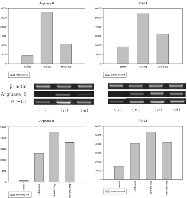
MPS는PS와달리Arginase II, PD-L1의gene expression
에영향
Arginase II
는macrophage
에서생성되는또하나의enzyme
으Fig. 6 −
Effect of Arginase II, PD-L1 gene expression on macrophagec by LPS, MPS and PS. 5×10
5RAW264.7 cells were incubated with MPS
or PS for 24 hr and then the gene expression of Arginase II and PD-1L were measured by RT-PCR. RT-PCR products were
electrophorased in 1% agarose gel and the bands were analyzed by UVIDoc Mw program. (i) control, (ii) PS 5 mg, (iii) MPS 5 mg,
(iv) control, (v) only LPS-treated, (vi) LPS+PS 5 mg, (vii) LPS+MPS 5 mg.
로
, NOS
와기질경쟁적으로작용하여NO
의생성을막고proline
전구체를생성하여조직회복에도움을주는것으로알려져있다
. PD-L1
은macrophage
를포함한antigen presenting cell(APC)
에 발현하는surface molecule
로costimulatory signal
을생성하여T cell
에inhibition signal
을전달하는것으로알려져있으나아직그기전에대해서는정확하게밝혀진바가없다
. RT-PCR
을수행하여그발현정도를측정하였으며
, Arginase II, PD-L1
모두
MPS
처리군에서는거의발현의변화가없는반면PS
처리 군에서는발현이증가하는것을볼수있었다(Fig. 6).
결 론
Macrophage
는 백혈구(Neutrophills),
자연살해세포(Natural Killer cells)
와더불어외부의병원균에대해서host defence
의 제1
선에서중요한역할을한다.
감염과염증이생성되면(L)- arginine
는nitric oxide synthase(iNOS)
에의해NO
로생산되는데활성화된
macrophage
는NO
와pro-inflammatory cytokine
을 분비하여다른면역세포와조직에영향을준다.
16)iNOS
에의해생성된적은농도의
NO
는항미생물작용을나타내나고농도의NO
는inflammation
과carcinogenesis
에중요한역할을하며17) 세포사멸및조직손상또한유발한다.
7)따라서이러한인자의생성을조절할수있는물질은
immune tolerance
를유도함으로써질병에대한방어능력을키울수있을것이다
.
본연구에 서는면역세포의활성을효과적으로증가시키는PS
와항염및항암작용을하는것으로알려진사포닌의혼합물
(MPS)
을처리하여각각의효능을비교평가하였다
. MPS
는macrophage
의NO, IL-1
β, TNF-
α, IL-6
의 생성을 증가시켰는데 이는MPS
가macrophage
활성을증가시켜면역력을증가시켰음을추측할수있다
.
그러나, PS
에의한macrophage
의NO, IL-1
β, TNF-
α, IL- 6
의증가비율은크지않았다.
반면LPS
에의이미활성화된macrophage
에PS
를 처리한 경우NO
와pro-inflammatory cytokines
증감이나타나지않았으나MPS
를처리한경우IL-6
의 생성이 감소되었다
.
이는MPS
가 지나치게 활성화된macrophage
를근절함으로써정상세포에대한손상을억제하는효과를나타낼것으로추측된다
. Arginase
효소는iNOS
와기질 경쟁적으로작용하여NO
의생성을억제하는효소이다. PD-L1 (programed death-ligand 1)
은항원제시세포에서발현하여활성 화된T
림프구에서발현하는PD-1(programed death-1)
과결합 하여T
림프구의inhibitory signal
을전달하는것으로알려져잇는데
, LPS
로활성화된macrophage
에서는Arginase
와PD-L1
의 발현이MPS
의해서는크게변하지않으나PS
에의해발현이증 가되었으며이는saponin
의영향인것으로추측된다.
이런면역조절능력에대한기전적연구가앞으로더수행되어져야할것 이다
.
감사의 말씀
이논문은
2006
년도중앙대학교학술연구비지원에의한것임.
참고문헌
1) Jung, I. S., Chung, H. Y., Yun, Y. S., Kim, K. H. and Lee, I. R. : The pattern of cytokine mRNA expression induced by polysaccharide from Panax ginseng C. A. Meyer. J. Ginseng Res.
22(4), 324 (1998).
2) Kim, M. H., Byon, Y. Y., Ko, E. J., Song, J. Y., Yun, Y. S., Shin, T. K. and Joo, H. G. : Immunomodulatory activity of ginsan, a polysaccharede of Panax ginseng, on dendritic cells. Korean J.
Physiol. Pharm.
13(3), 169 (2009).
3) Han, S. K., Song, J. Y., Yun, Y. S. and Yi, S. Y. : Ginsan improved Th1 immune response inhibited by gamma radiation. Arch.
Pharm. Res.
28(3), 343 (2005).
4) Nah, S. Y., Bhatia, K. S., Lyles, J., Ellinwood, E. H. and Lee, T. H. : Effects of ginseng saponin on acute cocaine-induced alterations in evoked dopamine release and uptake in rat brain nucleus accumbens. Brain Res.
1248, 184 (2009).
5) Shine, H. J., Kwon, Y. M., Jung, Y. E., Lee, D. H., Lee, J. C., Han, S. S., Kim, H. T., Park, J. H., Sung, E. G. and Lee, Y. C. : Antioxidative effect of ginseng saponin on cardiac endothelial cells in culture. Kor. J. Anat.
33(1), 77 (2000).
6) Hamilton, T. A. and Adams, D. O. : Molecular mechanisms of signal transduction in macrophage activation. Immunol. Today
8