2021 The Korean Society of Veterinary Science.
This is an open-access article distributed under the terms of the Creative Commons Attribution Non-Commercial license (http://creativecommons.
org/licenses/by-nc/4.0/), which permits unrestrict- ed non-commercial use, distribution, and repro- duction in any medium, provided the original work is properly cited.
The most common types of canine lymphomas are aggressive and high-grade.
However, recognizing indolent lymphoma is significant because it is associated with longer survival time. The most common histopathological subtypes of canine indolent lymphomas are T-zone lymphoma (TZL), marginal zone lymphoma, and follicular lymphoma. However, diagnoses of these lymphomas is mostly based on histopathological examination; thus, canine indolent lymphomas are probably un- derdiagnosed [1,2]. Recent reports demonstrated that a T-cell lymphoma with sig- nificant CD45 negative neoplastic cells could be classified as TZL [3-5]. However, reports describing clinical presentation, immunophenotype, molecular analysis, and histopathologic analysis of TZL in dogs remain scarce.
An 11-year-old, spayed female Golden Retriever presented lymphadenopathy, which was found upon physical examination (bilateral submandibular, prescapu- lar, and popliteal lymph nodes), radiography and abdominal ultrasound (bilateral retropharyngeal, medial iliac, and jejunal lymph nodes). Fine-needle aspiration cytology of the lymph nodes and blood smear evaluation of peripheral lymphocy- tosis were performed to evaluate lymphoid neoplasia. A homogenous lymphoid population comprising small to intermediate lymphocytes that frequently had a unipolar cytoplasmic extension, “hand-mirror cells” were identified from the lymph nodes with a low mitotic rate. Laboratory analysis showed lymphocytosis (lymphocyte count, 11,890 cells/µL [reference interval, 1,050-5,100 cells/µL]) with mild leukocytosis (white blood cell count, 17,400 cells/µL [reference interval,
CD45-/CD79a-/TCRαβ+/TCRγδ-/MHCII+
T-zone lymphoma in a dog with generalized lymphadenopathy: a case report
Sun Woo Shin 1 , Yu jin Lim 2 , Hyeona Bae 1 , Jihu Kim 1 , ARom Cho 1 , Jinho Park 3 , Dongbin Lee 1 , Dong-In Jung 1 , Sang-ki Kim 2,* , DoHyeon Yu 1,*
1
College of Veterinary Medicine, Gyeongsang National University, Jinju 52828, Korea
2
College of Industrial Science, Kongju National University, Yesan 32439, Korea
3
College of Veterinary Medicine, Jeonbuk National University, Iksan 54596, Korea pISSN 2466-1384 · eISSN 2466-1392
Korean J Vet Res 2021;61(3):e21 https://doi.org/10.14405/kjvr.2021.61.e21
*Corresponding author:
Sang-ki Kim
College of Industrial Science, Kongju National University, 54 Daehak-ro, Yesan- eup, Yesan 32439, Korea
Tel: +82-41-330-1525 Fax: +82-41-330-1529 E-mail: [email protected] DoHyeon Yu
College of Veterinary Medicine, Gyeongsang National University, 501 Jinju-daero, Jinju 52828, Korea
Tel: +82-55-772-2368 Fax: +82-55-772-2330 E-mail: [email protected] ORCID:
https://orcid.org/0000-0003-0292-8200 https://orcid.org/0000-0001-7645-6926 Conflict of interest:
The authors declare no conflict of interest.
Received: June 28, 2021 Revised: August 4, 2021 Accepted: August 9, 2021
Case Report
Canine T-zone lymphoma (TZL) is a mature T-cell lymphoma in dogs. The diagnosis and sub-classification are impossible without biopsy or immunophenotyping by flow cytometry. An 11-year-old, spayed, female Golden Retriever presented with lymph node enlargement. Clinical examination was consistent with canine multicentric lym- phoma. However, immunophenotyping revealed positive for CD3, CD4, CD5, CD8, CD21, TCRαβ, and MHCII but negative for CD34, CD45, CD79a, and TCRγδ. Histopa- thology revealed lymphocytes expanding to the cortex-preserving architecture and thinning of the nodal capsule, and CD3 positive but PAX-5 negative. Owing to the in- dolent nature of TZL, careful monitoring approach without clinical intervention was utilized.
Keywords: lymphoma; flow cytometry; immunophenotyping; dog; case report
5,050-16,760 cells/µL]) in the peripheral blood. Peripheral blood lymphocytes were also small to intermediate in size (Fig.
1). These findings were consistent with that of multicentric lymphoma with possible bone marrow involvement (World Health Organization [WHO] stage Va) but further confirmato- ry tests were needed since the small to intermediate lympho- cytes were predominant, and the dog only had subclinical man- ifestations.
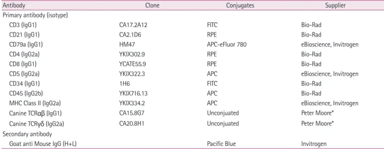
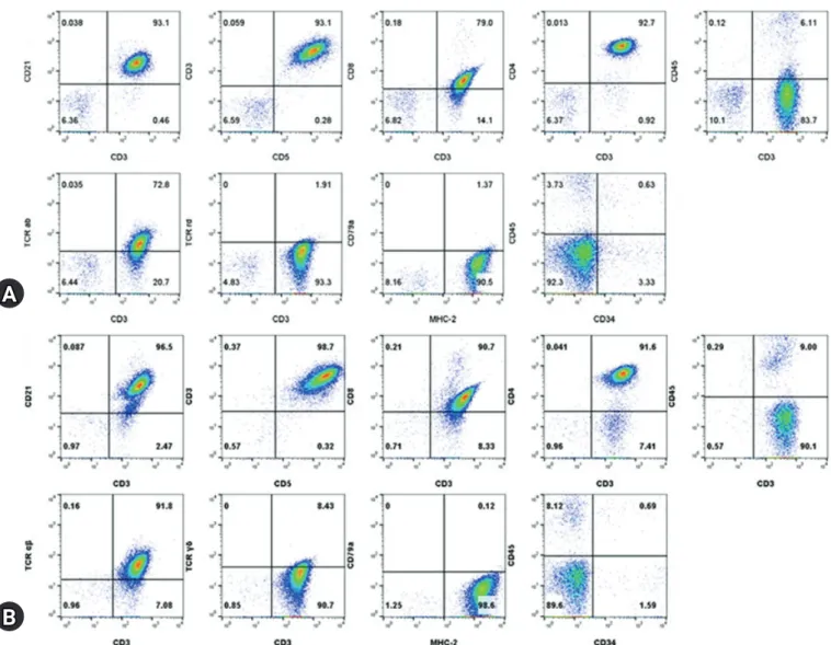
Immunophenotyping by flow cytometric analysis was per- formed on the lymph node aspirates and peripheral blood mononuclear cell isolates using antibodies (Table 1). Lympho- cytes from the nodes and blood showed a homogenous popula- tion with both CD3+ and CD21+ but CD79a-. The CD3+ cells
also showed CD4+/CD8+/CD5+/CD34-/TCRαβ+/TCRγδ-/
MHCII+ but were ultimately CD45- (Fig. 2). PARR assay using the peripheral lymph node, which was performed by the Clini- cal Immunopathology Laboratory at Colorado State University via the IDEXX reference laboratory, showed clonal rearrange- ment of both TCR and immunoglobulin genes. The immuno- phenotyping results of CD3+/CD5+/CD21+/CD45- cells were consistent with the findings of TZL. An additional bone mar- row examination showed orderly maturated myeloid and eryth- roid series, less than 2% of blast cells, and an estimated 1.5:1 ra- tio of myeloid to erythroid.
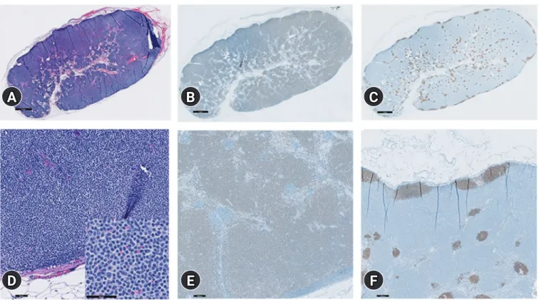
Histopathologic examination of the right prescapular lymph node confirmed TZL based on the WHO classification of ca-
Fig. 1. Fine-needle aspiration cytology of the lymph nodes (A), peripheral blood (B), and bone marrow (C). A homogenous lymphoid pop- ulation comprising small to intermediate lymphocytes in the lymph nodes (A) with low mitotic rate. Lymphocytes from peripheral blood (B) are also small to intermediate in size. In bone marrow cytology, both myeloid and erythroid series appear complete and display an orderly maturation. The myeloid:erythroid ratio is 1.5:1, and the number of blast cell are less than 2% of the nucleated cell numbers (C). Diff- Quik stain, scale bar: (A, B) 50 μm, (C) 100 μm.
Table 1. Antibodies for flow cytometry analysis used in the case
Antibody Clone Conjugates Supplier
Primary antibody (isotype)
CD3 (IgG1) CA17.2A12 FITC Bio-Rad
CD21 (IgG1) CA2.1D6 RPE Bio-Rad
CD79a (IgG1) HM47 APC-eFluor 780 eBioscience, Invitrogen
CD4 (IgG2a) YKIX302.9 RPE Bio-Rad
CD8 (IgG1) YCATE55.9 RPE Bio-Rad
CD5 (IgG2a) YKIX322.3 APC eBioscience, Invitrogen
CD34 (IgG1) 1H6 FITC Bio-Rad
CD45 (IgG2b) YKIX716.13 APC Bio-Rad
MHC Class II (IgG2a) YKIX334.2 APC eBioscience, Invitrogen
Canine TCR αβ (IgG1) CA15.8G7 Unconjuated Peter Moore*
Canine TCR γδ (IgG2a) CA20.8H1 Unconjuated Peter Moore*
Secondary antibody
Goat anti Mouse IgG (H+L) Pacific Blue Invitrogen
Bio-Rad (Hercules, CA, USA), eBioscience (San Diego, CA, USA), Invitrogen (Waltam, MA, USA).
FITC, fluorescein isothiocyanate; RPE, r-phycoerythrin; APC, allophycocyanin.
*Leukocyte Antigen Biology Laboratory, Universitiy of California, CA, USA.
A B C
50um 50um 100um