Abstract. Background: Resistance to gemcitabine is a major obstacle in the treatment of advanced pancreatic cancer. Previous exploration of protein kinase inhibitors demonstrated that blocking transforming growth factor-β (TGFβ) signal enhances the efficacy of gemcitabine in pancreatic cancer cells. Materials and Methods: We analyzed the cell viability after combinational treatment of TGFβ receptor I (TβRI) inhibitors, SB431542 and SB525334 with gemcitabine in pancreatic cancer cells. In addition, apoptotic cell death and cell migration were measured.
Results: Combination with TβRI inhibitors significantly augmented the cytotoxicity of gemcitabine in both parental and gemcitabine resistant pancreatic cancer cells. SB525334 significantly increased apoptotic cell death in gemcitabine- resistant cells. Treatment of SB525334 also affected the AKT signalling pathway, which plays a crucial role in gemcitabine resistance. Migration assay also revealed that blocking TβRI reduces cell migration. Conclusion:
Chemotherapeutic approaches using SB525334 might enhance the treatment benefit of the gemcitabine-containing regimens in the treatment of pancreatic cancer patients.
Pancreatic cancer is one of the most lethal malignancies and five-year survival of pancreatic cancer patients is less than 5% (1), since the majority of patients are diagnosed with disease at unresectable stages and do not receive benefit from curative surgery (2). Moreover, up to 50% of cases are associated with metastasis, which shortens the survival benefit of conventional chemotherapy (3). The innate chemoresistance in human malignancies may have multiple mechanisms, such as decreased intracellular drug accumulation, facilitation of drug detoxification mechanisms, and increased DNA repair capacity (4, 5).
To circumvent gemcitabine resistance various attempts using combinational therapy have been tried (6). However, most trials were not very promising except the combination with epidermal growth factor receptor (EGFR) inhibitor, which significantly but not satisfactorily extends overall survival compared to single treatment of gemcitabine (7).
Therefore discovering new therapeutic targets to overcome gemcitabine resistance is an urgent and essential task for treatment of pancreatic cancer.
Recent reports demonstrated that activation of several biochemical pathways induces acquired drug resistance during drug treatment (8, 9). Thus, exploration of kinome for sensitization of pancreatic cancer cells to gemcitabine can be a useful tool in order to isolate new target kinases. Recent developments in small molecule inhibitors for numerous protein kinases have enabled us to explore the protein kinase targets in pancreatic cancer cells. Through the screening of a series of protein kinase inhibitors (PKIs), we found that several PKIs show synergism in combination with gemcitabine (unpublished data). Among these newly found PKIs, a specific inhibitor for transforming growth factor-β (TGFβ) receptor I (TβRI), SB525334, exhibited substantial potency in reducing gemcitabine resistance.
TGFβ is a potent regulator of cell proliferation and differentiation (10). TGFβ preferentially binds to TβRII then the ligand-bound TβRII forms a heteromeric receptor This article is freely accessible online.
*These authors contributed equally to this work.
Correspondence to: Yeon-Sun Seong, MD, Ph.D., WCU Research Center of Nanobiomedical Science, Dankook University, San 29, Anseo-Dong, Cheonan, 330-714, Korea. Tel: +82 415503875, Fax:
+82 415501149, e-mail: [email protected] and Insoo Bae, Ph.D., Department of Oncology, Lombardi Comprehensive Cancer Center, Georgetown University, 3970 Reservoir Road, NW, Washington, DC 20057, U.S.A. Tel: +1 2026875267, e-mail: [email protected]
Key Words: Pancreatic cancer, gemcitabine, transforming growth factor-β receptor (TβR), protein kinase inhibitor, SB525334, MiaPaCa2, AsPC1 cells.
Transforming Growth Factor Beta Receptor I Inhibitor Sensitizes Drug-resistant
Pancreatic Cancer Cells to Gemcitabine
YEON JEONG KIM1*, JAE SEOK HWANG2,3*, YOUNG BIN HONG1,3, INSOO BAE1,3,4and YEON-SUN SEONG1
1
WCU Research Center of Nanobiomedical Science, Dankook University, Cheonan, Korea;
2
Department of Internal Medicine, Keimyung University College of Medicine, Daegu, Korea;
Departments of
3Oncology and
4Radiation Medicine, Lombardi
Comprehensive Cancer Center, Georgetown University, Washington, DC, U.S.A.
complex with TβRI, which triggers downstream signals (11). Among the seven isoforms of TβRIs, activin receptor- like kinase 5 (ALK5) is the predominant in most cell types.
As a canonical pathway, activated ALK5 recruits and phosphorylates SMAD2 and SMAD3, which translocate to the nucleus for the activation or repression of target genes after heteromeric complex formation with SMAD4 (reviewed in 11). Although TGFβ signaling has been regarded as tumor suppressive, there is accumulating evidence that aberrant TGFβ signaling initiates cancer and promotes tumor progression (reviewed in 12). In addition, TGFβ plays a crucial role in epithelial to mesenchymal transition (EMT)-mediated tumor progression (13).
Activation of SMAD2 and SMAD3, downstream of TGFβ has also been reported to induce EMT and metastasis of skin and breast cancer (14, 15).
The aberration of TGFβ signaling is also implicated in pancreatic cancer: immunohistochemical analysis revealed that elevated levels of TGFβ isoforms are negatively correlated with patient survival in pancreatic cancer (16).
Accordingly, several approaches have been tried to target TβRs in pancreatic cancer. Expression of soluble TβRII in pancreatic cancer cells exhibited a marked decrease in invasive capacity (17). Melisi et al. (18) demonstrated that a dual inhibitor of TβRI and TβRII, LY2109761, significantly reduced metastasis of pancreatic cancer cells in vivo.
Application with SB431542, a TβRI inhibitor, also exhibited efficacy in tumor cell sensitization to gemcitabine, which was evaluated in novel 3D culture of various pancreatic cancer cell lines (19). Therefore, there are quite substantial pre-clinical indications that targeting the TGFβ signal could reduce resistance to gemcitabine.
In order to overcome gemcitabine resistance we have studied the mechanism of acquired gemcitabine resistance in pancreatic cancer cells. Based on previous observations that TβRI may be a promising target for the enhancement of gemcitabine efficacy, in the present study, we evaluated the efficacy of SB525334, a TβRI inhibitor (20), in gemcitabine- resistant pancreatic cancer cells.
Materials and Methods
Cell culture and reagents. Human pancreatic cancer cell lines, MiaPaCa2 and AsPC1, were obtained from the American Type Culture Collection (ATCC, Manassas, VA, USA). MiaPaCa2 cells were cultured in Dulbecco’s modified Eagle’s medium (DMEM) containing 2.5% horse serum and 10% fetal bovine serum. AsPC1 cells were cultured in Roswell Park Memorial Institute (RPMI)- 1640 media supplemented with 20% fetal bovine serum. TβRI inhibitors, SB431542 and SB525334, were purchased from Selleck Chemicals (Houston, TX, USA).
Generation of gemcitabine resistant cells. For creating gemcitabine- resistant cells, MiaPaCa2 and AsPC-1 cells were exposed to incrementally increasing doses (starting at 0.1 μM) of gemcitabine.
When the cells adapted to a dose, the gemcitabine concentration was increased by 0.1 μM. After three months of selection, MiaPaCa2 and AsPC1 cells surviving at 1.5 μM and 1.0 μM of gemcitabine, respectively, were generated.
Cell viability and drug combination. To determine cell viability, cells were plated onto 96-well plates. After 72 h treatment with gemcitabine and/or TβRI inhibitors, cell viabilities were measured using (3-(4,5-Dimethylthiazol-2-yl)-2,5-diphenyltetrazolium bromide (MTT) assays. The half maximal effective concentration (EC50) of each drug and the combination index at half maximal effective concentration (CI50) of each drug combination was determined using CompuSyn software (ComboSyn Inc., Paramus, NJ, USA) (21).
Western blotting. Standard western blotting was performed in order to measure the expression levels of proteins. Cells cultured with gemcitabine with or without SB525334 were harvested and the proteins in total cell extracts were generated using RIPA buffer supplemented with protease inhibitors. Nuclear fractions of SB525334 and gemcitabine-treated cells were prepared using the Nuclear Extraction Kit (Affymetrix, Santa Clara, CA, USA) according to the manufacturer’s protocol. Protein samples were separated by sodium dodecyl sulfate polyacrylamide gel electrophoresis (SDS-PAGE) then transferred to polyvinylidene fluoride (PVDF) membranes. Anti-poly (ADP-ribose) polymerase 1 (PARP1), anti-X-linked inhibitor of apoptosis protein (XIAP), anti-B-cell lymphoma 2 (BCL2) (BD Bioscience Inc., Bedford, MA, USA), anti-AKT, anti-phospho-AKT, anti-BCL2-associated death promoter (BAD), anti-phospho-BAD, anti- β catenin (Cell Signaling Technology, Inc., Boston, MA), anti-lamin B (Abcam, Inc., Cambridge, MA, USA), anti-vimentin, anti-E- cadherin and anti-β-actin antibodies (Santa Cruz Biotechnology Inc., Santa Cruz, CA, USA) were used as primary antibodies. Anti-mouse, anti-goat and anti-rabbit IgG–peroxidase antibodies (Sigma, St. Louis, MO, USA) were used for secondary antibodies and enhanced chemiluminescence (ECL) solution (Santa Cruz Biotechnology, Inc.) was used for detection.
Caspase 3/7 assay. Protein extracts used for western blotting were analyzed for caspase activity. Fifty micrograms of proteins were incubated with substrate containing Caspase-Glo®3/7 Assay buffer (Promega, Madison, WI, USA) for thirty minutes. Caspase activities were calculated after detection of luminescence by a luminometer (Victor 2; Perkin Elmer, Walthan, MA, USA).
Transfection of siRNA. One hundred nanomoles of AKT-specific (5’- CCUUUUCGACGCUUAACCU-3’; Bioneer Inc., Korea) or control- siRNA (5’-GACGAGCGGCACGUGCACA-3’; Dharmacon, Lafayette, CO, USA) were transfected into MiaPaCa2-GR cells using Lipofectamine 2000 (Invitrogen, Carlsbad, CA, USA). After 48 h, cells were transferred into 6-well and 96-well plates for western blotting and MTT assay, respectively. Cells were further transfected with siRNAs for an additional 24 h then treated with gemcitabine for 72 h in the presence of siRNAs.
Cell migration assay. MiaPaCa2-GR cells were grown on 6-well plates until they became confluent. Then artificial wounds were generated by scratching of the monolayer of cells with a sterile plastic micropipette tip. Culture media were replaced with 1 or 10 μM of SB525334-containing media. Wound closure was monitored by phase-contrast microscopy (Olympus, Tokyo, Japan) and digital images were obtained after 24 and 48 h post-wounding.
Results
TβRI inhibitors enhance the cytotoxicity of gemcitabine. In order to evaluate the combinatorial effect of TβRI inhibitors with gemcitabine, we measured the cell viability after treatment of cells with gemcitabine and TβRI inhibitors. Combined treatment of gemcitabine and SB431542 significantly reduced cell viability of MiaPaCa2 (Figure 1A) and AsPC1 (Figure 1C).
Similarly, combined treatment of SB525334 with gemcitabine also effectively reduced the viability of both cell lines compared to single treatment with gemcitabine or SB525334 (Figure 1B and D).
TβRI inhibitors abrogate gemcitabine resistance. Since TβRI inhibitors efficiently enhanced the cytotoxicity of gemcitabine, we further evaluated the combinatorial effects in gemcitabine-
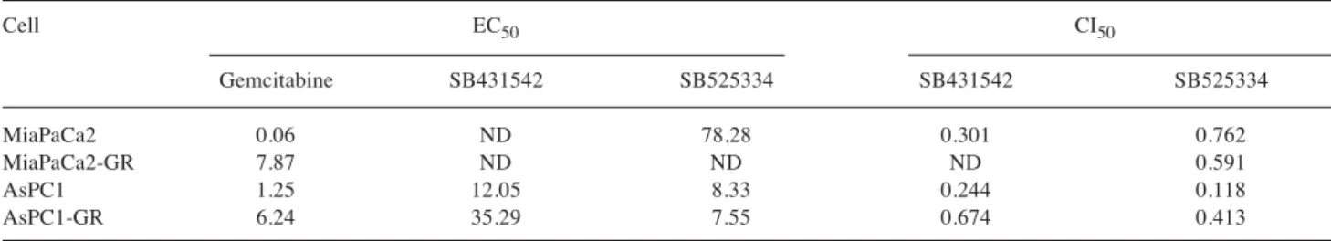
resistant cells. MTT assay revealed that TβRI inhibitors effectively reduced the cell viability of MiaPaCa2-GR cells in combination with gemcitabine as a 20:1 molar ratio (Figure 2A and B). Combinational treatment using SB431542 or SB525334 also dramatically sensitized AsPC1-GR cells to gemcitabine (Figure 2C and D). To determine the synergism between TβRI inhibitors and gemcitabine, we analyzed CI50 in pancreatic cancer cells (Table I). SB431542 showed synergistic effect in combination with gemcitabine in three cell lines, although CI50could not be determined in MiaPaCa2-GR cells due to low cytotoxicity. SB525334 also showed synergism in all of the cell lines.
Apoptotic cell death was increased by combination of SB525334 and gemcitabine. To further characterize the synergistic effect, we measured apoptotic cell death.
Figure 1. Combination of transforming growth factor-β receptor I inhibitors with gemcitabine efficiently reduces the viability of pancreatic cancer cells. MiaPaCa2 (A and B) and AsPC1 (C and D) cells were exposed to SB431542 (A and C), SB525334 (B and D) in combination with gemcitabine for 72 h then cell viability was determined by MTT assay as described in Materials and Methods. Data are expressed as mean±SD. Student’s t-test was applied for statistical analysis for comparison between gemcitabine treatment and combined treatment. *p<0.05; **p<0.01; and ***p<0.001.
Western blotting revealed that combination of gemcitabine and SB525334 increased PARP1 cleavage in MiaPaCa2- GR cells (Figure 3A). Drug combination also effectively reduced the expression of anti-apoptotic proteins such as XIAP and BCL2. To further confirm the increase in
apoptosis, we measured caspase-3 activity in MiaPaCa2- GR cells. Combinations of drugs significantly enhanced caspase-3 activity compared to single treatment of gemcitabine in both low- and high-dose combinations (Figure 3B).
Figure 2. Transforming growth factor-β receptor I inhibitors reduce gemcitabine-resistance. Gemcitabine resistant MiaPaCa2-GR (A and B) and AsPC1-GR (C and D) cells were incubated with TβRI inhibitors or gemcitabine for 72 h. Data are expressed as mean±SD. Student’s t-test was applied for statistical analysis for comparison between gemcitabine treatment and combined treatment. *p<0.05; **p<0.01; and ***p<0.001.
Table I. Synergism of gemcitabine and transforming growth factor-β receptor inhibitors.
Cell EC50 CI50
Gemcitabine SB431542 SB525334 SB431542 SB525334
MiaPaCa2 0.06 ND 78.28 0.301 0.762
MiaPaCa2-GR 7.87 ND ND ND 0.591
AsPC1 1.25 12.05 8.33 0.244 0.118
AsPC1-GR 6.24 35.29 7.55 0.674 0.413
EC50, Half maximal effective concentration; CI50, combination (gemcitabine and each TβR inhibitor) index at half maximal effective concentration.
EC50and CI50were calculated as described under Materials and Methods.
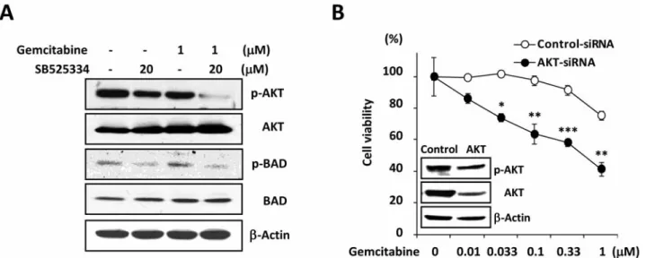
SB525334 suppresses AKT pathways in gemcitabine-resistant cells. To characterize the gemcitabine sensitization by TβRI inhibitors, we analyzed the change of cell survival pathways.
Since the phosphoinositide 3-kinase (PI3K)/AKT pathway is the most well-known pathway directing gemcitabine resistance (22), we measured the changes in AKT activation.
Western blotting demonstrated that incubation of cells with SB525334 significantly reduced phosphorylation of AKT and BAD without changes of total amounts of protein in gemcitabine-resistant cells (Figure 4A). To further confirm that inactivation of AKT by TβRI inhibitor is crucial for the loss of gemcitabine resistance, we measured the change of
cell viability after knockdown of AKT. Abrogation of AKT significantly reduced gemcitabine resistance of MiaPaCa2- GR cells (Figure 4B). These data indicate that one of the mechanisms of TβR inhibitor in gemcitabine resistance is through inactivation of AKT mediated cell survival signals.
EMT characteristics in gemcitabine-resistant cells are abrogated by inhibition of TβRI. EMT is a distinct trait in cells with acquired drug resistance. To determine whether TβRI inhibitor reduces markers of EMT, we measured several markers of EMT after 24 h treatment with SB525334.
Expression levels of vimentin were reduced in a dose- dependent manner, while the levels of E-cadherin were not changed by the incubation of cells with SB525334 (Figure 5A). The levels of nuclear localized β-catenin, an EMT marker, also increased. Cell migration assay also supported the changes of EMT markers. Incubation with SB525334 dramatically reduced the migratory activity of MiaPaCa2-GR cells (Figure 5B). Thus inhibition of TβRI effectively reduced expression of markers of EMT and migratory activity in gemcitabine-resistant cells.
Discussion
Based on the observation that TGFβ prevents proliferation of normal epithelial cells and cancer cells at early stages of tumorigenesis, TGFβ was regarded as a tumor suppressor (10).
Inactivation of TβRI, TβRII, SMAD2 and SMAD4 through mutation or loss of heterozygosity correlates to tumorigenesis (23). Transcriptional inactivation of TβRI and TβRII was also observed in various types of cancer (11). Besides the loss of the tumor suppressor function of TGFβ signaling, however, considerable evidence indicates that perturbation of TGFβ signaling enhances disease malignancy (12).
In addition to SMAD-mediated transcription, TGFβ can activate other signaling cascades Smad-independently (10).
Chow et al. revealed that TGFβ down-regulates phosphatase and tensin homolog (PTEN) through activation of nuclear factor κB (NFκB) (24), which results in the activation of AKT signal (25). In this study, we observed that inhibition of TβRI reduces phosphorylation of AKT and activation of downstream BAD in gemcitabine-resistant cells. Subsequent cell viability measurements also demonstrated that abrogation of AKT sensitizes resistant cells to gemcitabine. Previously, we observed that AKT-specific inhibitors significantly increased gemcitabine sensitivity in parental and gemcitabine- resistant cells (data not shown). Therefore, the synergism of SB525334 and gemcitabine may, in part, be derived from the inactivation of AKT and the consequent incapability of activation of BAD in gemcitabine-resistant cells.
Recent findings indicate that TGFβ signaling pathways facilitate metastasis of tumor cells (26). To initiate cell migration, cell-cell junctions need to be weakened. During EMT, TGFβ down-regulates claudins and occludins, which are Figure 3. Transforming growth factor-β receptor inhibitor enhances
apoptotic cell death in gemcitabine-resistant cell. A: MiaPaCa2-GR cells were incubated with drugs for 72 h then expression levels of apoptosis markers were measured by western blotting. B: Caspase-3/7 activities in same samples were determined as described in Materials and Methods. Student’s t-test was applied for statistical analysis.
*p<0.05; **p<0.01; and ***p<0.001.
involved in tight junctions. In addition, TGFβ also induced snai1 and snai2, zinc finger E-box-binding homeobox 1 (ZEB1) and ZEB2, resulting in suppression of E-cadherin, an adherens junction protein. Loss of E-cadherin induces the release of β-catenin (27), which translocates into the nucleus and transactivates EMT-related proteins, such as vimentin, after binding to T-cell factor (TCF)/lymphoid enhancer factor 1 transcription factor family (28). Therefore, our observation that SB525334 reduced the major mesenchymal markers (e.g.
β-catenin and vimentin) indicates that inhibition of TβRI
efficiently reduces the EMT trait in gemcitabine-resistant cells.
Although we did not observe significant up-regulation of E- cadherin by SB525334, further confirmation with the cell migration assay supports the efficacy of TβRI inhibitor against metastasis of pancreatic cancer cells.
The significance of EMT is also implied in cancer stem cell-(CSC) associated drug resistance. Currently the presence of CSCs in human tumors and the correlation of CSCs with drug resistance are proved and accepted by numerous researchers (29). Several groups demonstrated the presence Figure 4. Inactivation of AKT by SB525334 renders MiaPaCa2-GR cells sensitive to gemcitabine. A: MiaPaCa2-GR cells were exposed to SB525334 (20 μM) or gemcitabine (1 μM) for 24 h then AKT signaling was measured by western blotting. B: MiaPaCa2-GR cells were transfected with control or AKT specific siRNA for 72 h, then exposed to gemcitabine for an additional 72 h in the presence of siRNA before MTT assay. Inset: Changes of AKT level after AKT knockdown as confirmed by western blotting. Student’s t-test was applied for statistical analysis. *p<0.05; **p<0.01; and
***p<0.001.
Figure 5. SB525334 reduces epithelial to mesenchymal transition trait in gemcitabine-resistant cells. A: MiaPaCa2-GR cells were treated with SB525334 for 24 h then changes in expression levels of epithelial and mesenchymal markers were analyzed by western blotting. B: Influence of SB525334 on the migration capacity of MiaPaCa2-GR cells was monitored as described in the Materials and Methods.
of CSCs in pancreatic cancer by isolation of the side population (SP), which has the stem cell property of tumorigenicity when transplanted into immune-compromised mice (30, 31). These SPs are also reported to exhibit an increased resistance to gemcitabine in pancreatic cancer (32).
There are several lines of evidence that EMT is linked to drug resistance. Witta et al. demonstrated that there is positive correlation between expression level of E-cadherin and drug sensitivity (33). Li et al. also reported that cells possessing CSC features are resistant to neoadjuvant chemotherapy (34).
The crucial role of EMT and CSCs in drug resistance and cancer metastasis is also regarded as a key characteristic of the malignancy of pancreatic cancer (35). CSCs and EMT share common biochemical pathways such as Wnt, Notch and Hedgehog (36). For example, CD44high, a common marker of CSCs in various types of cancer, is transcriptionally regulated by β-catenin/TCF. Collectively, there are significant intercorrelations among CSCs, EMT and drug resistance, and TGFβ signaling stands in the center of these.
Although we have failed to demonstrate that TGFβ directly induces gemcitabine resistance (data not shown), our data clearly demonstrate that inhibition of TGFβ signaling by SB525334 significantly reduces gemcitabine resistance through inhibition of AKT-mediated cell survival signals.
Enhancement of chemotherapy may require combinatorial treatments (36): a conventional cytotoxic drug for killing the bulk the tumor and a specific inhibitor for reduction of CSCs/EMT. In this context, our approach, targeting TGFβ signaling by SB525334 in combination with gemcitabine, might be a good strategy for further clinical evaluation in pancreatic cancer.
Acknowledgements
This work was supported by the National Institute of Health (1R03CA152530) and by the World Class University program through the National Research Foundation of Korea funded by the Ministry of Education, Science, and Technology (R31-10069).
References
1 Jemal A, Siegel R, Ward E, Hao Y, Xu J, Murray T and Thun MJ: Cancer statistics, 2008. CA Cancer J Clin 58: 71-96, 2008.
2 Heinemann V: Gemcitabine in the treatment of advanced pancreatic cancer: a comparative analysis of randomized trials.
Semin oncol 29: 9-16, 2002.
3 Kang SP and Saif MW: Pharmacogenomics and pancreatic cancer treatment. Optimizing current therapy and individualizing future therapy. J Pancreas 9: 251-266, 2008.
4 Itamochi H, Kigawa J and Terakawa N: Mechanisms of chemoresistance and poor prognosis in ovarian clear cell carcinoma. Cancer Sci 99: 653-658, 2008.
5 Coley HM: Mechanisms and strategies to overcome chemotherapy resistance in metastatic breast cancer. Cancer Treat Rev 34: 378- 390, 2008.
6 Hidalgo M: Pancreatic cancer. N Engl J Med 362: 1605-1617, 2010.
7 Moore MJ, Goldstein D, Hamm J, Figer A, Hecht JR, Gallinger S, Au HJ, Murawa P, Walde D, Wolff RA, Campos D, Lim R, Ding K, Clark G, Voskoglou-Nomikos T, Ptasynski M and Parulekar W: Erlotinib plus gemcitabine compared with gemcitabine alone in patients with advanced pancreatic cancer: a phase III trial of the National Cancer Institute of Canada Clinical Trials Group. J Clin Oncol 25: 1960-1966, 2007.
8 Duxbury MS, Ito H, Zinner MJ, Ashley SW and Whang EE:
Inhibition of SRC tyrosine kinase impairs inherent and acquired gemcitabine resistance in human pancreatic adenocarcinoma cells. Clin. Cancer Res 10: 2307-2318, 2004.
9 Giroux V, Iovanna J and Dagorn JC: Probing the human kinome for kinases involved in pancreatic cancer cell survival and gemcitabine resistance. FASEB J 20: 1982-1991, 2006.
10 Derynck R and Zhang YE: Smad-dependent and Smad- independent pathways in TGF-β family signalling. Nature 425:
577-584, 2003.
11 Bierie B and Moses HL: Tumour microenvironment: TGFβ: the molecular Jekyll and Hyde of cancer. Nat Rev Cancer 6: 506- 520, 2006.
12 Ikushima H and Miyazono K: TGFβ signalling: a complex web in cancer progression. Nat Rev Cancer 10: 415-424, 2010.
13 Moustakas A and Heldin CH: Signaling networks guiding epithelial-mesenchymal transitions during embryogenesis and cancer progression. Cancer Sci 98: 1512-1520, 2007.
14 Oft M, Akhurst RJ and Balmain A: Metastasis is driven by sequential elevation of H-ras and Smad2 levels. Nat Cell Biol 4:
487-494, 2002.
15 Petersen M, Pardali E, van der Horst G, Cheung H, van den Hoogen C, van der Pluijm G and Ten Dijke P: Smad2 and Smad3 have opposing roles in breast cancer bone metastasis by differenti-ally affecting tumor angiogenesis. Oncogene 29: 1351- 1361, 2010.
16 Friess H, Yamanaka Y, Buchler M, Ebert M, Beger HG, Gold LI and Korc M: Enhanced expression of transforming growth factor beta isoforms in pancreatic cancer correlates with decreased survival. Gastroenterology 105: 1846-1856, 1993.
17 Rowland-Goldsmith MA, Maruyama H, Kusama T, Ralli S and Korc M: Soluble type II transforming growth factor β (TGFβ) receptor inhibits TGFβ signaling in COLO-357 pancreatic cancer cells in vitro and attenuates tumor formation. Clin Cancer Res 7: 2931-2940, 2001.
18 Melisi D, Ishiyama S, Sclabas GM, Fleming JB, Xia Q, Tortora G, Abbruzzese JL and Chiao PJ: LY2109761, a novel transforming growth factor beta receptor type I and type II dual inhibitor, as a therapeutic approach to suppressing pancreatic cancer metastasis. Mol Cancer Ther 7: 829-840, 2008.
19 Sempere LF, Gunn JR and Korc M: A novel 3-dimensional culture system uncovers growth stimulatory actions by TGFβ in pancreatic cancer cells. Cancer Biol Ther 12: 198-207, 2011.
20 Laping NJ, Everitt JI, Frazier KS, Burgert M, Portis MJ, Cadacio C, Gold LI and Walker CL: Tumor-specific efficacy of transforming growth factor-beta RI inhibition in Eker rats. Clin Cancer Res 13: 3087-3099, 2007.
21 Chou TC: Theoretical basis, experimental design, and computerized simulation of synergism and antagonism in drug combination studies. Pharmacol Rev 58: 621-681, 2006.
22 Ng SSW, Tsao MS, Chow S and Hedley DW: Inhibition of phosphatidylinositide 3-kinase enhances gemcitabine-induced apoptosis in human pancreatic cancer cells. Cancer Res 60:
5451-5455, 2000.
23 Levy L and Hill CS: Alterations in components of the TGF-beta superfamily signaling pathways in human cancer. Cytokine Growth Factor Rev 17: 41-58, 2006.
24 Chow JY, Ban M, Wu HL, Nguyen F, Huang M, Chung H, Dong H and Carethers JM: TGFβ downregulates PTEN via activation of NFκB in pancreatic cancer cells. Am J Physiol Gastrointest Liver Physiol 298: G275-282, 2010.
25 Chow JY, Quach KT, Cabrera BL, Cabral JA, Beck SE and Carethers JM: RAS/ERK modulates TGFβ-regulated PTEN expression in human pancreatic adenocarcinoma cells.
Carcinogenesis 28: 2321-2327, 2007.
26 Wendt MK, Tian M and Schiemann WP: Deconstructing the mechanisms and consequences of TGF-β-induced EMT during cancer progression. Cell Tissue Res 347: 85-101, 2012.
27 Novak A, Hsu SC, Leung-Hagesteijn C, Radeva G, Papkoff J, Montesano R, Roskelley C, Grosschedl R and Dedhar S: Cell adhesion and the integrin-linked kinase regulate the LEF-1 and β-catenin signaling pathways. Proc Natl Acad Sci USA 95:
4374-4379, 1998.
28 Gilles C, Polette M, Mestdagt M, Nawrocki-Raby B, Ruggeri P, Birembaut P and Foidart JM: Transactivation of vimentin by β- catenin in human breast cancer cells. Cancer Res 63: 2658-2664, 2003.
29 Donnenberg VS and Donnenberg AD: Multiple drug resistance in cancer revisited: the cancer stem cell hypothesis. J Clin Pharmacol 45: 872-877, 2005.
30 Zhou J, Wang CY, Liu T, Wu B, Zhou F, Xiong JX, Wu HS, Tao J, Zhao G, Yang M and Gou SM: Persistence of side population cells with high drug efflux capacity in pancreatic cancer. World J Gastroenterol 14: 925-930, 2008.
31 Wang YH, Li F, Luo B, Wang XH, Sun HC, Liu S, Cui YQ and Xu XX: A side population of cells from a human pancreatic carcinoma cell line harbors cancer stem cell characteristics.
Neoplasma 56: 371-378, 2009.
32 Yao J, Cai HH, Wei JS, An Y, Ji ZL, Lu ZP, Wu JL, Chen P, Jiang KR, Dai CC, Qian ZY, Xu ZK and Miao Y: Side population in the pancreatic cancer cell lines SW1990 and CFPAC-1 is enriched with cancer stem-like cells. Oncol Rep 23: 1375-1382, 2010.
33 Witta SE, Gemmill RM, Hirsch FR, Coldren CD, Hedman K, Ravdel L, Helfrich B, Dziadziuszko R, Chan DC, Sugita M, Chan Z, Baron A, Franklin W, Drabkin HA, Girard L, Gazdar AF, Minna JD and Bunn PA Jr.: Restoring E-cadherin expression increases sensitivity to epidermal growth factor receptor inhibitors in lung cancer cell lines. Cancer Res 66: 944-950, 2006.
34 Li X, Lewis MT, Huang J, Gutierrez C, Osborne CK, Wu MF, Hilsenbeck SG, Pavlick A, Zhang X, Chamness GC, Wong H, Rosen J and Chang JC: Intrinsic resistance of tumorigenic breast cancer cells to chemotherapy. J Natl Cancer Inst 100: 672-679, 2008.
35 Sarkar FH, Li Y, Wang Z and Kong D: Pancreatic cancer stem cells and EMT in drug resistance and metastasis. Minerva Chir 64: 489-500, 2009.
36 Singh A and Settleman J: EMT, cancer stem cells and drug resistance: an emerging axis of evil in the war on cancer.
Oncogene 29: 4741-4751, 2010.
Received December 22, 2011 Revised January 25, 2012 Accepted January 26, 2012