ω3-Polyunsaturated Fatty Acids-induced Inhibition of Tumorigenicity and Invasion by Suppression of COX-2/MMPs/VEGF through NF-kB in Colon Cancer Cells
Soyeon Shin
1,3†, Yong-Jo Kim
1†, Seung-Hyeon Han
1,2, Prashanta Silwal
1,2, Jun-Young Heo
1,2, Young-Joo Jeon
1,2, Seung-Kiel Park
1, Gi-Ryang Kweon
1,2, Jong-Il Park
1and Kyu Lim
1,2,3*
1Department of Biochemistry, Chungnam National University, Daejeon 35015, Korea
2Department of Medical Science, College of Medicine, Chungnam National University, Daejeon 35015, Korea
3Cancer Research Institute, Chungnam National University, Daejeon 35015, Korea Received June 7, 2017 /Revised July 24, 2017 /Accepted July 24, 2017
Epidemiology studies have reported a reduced incidence of colon cancer among populations that con- sume a large quantity of ω3-polyunsaturated fatty acids (ω3-PUFAs) of marine origin. Herein, we demonstrated a mechanism of anticancer action of ω3-PUFAs, showing that they suppressed invasion and tumorigenicity in colon cancer cells. Docosahexaenoic acids (DHA) inhibited the cell growth of HT29 cells. This action likely involved apoptosis, given that the DHA treatment increased the cleaved form of PARP and sub G1 cells. Moreover, the invasiveness of HT29 cells was inhibited following DHA treatment, whereas arachidonic acid (AA) had no effect. The levels of Matrix-metalloproteinase-9 (MMP-9) and MMP-2 mRNA decreased after DHA pretreatment. DHA treatment inhibited MMP-9 and MMP-2 promoter activities and reduced VEGF promoter activity. DHA pretreatment also inhibited the activities of prostaglandin-2 (PGE2)-induced MMPs and the VEGF promoter. Cyclooxygenase-2 (COX-2) overexpression increased the activity of MMPs and that of the Vascular endotherial growth factor (VEGF) promoter in HT29 cells, and DHA inhibited NF-kB and COX-2 promoter reporter activities. As shown by in vivo experiments, when mouse colon cancer cells (MCA38) were implanted into Fat-1 and wild-type mice, both the tumoral size and volume were dramatically inhibited in Fat-1 transgenic mice. Furthermore, TUNEL-positive cells increased in tumors from Fat-1 mice compared with wild mice. In immunohistochemistry, the intensity of CD31 in Fat-1 tumors was weaker. These findings suggest that ω3-PUFAs may inhibit tumorigenicity and angiogenesis as well as cancer cell invasion by suppression of COX-2, MMPs and VEGF via the reduction of NF-kB in colon cancer.
Key words : COX-2, docosahexaenoic acid, invasion, matrix metalloproteinase, ω3-polyunsaturated
fatty acids
†Aurhors contributed equally.
*Corresponding author
*Tel : +82-42-580-8223, Fax : +82-42-580-8121
*E-mail : [email protected]
This is an Open-Access article distributed under the terms of the Creative Commons Attribution Non-Commercial License (http://creativecommons.org/licenses/by-nc/3.0) which permits unrestricted non-commercial use, distribution, and reproduction in any medium, provided the original work is properly cited.
Journal of Life Science 2017 Vol. 27. No. 9. 1020~1030 DOI : https://doi.org/10.5352/JLS.2017.27.9.1020
서 론
필수지방산에는 오메가-3 지방산(이하 오메가-3) 및 오메가 -6 지방산(이하 오메가-6)이 있는데 이들은 인지질의 구성성분 으로 세포막을 이루고 있으며 phospholipase A2에 의해 절단 되며, prostaglandin을 포함한 eicosanoids합성의 전단계 물질 로써 중요하다[14, 19]. 중요한 오메가-6에는 arachidonic acid (AA), 오메가-3에는 eicosapentaenoic acid (EPA) 및 docosa- hexaenoic acid (DHA) 등이 있으며 이들은 그 생리적인 기능 이 다르다고 알려져 있는데, 오메가-6는 암의 형성, 성장, 전이
등을 증가시키는 반면에 오메가-3는 이들 효과를 억제한다고 [6, 19, 32] 한다. 따라서 오메가-6 및 오메가-3의 절대량 보다 오메가-6/오메가-3의 비율이 중요하며 이들 비율의 감소가 암 을 포함한 염증 관련 질환의 예방 뿐만 아니라 치료에 중요하 다고[1, 13, 21, 28, 36, 44] 한다. 서양식이는 오메가-6 함량이 많아 오메가-6/오메가-3 비율이 약 30:1로 매우 높아 오메가-6 를 많이 함유한 식이가 염증을 유발하고 따라서 암의 발생 빈도를 높이는 반면에, 생선을 섭취하면 생선에 다량 함유된 오메가-3 섭취를 높여 오메가-6/오메가-3의 비율이 낮아지며, 따라서 암을 예방할 수 있으리라 생각되고 있다[16, 19]. 그린 랜드 에스키모인들이 서양인들에 비해 대장암 발생 빈도가 낮다고 보고[7]되고 있는데, 이는 이들의 식이에 오메가-3 함 량이 서양식이 보다 높기 때문[33] 이라고 한다.
오메가-3의 항암 기전에 대해서는 본 연구진을 비롯한 많은
연구자들에 의해 보고 되어 왔는데, 즉 간암 및 간담도암에서
는 오메가-3가 Wnt/β-catenin 신호전달 경로를 차단[24, 25],
COX-2 억제[24, 25], 유방암에서는 전사인자인 NF-kB[34, 46],
AP-1[27, 46] 등을 통한 혈관신생 및 전이 억제, 폐암 등에서
reactive oxygen species (ROS)에 의한 MAPK 활성유도[15], 자궁경부암 및 전립선암 등에서는 자가포식 유도[18], ubiq- uitin/proteasome 활성화 유도[17] 등을 보고 하였으며, hu- man umbilical vein endothelial cell (HUVEC)에서 오메가-3 가 cyclooxygenase-2 (COX-2) 억제에 의해 혈관신생을 조절할 수 있다고 하였다[37, 39].
대장암은 가장 흔한 악성종양의 하나로 최근 점차 발생빈도 가 증가하여 미국에서는 악성종양 중 제3위의 발생 및 사망 빈도를 보이는 질환이며[35], 최근 국내에서도 발생 빈도가 높 은 암으로 알려져 있다. 진단법과 수술기법의 발전 등으로 빠 른 절제율이 약 80-90%에 이르고 있지만 과반수에 이르는 환 자들이 암의 침윤에 의한 국소재발 또는 원격전이에 의해 사 망하고 있음으로[5, 26] 암전이의 예방은 암전문의 및 연구자 들의 최종 목표가 되고 있다.
최근의 역학 조사에 의하면 일본인들은 대장암에 대한 발생 빈도가 상대적으로 낮았으나 하와이의 일본인 이민자에서는 점차 증가함으로, 육류가 많이 포함되어 있는 서양식이와 대장 암 발생빈도가 밀접한 상관관계가 있다고 보고되고 있다[22].
Kang 등은 Caenorhabditis elegans의 Fat-1 (ω3-desaturase) 유전자를 삽입한 transgenic mouse (Fat-1 mouse)를 확립하였 으며 이는 전체 조직에서 오메가-3 함량이 wild mouse 비해 현저히 높다고 보고하였다[20,]. 이 Fat-1 mice는 melanoma [43], 췌장암[37], 전립선암[1], 간세포암[24] 등에 대한 종양형 성능을 억제하였다. 그러나 대장암의 침윤, 혈관 신생에 대한 오메가-3의 억제 기전에 대해서는 명확하게 밝혀져 있지 않으 며 특히 Fat-1 mice를 이용한 종양형성능에 대한 연구는 전혀 시도되지 않았다.
이에 본 연구에서는 먼저 HT29 등 대장암세포에서 DHA에 의한 세포독성 기전을 확인하고, DHA의 침윤능 및 혈관신생 의 억제뿐만 아니라 Fat-1 mice를 이용한 동물모델에서 종양 형성능 억제기전을 규명하여 약간의 지견을 얻었기에 보고하 는 바이다.
재료 및 방법
실험재료
Agarose와 RPMI 1640, fetal bovine serum 등 세포배양에 필요한 시약들은 GIBCO-BRL사(Gaithersburg, MD, USA)에 서, caspase-3, PARP, β–actin 등의 항체는 Santa Crutz사 (Santa Cruz Biotechnology Inc., TX, USA), DHA과 AA는 Cayman Chemical사(Michigan, USA), sodium dodecyl sul- fate (SDS) 등 기타 시약은 Sigma사(MO, USA)의 것을 사용하 였다.
대장암 세포주의 배양
인체 대장암 세포주 HT29 및 C57BL6에서 유래된 대장암
세포주인 MCA38의 배양은 10% heat-inactivated fetal bovine serum (FBS), penicillin (100 units/ml), streptomycin (100 μg/
ml)을 첨가한 RPMI1640 및 Dulbecco’s Modified Eagle Medium (DMEM)을 배양액으로 5% CO
2, 37℃ 배양기에서 배양하고 세포의 밀도가 높아지면 수분간의 trypsin-EDTA (0.05% trypsin, 0.02% EDTA in Hank’s balanced salt solution without calcium and magnesium)로 처리하고 실험 시 지수기 에 있도록 계대배양 하였고, 모든 실험 전에는 FBS가 포함되지 않은 배지로 serum deprivation을 24시간 시킨 후 사용하였다.
세포 증식능 검색
대장암 세포주의 증식능에 대한 DHA 등의 영향은 colori- metric MTT assay를 이용해 측정하였다. 즉 96 well microtiter tissue culture plate에 대장암 세포를 5~10×10
3cells/well로 plating하고 하룻밤 배양 후 FBS가 포함되지 않은 배지로 교환 한 다음 24시간 배양하고 DHA 등을 농도별로 처리하였다.
24시간 배양한 다음 세포의 배양액을 제거하고 MTT (0.5 mg/ml) 용액으로 37℃에서 2시간 반응시키고 상청액을 제거 한 후 DMSO에 녹여 570 nm에서 automatic ELISA plate reader로 흡광도를 측정하였다.
유세포 계측 (Flow cytometric analysis)
유세포 계측은 DHA를 일정시간 동안 처리하여 배양한 세 포를 trysin-EDTA로 처리한 후 세포를 모아 원심분리 하여 pellet를 PBS에 부유시킨 다음 70% ethanol로 -20℃에 사용 전까지 보관하였다. 유세포 계측은 ethanol로 고정된 세포에 RNase A (0.25 μg/ml)을 처리한 후 propidium iodide (50 μg/
ml)를 이용하여 염색하고, spectra/Mesh Nylon로 filter하여 측정하였다. 세포주기별 분포율은 FACscan (Becton Dickin- son)의 FL-2A와 FL-2W linear plot에 근거하여, Cell Quest software를 이용한 histogram으로 계산하였다.
Western blot
Western blot은 SDS-PAGE 후에 분리된 단백을 전기적 전 사법으로 nitrocellulose membrane에 옮긴 후 전사된 nitro- cellulose membrane을 5% skim milk로 실온에서 1시간 blocking한 다음 동일 용액으로 PARP, caspase-3, β-actin 등 의 antibody (1:1,000)로 4℃에서 하룻밤 반응시켰다. 이 후 TBS-T로 수회 세척하고 peroxidase conjugated secondary an- tibody를 5% skim milk/PBS-T용액에 1:10,000으로 희석하여 1시간 반응시키고 TBS-T로 수회 세척한 후 ECL Western blot- ting detection system을 이용하여 단백질 band를 확인하였다.
Transfection and luciferase reporter 활성 측정
대장암 세포를 6-well culture plate에 80% confluent하게
plating한 후 0.5 μg/well로 COX-2 reporter plasmid 등을 lip-
ofectamine을 이용하여 transfection 하였다. Transfection된 세포에 DHA등의 약물을 처리하여 일정시간 배양한 후 세포 를 1X reporter lysis buffer로 lysis 시키고 luciferase assay system (Promega사)을 이용하여 반응시킨 후 Luminometer (Thermo사)로 luciferase 활성을 측정하였다. Luciferase활성 의 측정은 효소액의 단백질 양을 측정한 후 동일한 단백질 양으로 조절한 다음 시행하였다.
In vitro 침윤능
먼저 matrigel drop assays는 Szymczak 등[39]의 방법을 변 개하여 시행하였다. 즉, HT29 세포를 trypsin-EDTA 처리 후 FBS가 포함되지 않은 배지로 세척하고 1×10
6cells/ml로 세포 수를 조절한 다음 matrigel (BD)과 1:1로 혼합하였다. 이를 20 μl (1×10
4cells)씩 6-well plate상에 떨어뜨리고 37℃ CO
2in- cubator에서 5분 동안 방치하여 matrigel을 굳힌 다음 2% FBS 가 포함된 배지에서 24시간 배양한 후 Matrigel droplet 밖으 로 이동된 세포 수를 측정하였다.
Transwell chamber를 통한 침윤능의 측정은 Yoon 등[45]
의 방법에 따라 시행하였다. 즉, 6.5 mm 직경의 8 μm pores를 가진 polycarbonate filter를 내장한 transwell culture cham- ber (Costa, Cambridge, MA)를 이용하였다. 즉 침윤능 검색을 위하여 filter위에 Matrigel을 도포시킨 후 37℃에서 2시간 배 양하여 젤화 되도록 한 다음 세포현탁액 200 μl (2×10
5cells)를 upper chamber에 넣고 lower chamber에는 DHA가 포함된 배양액을 넣은 후 일정시간 배양시켰다. 배양이 끝나면 filter 윗면의 세포들을 면봉을 이용해 제거한 후 filter를 분리해 hematoxylin-eosin으로 염색한 후 현미경을 이용해 filter 아랫 면의 세포 수를 검경 계수하여 판독하였다.
종양 형성능 측정
실험에 사용한 생쥐는 특정 미생물 미감염(SPF) 하에서 유 지하였으며, 충남대학교 실험동물위원회의 가이드라인에 따 라 시행 하였다(동물승인번호: CNU-00552). Fat-1 mice는 Harvard Universitydml Dr. Jing X. Kang 으로부터 분양 받았 다. Fat-1 mice와 wild type mice의 종양 형성능의 차이를 검색 하기 위하여 C57BL6에서 유래된 대장암 세포주인 MCA38을 배양하여 3×10
6cells/100 μl로 조절하여 6-7주령된 Fat-1 mice 와 C57BL6 wild mice의 오른쪽 옆구리의 피하에 각각 주입하 고 종양의 발생 및 성장에 미치는 영향을 검색하였다. 종양형 성이 육안으로 확인된 날부터 2주동안 종양의 크기 및 용적계 측을 시행하였다. 종양의 용적은 다음의 공식으로 계산하였다.
Tumor volume = L × W
2× 0.5
Total RNA 조제
Total RNA는 Ultraspec Ⅱ Kit (Biotecx Lab. Inc, USA)을 이용하여 조제하였다. 즉, 각 약물을 처리한 HT29 세포를 일정
시간 배양한 후에 배지를 제거하고 PBS로 2회 씻은 다음 PBS 1 ml씩을 가해 세포를 수집한 후 여기에 Ultraspec Ⅱ 용액을 일정량 가하여 세포를 용해시켜 4℃에서 5분간 방치한 후 0.1 배 용량의 chloroform용액을 가하고 진탕, 혼합하였다. 이를 다시 4℃에서 5분간 방치하고, 4℃에서 12,000 rpm으로 15분 간 원심분리하였다. 원심분리 후 상청액을 취하고 이에 0.5배 용량의 isopropyl alcohol을 가하고 다시 Ultraspec Ⅱ resin을 0.05배 용량 가하여 진탕혼합하고 12,000 rpm에서 1분간 원심 분리 하였다. 이때 얻은 침전물을 70% ethanol로 2회 씻고 침 전물을 녹여 resin에 결합되어 있는 RNA를 용출시켰다. 이때 얻은 total RNA농도를 260 nm에서 측정하고 사용할 때까지 50% ethanol 용액에서 -70℃에 보관하였다.
Reverse Transcriptase Polymerase Chain Reaction (RT-PCR)
DHA가 처리된 세포와 control 세포로부터 각각 조제한 to- tal RNA 1 μg, 8 μl의 5x RT buffer (250 mM Tris-HCl, pH 8.3, 250 mM KCl, 50 mM MgCl
2, 2.5 mM spermidine, 50 mM DTT), 2.5 mM dNTP 4μl, oligo-dT (100 pmol/μl) 1 μl, RNase inhibitor (4 unit/μl) 2 μl, AMV reverse transcriptase (5 unit/
μl) 2 μl를 42℃에서 1시간 동안 반응하여 single-strand cDNA 를 합성하였다. 이를 MMP-9 및 MMP-2 유전자의 primer (MMP-2, Forward: 5’-GGCCCTGTCACTCCTGAGAT-3’, Re- verse: 5’-GGCATCCAGGTTATCGGGGA-3’; MMP-9, For- ward: 5’-TGGACGATGCCTGCAACGTG-3’, Reverse: 5’-GT CGTGCGTGTCCAAAGGCA-3’)와 dNTP, Taq polymerase, Taq buffer등과 섞어 94℃ 1분, 65℃ 1분, 72℃ 1분씩 30 cycle 을 반응시킨 후 1.5% agarose gel에 전기영동하여 그 변동을 확인하였다.
Immunocytochemistry
HT29세포를 5×10
4cells/well로 cover glass가 들어있는 24-well plate에 plating한 후 DHA 처리하여 24시간 동안 배양 하였다. 이들 세포를 PBS로 2회 washing한 다음 4% paraf- ormaldehyde로 상온에서 20분간 고정시킨 다음 0.1% TritonX- 100으로 permeabilization 시킨 후 NF-kB subunit인 p65에 대 한 anti-p65 antibody로 4℃에서 18시간 동안 반응시켰다. 이 들 세포를 Tween 20이 포함된 PBS로 3회 washing한 후 FITC 가 conjugation된 mouse 2차 antibody와 반응 시킨 다음 RNAase (5 μg/ml)가 포함된 PI로 핵을 염색한 후 confocal microscopy로 검경하였다.
Immunohistochemistry
Wild type 및 Fat-1 mice의 종양 조직으로부터 얻은 paraf-
fin-embedded tissue section을 deparaffinized와 rehydrated
후 상온에서 30분간 blocking한 다음 anti-CD31 antibody와
A B
C D
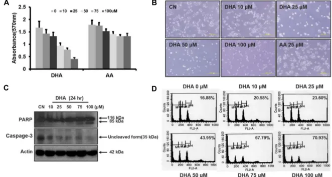
Fig. 1. DHA induces apoptosis in HT29 cells. A. HT29 were incubated with different concentrations of DHA, and AA for 24 hr, and cell growth was determined using the MTT assay as described in Materials and Methods. B. Representative images of HT29 cells exposed to DHA for 24 hr (scale bar, 200 μm). C. Levels of cleaved PARP and uncleaved caspase-3 in response to DHA. HT29 cells were incubated with 0-100 μM DHA for 24 hr, and cleaved PARP and uncleaved caspase-3 were monitored by Western blot analysis. D. Cell cycle analysis of HT29 cells exposed to DHA. HT29 cells were incubated with 0-100 μM DHA for 24 hr and then determined to flow cytometry after staining with PI.
1시간 동안 반응시켰다. 이 후 TBS/T로 3회 세척하고 2차 an- tibody (TRITC-conjugated rabbit secondary antibody)와 반 응시킨 후 hematoxylin-eosin으로 counterstaining하여 wild type과 Fat-1 mice의 종양조직의 시료에서 CD31의 염색강도 차이를 confocal microscopy로 검경하였다.
통계분석
표시된 결과는 3번 이상의 독립적인 실험 결과이며, 실험 결과의 통계 처리는 student's t-test에 준하여 처리하였고,
p-value가 최대치 0.05(p<0.05) 이하인 경우를 유의한 것으로판정하였다.
결과 및 고찰
인체 대장암 세포의 증식에 미치는 오메가-3 지방산의 영향 오메가-3 지방산은 유방암을 비롯한 수종의 암세포주에 대 해 세포독성을 나타낸다[16, 19]고 알려져 있다. 이에 대장암 세포주 HT29에 대한 오메가-3의 세포 독성을 오메가-6와 비교 하였다. 이때 오메가-3로는 DHA를, 오메가-6로는 AA를 사용 하였다. 96 well tissue plate에 HT29세포를 0.5~1×10
4cells/
well 로 배양한 다음 무혈청 배지로 24시간 배양하고 0~100 μM의 DHA를 처리 하고 24시간 후 MTT로 세포 증식능을 검색하였다. DHA는 HT29세포의 증식을 농도 의존적으로 억
제하였으나 AA는 거의 영향이 없었다(Fig. 1A). HT29에 DHA 처리 후 세포의 모양을 inverted microscope으로 관찰 했을 때도 세포의 모양이 변형되고 성장이 억제 되었다(Fig. 1B).
이는 HT29 세포에 대해 오메가-3인 DHA가 세포 독성이 있으 며 이 독성은 오메가-3 특이 작용임을 시사한다.
오메가-3는 수종암세포의 apoptosis를 유도한다[9, 24, 25]
고 알려져 왔으며 최근 Yun 등[46]은 유방암 세포에서 DHA 및 EPA가 apoptosis를 유도한다고 하였다. 이에 본 연구에서 DHA가 대장암세포의 apoptosis를 유도하는지 확인하기 위해 먼저 HT29세포에 DHA를 0~100 μM로 처리하여 PARP, cas- pase-3등의 cleavage를 Western blot으로 검색하였다. DHA의 농도의존적으로 cleaved PARP (85 kDa)의 단백질 양이 증가 하였고, caspase-3의 uncleaved form (35 kDa)이 점차 감소하 였다(Fig. 1C). 따라서 DHA에 의해 HT29 세포의 apoptosis가 유도됨을 알 수 있었다.
상기 실험에서 DHA에 의해 세포증식이 억제되고, apop- totic marker인 PARP, caspase-3의 cleavage도 증가 되었음으 로 apoptosis를 확증하기 위해 FACS analysis 를 통해 sub G
1population을 측정하였다. 즉 DHA에 의한 대장암 세포의 세
포주기에 미치는 영향을 알아보기 위해 HT29 세포를 FBS가
포함되지 않은 배지로 24시간 배양한 다음 DHA를 0~100 μM
로 24시간 처리하여 70% ethanol로 고정 후 PI로 염색하고
FACS analysis를 통해 sub G
1population, cell cycle arrest
A B
Fig. 2. Effect of DHA on invasiveness of HT29 cells. A, matrigel drop assays. HT29 cells (1×10⁶ cells/ml) were mixed with the solution of matrigel (1:1) with absence or presence of DHA, and then 20 μl droplet containg 104cells was applied to the surface of a 6-well culture plate. The droplet incubated at 37℃ for 2 hr and filled the each well with RPMI containing 2% FBS. After 48 hr, the migrated cells out of the droplet were counted under phase-contrast microscopy. Upper panel, Microphotographs of filters of matrigel invasion assay are shown. Lower panel, numbers of invasive cells (mean ± SD) counted in ten random fields. CN, control; AA, arachidonic acid; bars, SD; ***, p<0.001 compared with control. B, transwell chamber assay. The cells were loaded onto matrigel-coated upper chamber transwell as described in Materials and Methods.
The cells were treated with DHA for 48 hr and the filtrated cells were stained. Upper panel, microphotographs of filters of matrigel chamber invasion assay. Lower panel, numbers of invasive cells (mean ± SD) counted in ten random fields.
CN, control; bars, SD; ***, p<0.001 compared with control.
등 세포주기 변동을 분석하였는데, DHA 처리 후 subG
1pop- ulation이 농도에 의존적으로 현저히 증가하였다(Fig. 1D). 이 는 DHA에 의해 HT29 세포의 증식억제에 apoptosis에 의한 세포사멸이 관련되어 있으며 이는 오메가-3의 특이 작용임을 강력히 시사하는 것이다.
HT29 세포의 침윤능에 미치는 DHA의 영향
Yun 등[46]은 오메가-3가 유방암세포의 침윤능을 억제한다 고 하였다. 이에 DHA의 대장암세포에 미치는 영향이 세포독 성 유도 외에 세포 침윤능 억제작용도 있는가를 밝히기 위해 HT29세포의 침윤능을 검색하였는데 이 때 세포증식에 거의 영향을 미치지 않는 농도인 10, 25 μM의 DHA를 사용하였다.
먼저 Matrigel drop assay로 HT29 세포의 matrigel을 통한 침 윤능을 시행 하였을 때 DHA에 의한 matrigel을 통한 HT29 세포의 이동은 농도의존적으로 감소하였으나 오메가-6인 AA 에 의해서는 거의 영향이 없었다(Fig. 2A). 또한 HT29세포를 transwell chamber에 5x10
5cells/well로 조절하고 10, 25 μM 의 DHA로 처리한 다음 24~48시간 후에 filter를 통해 이동한 세포를 측정하여 침윤능을 검색하였을 때도 DHA에 의한 HT29 (Fig. 2B)의 침윤능은 억제되었다. 이는 DHA가 대장암
세포의 침윤을 억제할 수 있음을 시사한다.
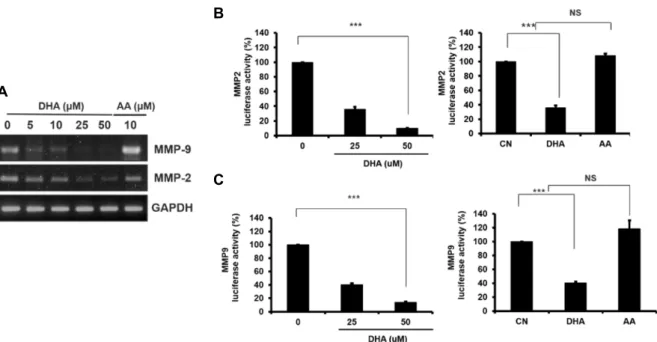
암세포의 침윤에는 MMP-9 등 metalloproteinase들의 활성 이 중요하며[12, 29, 30, 38] 최근 Yun 등[46]은 유방암 세포에 서 DHA에 의해 MMP-9 및 MMP-2 유전자 발현이 억제된다 고 하였다. 이에 대장암 HT29 세포에서 DHA에 의한 MMP-9 및 MMP-2 발현을 검색하기 위하여 DHA 처리 후 total RNA 를 조제하여 RT-PCR을 시행한 바 농도의존적으로 MMP-9 및 MMP-2 mRNA양이 감소하였으나 AA에 의해서는 영향이 없 었다(Fig. 3A). 이는 HT29 세포에서 DHA에 의한 MMP-9 및 MMP-2 유전자 발현의 억제가 오메가-3의 특이 작용임을 시사 하는 것이다.
상기 실험에서 DHA에 의해 MMP-2 및 MMP-9 mRNA양
이 각각 감소하였는데 이들의 억제가 전사 단계에서 조절되는
지 확인하기 위해 DHA 처리한 후 MMP-2 및 MMP-9의 pro-
moter 활성을 측정하였다. 6-well plate에 HT29를 배양하여
MMP-2 및 MMP-9 promoter 유전자가 포함된 reporter plas-
mid를 transient transfection 후 그 활성에 대한 DHA의 영향
을 lucifersae 활성을 측정함으로 검색하였다. 먼저 MMP-2
promoter의 luciferase 활성은 HT29 세포에서 DHA 처리 후
억제되었으며 이는 농도의존적으로 나타났다. 그러나 AA는
A
B
C
Fig. 3. DHA reduces in vitro cell invasion by modulating MMP-2 and MMP-9 expression. A. HT29 cells were treated with increased dose of DHA (0-50 μM) for 24 hr, and the levels of MMP-2 and MMP-9 mRNA were analysed by RT-PCR as described in Materials and Methods. B and C. HT29 cells were plated and MMP-2 (B) or MMP-9 (C) luciferase reporter gene was transfected by using lipofectamin reagent as described in Materials and Methods. After the transfection performed, the cells were treated with DHA for 24 hr, and then the luciferase activity was measured using the dual luciferase assay system (Promega) in luminometer (Thermo). CN, control; columns, mean of four independent experiments; bars, SD; ***, p<0.001 compared with control.
A B
Fig. 4. DHA suppresses COX-2 and VEGF promotor activity. HT29 cells were transfected with luciferase reporter containing COX-2 (A) and VEGF (B) promoter and treated with DHA for 24 hr. Then, the cells were lysed and the luciferase activity was measured with the dual luciferase assay. ***, p<0.001 compared with control, Student’s t test, significantly different from the control.
MMP-2 활성에 영향을 미치지 않았다(Fig. 3B). 또한 MMP-9 promoter 활성에 대한 DHA의 영향을 luficerase 활성을 측정 함으로 검색한 바 DHA 처리 후 MMP-9 promoter 활성이 억 제되었고 이는 DHA 농도의존적으로 나타났으나, AA는 MMP-9 활성에는 영향을 미치지 않았다(Fig. 3C). 이는 DHA 는 MMP-2 및 MMP-9 전사를 억제할 수 있으며, DHA에 의한 MMP-2 및 MMP-9 전사 조절은 오메가-3의 특이 작용임을 시 사하는 것이다.
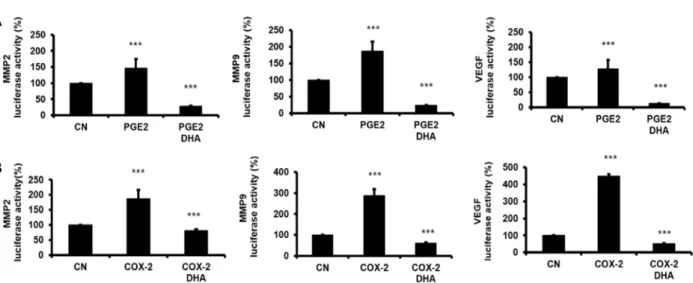
대장암세포에서 COX-2에 의해 증가된 MMP-2 및 MMP-9 활성에 대한 DHA의 영향
COX-2 발현은 많은 암에서 증가 되어 있고 AA로부터
COX-2에 의해 산생된 PGE2는 종양형성 뿐만 아니라 암세포
의 침윤에도 중요하다고 하며[16, 19], 또한 COX-2/PGE2는
혈관신생에 중요한 인자인 VEGF를 조절할 수 있다고 알려져
있다[41]. Lim 등[25]은 오메가-3가 간담도암 세포에서 COX-2
promoter활성을 억제한다고 하였으며 Yun 등[46]은 유방암
세포에서 오메가-3가 VEGF promotor 활성을 억제한다고 하
였다. 이에 먼저 대장암 HT29 세포에서 오메가-3가 COX-2
및 VEGF의 전사를 억제할 수 있는지 확인하기 위해 COX-2
및 VEGF promotor 활성에 대한 DHA의 영향을 검색한바
COX-2 및 VEGF promotor 활성이 각각 억제 되었다(Fig. 4).
A
B
Fig. 5. Effect of DHA on COX-2/PGE2-induced MMP-2, MMP-9 and VEGF promoter activity in HT29 cells. MMP-2, MMP-9 and VEGF luciferase reporter gene were transfected in HT29 cells and incubated. After 18 hr, transfected cells were pretreated with DHA (25 μM) for 2 hr, and then PGE2 (1 μM) were added. After 12 hr, the luciferase activity was measured using the dual luciferase assay system (Promega) in luminometer (Thermo). In case of COX-2 overexpression experiment, COX-2 plasmid was cotransfected with MMP-2, MMP-9 and VEGF luciferase reporter gene. After 18 hr, the luciferase activities were also measured. A, PGE2 effect; B, COX-2 overexpression effect. CN, control; columns, mean of four independent experi- ments; bars, SD; ***, p<0.001 compared with control
이는 오메가-3인 DHA가 COX-2 및 VEGF의 전사를 억제할 수 있음을 시사한다.
Pavlovic 등[31]은 macrophage에서 PGE
2에 의해 MMP-9의 활성이 증가된다고 하였고, Yun 등[46]은 유방암 세포에서 AA 및 COX-2/PGE2에 의해 MMP-2 및 MMP-9 promoter 활 성이 증가하며 이는 DHA에 의해 억제된다고 하였다. 이에 먼저 대장암 HT29 세포에서 transient transfection한 MMP-9, MMP-2 및 VEGF promoter 활성에 대한 PGE
2의 영향을 검색 한 바 MMP-9 promoter의 luciferase 활성이 PGE
2에 의해 증 가하였으며, 이는 DHA 전 처리에 의해 감소하였다(Fig. 5A).
Larkin 등[23]은 COX-2의 억제가 유방암세포의 침윤 및 MMPs의 발현을 억제한다고 하였다. 상기 실험에서도 PGE
2가 MMP-9 및 MMP-2 promoter 활성조절에 밀접히 관련되어 있 음으로 AA로부터 COX-2에 의해 합성된 PGE
2가 그 조절에 관련되어 있는지를 확인하기 위해 COX-2 유전자를 과발현 시킨 후 MMP-2, MMP-9 및 VEGF promoter의 reporter 활성 의 변동 및 DHA에 의한 조절을 검색하였다. HT29세포에서 COX-2유전자를 과발현 시켰을 때 MMP-9, MMP-2, 및 VEGF promoter의 활성이 증가 하였으며 이는 DHA 전처리 후 억제 되었다(Fig. 5B). 이는 DHA가 COX-2 억제에 따른 PGE
2신호 전달경로를 억제하여 MMP-9, MMP-2 및 VEGF promoter의 전사를 조절하였으리라 생각되며 따라서 침윤 및 혈관신생을 억제할 수 있음을 시사한다.
HT29 세포에서 NF-kB 활성에 대한 DHA의 영향 NF-kB는 전사인자로써 염증반응에 관련되어 있는 IL-1β,
TNF-α등 염증분자들의 조절에 관련되어 있을 뿐만 아니라[42, 47] 과발현시 암세포의 apoptosis를 억제하여 암세포의 증식 을 유도한다고 알려져 있으며[4], 최근에 Yun 등은 오메가-3인 DHA가 NF-kB활성을 감소시킨다[46]고 보고 하였다. 또한 MMP-9 [2, 11], VEGF [40], COX-2 [10] 등의 유전자 promoter 에는 공통적으로 NF-kB가 결합할 수 있는 cis element가 존재 한다고 한다. 이에 DHA에 의한 NF-kB 활성 변동을 검색하기 위해 먼저 NF-kB binding element를 가진 reporter plasmid를 transient transfection 후 DHA의 영향을 검색한 바 DHA 처리 후 NF-kB reporter 활성은 감소하였으며 이는 농도의존적으 로 나타났다(Fig. 6A). NF-kB report활성이 감소 하였음으로 핵속에서 NF-kB 단백질 양의 감소를 확인하기 위해 HT29 세 포를 DHA 처리 후 NF-kB subunit인 p65에 대한 면역 형광염 색을 시행하였다. HT29 세포를 10 μM DHA로 24시간 처리 후 control과 비교했을 때 NF-kB단백은 핵속에서 거의 검출되 지 않았으나 오메가-6인 AA처리 후에는 control과 큰 차이를 보이지 않았다(Fig. 6B). 이는 대장암 세포에서 DHA가 NF-kB 을 억제함으로 MMP-9, MMP-2 및 VEGF 전사를 억제하여 침윤 및 혈관신생을 억제할 수 있음을 시사한다.
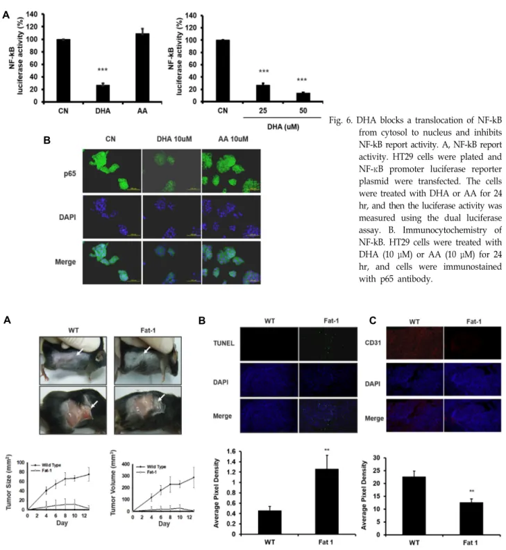
Fat-1 mice에서 생쥐 대장암 세포주 MCA-38의 종양 형 성능의 억제
오메가-3는 nude mouse에서 수종암세포의 종양형성능을
억제한다고[3, 8] 알려져 있으며, Xia 등[43]은 C. elegans의
Fat-1 유전자(ω3-desaturase)를 주입한 transgenic mice인 Fat-
1 mice에서 melanoma의 종양 형성능이 억제된다고 하였다.
A
B
Fig. 6. DHA blocks a translocation of NF-kB from cytosol to nucleus and inhibits NF-kB report activity. A, NF-kB report activity. HT29 cells were plated and NF-κB promoter luciferase reporter plasmid were transfected. The cells were treated with DHA or AA for 24 hr, and then the luciferase activity was measured using the dual luciferase assay. B. Immunocytochemistry of NF-kB. HT29 cells were treated with DHA (10 μM) or AA (10 μM) for 24 hr, and cells were immunostained with p65 antibody.
A B C
Fig. 7. ω3-PUFA-rich environment retards tumor growth in vitro. Fat-1 and C57BL/6 wild type mice were implanted with murine colon adenocarcinoma cell line MCA-38 cells. After inoculation, the animals were closely monitored for the development of subcutaneous tumor. Day of the implantation of the tumor cells was designated day 0. A. Cultured MCA38 cells were washed with HBSS and 3×10⁶ cells were injected s.c. into the right flank in each mouse. The tumor size was measured at indicated time with a caliper. The tumor’s greatest dimension and the one perpendicular to it were measured using calipers and calculated as length × width = tumor size and 0.5× (width)2 × length = tumor volume . WT, wild type. B. The animals were sacrificed at two weeks after implanted, and tumor tissues were fixed with formalin and TUNEL stain was performed.
The apoptotic index of tumor cells increased in Fat-1 mice compared with wild type mice (apoptotic cells appear fluorescent green. **, p<0.01 compared with WT; original magnification ×100) C. The xenograft tissue in tumor tissues were fixed and stained with endothelial marker CD31 antibody. The intensity of CD31 decreased in Fat-1 mice compared with wild type mice (Intensity of CD31 appears fluorescent red). **, p<0.01 compared with WT; original magnification x100).
Fig. 8. Proposed mechanism of ω3-PUFA-induced cell death in colon cancer cells. ω3-PUFAs may inhibit tumorigenicity and angio- genesis as well as cancer cell invasion by suppression of MMPs and VEGF via reduction of NF-kB in colon cancer.
이에 Fat-1 mice에서 대장암세포의 종양 형성능을 검색하기 위해 Fat-1 mice (n=5)와 wild type mice (n=5)에 C57BL6에서 유래된 대장암 MCA38세포 3x10
6cells/100 μl를 오른편 옆구 리에 피하 주사하여 4일 후부터 2일마다 종양의 크기를 측정 하였다. Fig. 7A에서 보는 것처럼 Fat-1 mice에서의 종양의 크 기 및 용적은 wild type mice에 비해 현저히 감소하였다. 이는 Fat-1 mice가 오메가-3가 wild type에 비해 높은 점을 감안할 때, 오메가-3가 대장암의 종양형성능을 억제할 수 있음을 시사 한다.
상기 실험에서 Fat-1 mice에서 MCA38 대장암 세포의 종양 형성능이 감소되었는 바 이의 기전을 밝히기 위해 형성된 종 양조직에서 TUNEL assay를 시행하여 apoptotic cells를 검색 하였다. Fat-1 mice에서는 wild type mice에 비해 apoptotic index가 현저히 증가 하였는데(p<0.01), 이는 Fat-1 mice의 종 양조직에서 apoptosis가 증가함에 따라 그 종양 형성능이 감 소한 것이라 생각된다(Fig. 7B).
혈관신생은 종양형성능과 밀접히 관련 되어 있으며, 혈관신 생에는 VEGF 등 혈관신생인자의 발현이 중요하다고 알려져 있다 Yun 등[46]은 Fat-1 mice에서 VEGF에 의한 혈관신생이 억제된다고 하였다. 상기 실험에서 DHA에 의해 VEGF pro- moter 활성이 억제 되었음으로, 이에 Fat-1 mice의 종양조직에 서 혈관신생의 억제 여부를 검색하기 위해 혈관 내피세포에 존재하는 혈관의 표식자인 CD31의 조직 형광면역염색을 시 행하였다. Fat-1 mice의 종양조직에서 CD31 intensity는 wild type mice에 비해 현저히 감소하였다(p<0.01). 이는 Fat-1 transgenic mice의 종양 형성능의 억제에 오메가-3지방산에 의한 혈관신생의 억제도 관련되어 있음을 시사한다(Fig. 7C).
이상의 결과로 오메가-3는 대장암 세포에서NF-kB 억제에 따른 COX-2와 MMP-2 및 MMP-9 등 matrix metallopro- teinase의 억제를 통한 침윤능의 억제, VEGF 억제를 통한 혈 관신생의 억제등 복합적 기전에 의해 항암작용을 나타내리라 생각되며(Fig. 8), 따라서 오메가-3의 복용은 대장암의 예방 및 치료 목적으로 유용하게 사용할 수 있으리라 시사된다.
감사의 글
본 연구는 2015년 충남대학교 학술연구비에 의해 지원 되었 음. 본 연구에 사용한 Fat-1 transgeic mice를 분양해 주신 Harvard University의 Jing X. Kang 박사에게 감사드립니다.
References
1. Berquin, I. M., Min, Y., Wu, R., Wu, J., Perry, D., Cline, J. M., Thomas, M. J., Thornburg, T., Kulik, G., Smith, A., Edwards, I. J., D'Agostino, R., Zhang, H., Wu, H., Kang, J. X. and Chen, Y. Q. 2007. Modulation of prostate cancer genetic risk by omega-3 and omega-6 fatty acids. J. Clin.
Invest. 117, 1866-1875.
2. Bond, M., Chase, A. J., Baker, A. H. and Newby, A. C. 2001.
Inhibition of transcription factor NF-kappaB reduces matrix metalloproteinase-1, -3 and -9 production by vascular smooth muscle cells. Cardiovasc. Res. 50, 556-565.
3. Boudreau, M. D., Sohn, K. H., Rhee, S. H., Lee, S. W., Hunt, J. D. and Hwang, D. H. 2001. Suppression of tumor cell growth both in nude mice and in culture by n-3 poly- unsaturated fatty acids: mediation through cyclooxygenase- independent pathways. Cancer Res. 61, 1386-1391.
4. Bours, V., Bentires-Alj, M., Hellin, A. C., Viatour, P., Robe, P., Delhalle, S., Benoit, V. and Merville, M. P. 2000. Nuclear factor-kappa B, cancer, and apoptosis. Biochem. Pharmacol.
60, 1085-1089.
5. Bresalier, R. S., Boland, C. R. and Kim, Y. S. 1984. Character- istics of colorectal carcinoma cells with high metastatic potential. Gastroenterology 87, 115-122.
6. Brown, M. D., Hart, C. A., Gazi, E., Bagley, S. and Clarke, N. W. 2006. Promotion of prostatic metastatic migration to- wards human bone marrow stoma by Omega 6 and its in- hibition by Omega 3 PUFAs. Br. J. Cancer 94, 842-853.
7. Byers, T. 1996. Nutrition and cancer among American Indians and Alaska Natives. Cancer 78, 1612-1616.
8. Calviello, G., Di Nicuolo, F., Gragnoli, S., Piccioni, E., Serini, S., Maggiano, N., Tringali, G., Navarra, P., Ranelletti, F. O.
and Palozza, P. 2004. n-3 PUFAs reduce VEGF expression in human colon cancer cells modulating the COX-2/PGE2
induced ERK-1 and -2 and HIF-1alpha induction pathway.
Carcinogenesis 25, 2303-2310.
9. Calviello, G., Serini, S. and Piccioni, E. 2007. n-3 poly- unsaturated fatty acids and the prevention of colorectal can- cer: molecular mechanisms involved. Curr. Med. Chem. 14, 3059-3069.
10. Charalambous, M. P., Lightfoot, T., Speirs, V., Horgan, K.
and Gooderham, N. J. 2009. Expression of COX-2, NF-kappaB-p65, NF-kappaB-p50 and IKKalpha in malig- nant and adjacent normal human colorectal tissue. Br. J.
Cancer. 101, 106-115.
11. Eberhardt, W., Huwiler, A., Beck, K. F., Walpen, S. and Pfeilschifter, J. 2000. Amplification of IL-1 beta-induced ma- trix metalloproteinase-9 expression by superoxide in rat glo- merular mesangial cells is mediated by increased activities of NF-kappa B and activating protein-1 and involves activa- tion of the mitogen-activated protein kinase pathways. J.
Immunol. 165, 5788-5797.
12. Eeckhout, Y. and Vaes, G. 1977. Further studies on the acti- vation of procollagenase, the latent precursor of bone collagenase. Effects of lysosomal cathepsin B, plasmin and kallikrein, and spontaneous activation. Biochem. J. 166, 21-31.
13. Gago-Dominguez, M., Yuan, J. M., Sun, C. L., Lee, H. P.
and Yu, M. C. 2003. Opposing effects of dietary n-3 and n-6 fatty acids on mammary carcinogenesis: The Singapore Chinese Health Study. Br. J. Cancer 89, 1686-1692.
14. Hardman, W. E. 2004. (n-3) fatty acids and cancer therapy.
J. Nutr. 134, 3427S-3430S.
15. Jeong, S., Jing, K., Kim, N., Shin, S., Kim, S., Song, K. S., Heo, J. Y., Park, J. H., Seo, K. S., Han, J., Wu, T., Kweon, G. R., Park, S. K., Park, J. I. and Lim, K. 2014. Docosahexae- noic acid-induced apoptosis is mediated by activation of mi- togen-activated protein kinases in human cancer cells. BMC Cancer 14, 481.
16. Jing, K. and Lim, K. 2013. Chapter 15. Potent Anticancer Actions of Omega-3 Polyunsaturated Fatty acids of Marine Nutraceuticals, In: Kim SK, ed. Marine Nutrceuticals–
Prospects and Perspectives-. CRC Press/Taylor Francis Group LLC. 199-232.
17. Jing, K., Shin, S., Jeong, S., Kim, S., Song, K. S., Park, J.
H., Heo, J. Y., Seo, K. S., Park, S. K., Kweon, G. R., Wu, T., Park, J. I. and Lim, K. 2014. Docosahexaenoic acid in- duces the degradation of HPV E6/E7 oncoproteins by acti- vating the ubiquitin-proteasome system. Cell Death Dis. 5, e1524.
18. Jing, K., Song, K. S., Shin, S., Kim, N., Jeong, S., Oh, H.
R., Park, J. H., Seo, K. S., Heo, J. Y., Han, J., Park, J. I., Han, C., Wu, T., Kweon, G. R., Park, S. K., Yoon, W. H., Hwang, B. D. and Lim, K. 2011. Docosahexaenoic acid in- duces autophagy through p53/AMPK/mTOR signaling and promotes apoptosis in human cancer cells harboring wild-type p53. Autophagy 7, 1348-1358.
19. Jing, K., Wu, T. and Lim, K. 2013. Omega-3 polyunsaturated fatty acids and cancer. Anticancer Agents Med. Chem. 13, 1162-1177.
20. Kang, J. X., Wang, J., Wu, L. and Kang, Z. B. 2004. Transgen-
ic mice: fat-1 mice convert n-6 to n-3 fatty acids. Nature 427, 504.
21. Kobayashi, N., Barnard, R. J., Henning, S. M., Elashoff, D., Reddy, S. T., Cohen, P., Leung, P., Hong-Gonzalez, J., Freedland, S. J., Said, J., Gui, D., Seeram, N. P., Popoviciu, L. M., Bagga, D., Heber, D., Glaspy, J. A. and Aronson, W.
J. 2006. Effect of altering dietary omega-6/omega-3 fatty acid ratios on prostate cancer membrane composition, cyclo- oxygenase-2, and prostaglandin E2. Clin. Cancer Res. 12, 4662-4670.
22. Kolonel, L. N., Altshuler, D. and Henderson, B. E. 2004. The multiethnic cohort study: exploring genes, lifestyle and can- cer risk. Nat. Rev. Cancer 4, 519-527.
23. Larkins, T. L., Nowell, M., Singh, S. and Sanford, G. L. 2006.
Inhibition of cyclooxygenase-2 decreases breast cancer cell motility, invasion and matrix metalloproteinase expression.
BMC Cancer 6, 181.
24. Lim, K., Han, C., Dai, Y., Shen, M. and Wu, T. 2009.
Omega-3 polyunsaturated fatty acids inhibit hepatocellular carcinoma cell growth through blocking beta-catenin and cyclooxygenase-2. Mol. Cancer Ther. 8, 3046-3055.
25. Lim, K., Han, C., Xu, L., Isse, K., Demetris, A. J. and Wu, T. 2008. Cyclooxygenase-2-derived prostaglandin E2 acti- vates beta-catenin in human cholangiocarcinoma cells: evi- dence for inhibition of these signaling pathways by omega 3 polyunsaturated fatty acids. Cancer Res. 68, 553-560.
26. Liotta, L. A. 1984. Tumor invasion and metastases: role of the basement membrane. Warner-Lambert Parke-Davis Award lecture. Am. J. Pathol. 117, 339-348.
27. Liu, G., Bibus, D. M., Bode, A. M., Ma, W. Y., Holman, R.
T. and Dong, Z. 2001. Omega 3 but not omega 6 fatty acids inhibit AP-1 activity and cell transformation in JB6 cells.
Proc. Natl. Acad. Sci. USA 98, 7510-7515.
28. Maillard, V., Bougnoux, P., Ferrari, P., Jourdan, M. L., Pinault, M., Lavillonniere, F., Body, G., Le Floch, O. and Chajes, V. 2002. N-3 and N-6 fatty acids in breast adipose tissue and relative risk of breast cancer in a case-control study in Tours, France. Int. J. Cancer 98, 78-83.
29. Nagase, H. and Woessner, J. F. Jr. 1999. Matrix metal- loproteinases. J. Biol. Chem. 274, 21491-21494.
30. Okada, Y., Gonoji, Y., Naka, K., Tomita, K., Nakanishi, I., Iwata, K., Yamashita, K. and Hayakawa, T. 1992. Matrix metalloproteinase 9 (92-kDa gelatinase/type IV collagenase) from HT 1080 human fibrosarcoma cells. Purification and activation of the precursor and enzymic properties. J. Biol.
Chem. 267, 21712-21719.
31. Pavlovic, S., Du, B., Sakamoto, K., Khan, K. M., Natarajan, C., Breyer, R. M., Dannenberg, A. J. and Falcone, D. J. 2006.
Targeting prostaglandin E2 receptors as an alternative strat- egy to block cyclooxygenase-2-dependent extracellular ma- trix-induced matrix metalloproteinase-9 expression by macrophages. J. Biol. Chem. 281, 3321-3328.
32. Rose, D. P. and Connolly, J. M. 2000. Regulation of tumor angiogenesis by dietary fatty acids and eicosanoids. Nutr.
Cancer 37, 119-127.
33. Roynette, C. E., Calder, P. C., Dupertuis, Y. M. and Pichard,
초록:오메가-3 지방산에 의한 COX-2/MMPs/VEGF 억제에 따른 대장암세포의 종양 형성 및 침윤 억제
신소연
1,3†․김용조
1†․한승현
1,2․프라산타
1,2․허준영
1,2․전영주
1,2․박승길
1․권기량
1,2․박종일
1․임규
1,2,3*
(1충남대학교 의과대학 생화학교실, 2충남대학교 의과학과, 3충남대학교 암연구소)
대장암은 미국 등 서양 국가뿐만 아니라 국내에서도 2번째로 많이 발병이 되는 암으로 알려져 있다. 역학조사에 의하면 오메가-3를 많이 섭취한 인종에서 대장암 발생빈도가 감소하고 최근 오메가-3는 수종의 암에 대해 항암작용 을 나타낸다고 한다. 이에 본 연구에서는 대장암에서 DHA의 항침윤, 항혈관 신생 및 항종양 형성능 억제의 기전을 규명하여 다음과 같은 결과를 얻었다. DHA는 인체 대장암 세포주 HT29 의 증식을 농도 의존적으로 억제하였으나 AA는 거의 영향이 없었다. FACS 분석에서 DHA 처리했을 때 Sub G
1phase의 세포가 DHA의 농도 의존적으로 증가 하였다. DHA 처리 후 cleaved PARP가 증가하고, uncelaved caspase-3가 감소 하였다. HT29 세포의 침윤능은 DHA 처리에 의해 억제 되었다. DHA 처리 후 MMP-9 및 MMP-2 mRNA양이 감소 되었을 뿐만 아니라 그 pro- moter의 reporter 활성도 억제하였으며 VEGF promoter 활성도 DHA에 의해 억제 되었다. NF-kB promoter 활성 및 핵으로의 이동도 DHA에 의해 억제 되었다. In vivo 동물실험에서 생쥐 대장암 세포주인 MCA38에 대한 Fat-1 transgenic mice에서의 종양 형성능은 현저히 억제 되었다. 면역형광염색법을 이용한 Fat-1 transgenic mice의 종양 조직에서의 TUNEL 양성세포는 wild type mice에 비해 현저히 증가하였으나 CD31의 형광강도는 감소 되었다. 이 상의 결과로 오메가-3는 대장암 세포에서 NF-kB 억제에 따른 COX-2, MMP-2 및 MMP-9 등 matrix mata- lloproteinase의 억제를 통한 침윤능의 억제, VEGF 억제를 통한 혈관신생의 억제등 복합적 기전에 의해 항암작용을 나타내리라 생각되며, 따라서 오메가-3는 대장암의 예방 및 치료에 유용하게 사용될 수 있으리라 생각된다.
C. 2004. n-3 polyunsaturated fatty acids and colon cancer prevention. Clin. Nutr. 23, 139-151.
34. Schwartz, S. A., Hernandez, A. and Mark Evers, B. 1999.
The role of NF-kappaB/IkappaB proteins in cancer: im- plications for novel treatment strategies. Surg. Oncol. 8, 143-153.
35. Siegel, R. L., Miller, K. D. and Jemal, A. 2017. Cancer Statistics, 2017. CA. Cancer J. Clin. 67, 7-30.
36. Simopoulos, A. P. 2002. The importance of the ratio of ome- ga-6/omega-3 essential fatty acids. Biomed. Pharmacother. 56, 365-379.
37. Song, K. S., Jing, K., Kim, J. S., Yun, E. J., Shin, S., Seo, K. S., Park, J. H., Heo, J. Y., Kang, J. X., Suh, K. S., Wu, T., Park, J. I., Kweon, G. R., Yoon, W. H., Hwang, B. D.
and Lim, K. 2011. Omega-3-polyunsaturated fatty acids sup- press pancreatic cancer cell growth in vitro and in vivo via downregulation of Wnt/Beta-catenin signaling. Pancreatol- ogy 11, 574-584.
38. Suzuki, K., Enghild, J. J., Morodomi, T., Salvesen, G. and Nagase, H. 1990. Mechanisms of activation of tissue pro- collagenase by matrix metalloproteinase 3 (stromelysin).
Biochemistry 29, 10261-10270.
39. Szymczak, M., Murray, M. and Petrovic, N. 2008. Modula- tion of angiogenesis by omega-3 polyunsaturated fatty acids is mediated by cyclooxygenases. Blood 111, 3514-3521.
40. Tong, Q., Zheng, L., Lin, L., Li, B., Wang, D., Huang, C., Matuschak, G. M. and Li, D. 2006. Participation of the PI-3K/Akt-NF-kappa B signaling pathways in hypoxia-in- duced mitogenic factor-stimulated Flk-1 expression in endo- thelial cells. Respir. Res. 7, 101.
41. Toomey, D. P., Murphy, J. F. and Conlon, K. C. 2009. COX-2, VEGF and tumour angiogenesis. Surgeon 7, 174-180.
42. Weldon, S. M., Mullen, A. C., Loscher, C. E., Hurley, L. A., and Roche, H. M. 2007. Docosahexaenoic acid induces an anti-inflammatory profile in lipopolysaccharide-stimulated human THP-1 macrophages more effectively than eicosa- pentaenoic acid. J. Nutr. Biochem. 18, 250-258.
43. Xia, S., Lu, Y., Wang, J., He, C., Hong, S., Serhan, C. N.
and Kang, J. X. 2006. Melanoma growth is reduced in fat-1 transgenic mice: impact of omega-6/omega-3 essential fatty acids. Proc. Natl. Acad. Sci. USA 103, 12499-12504.
44. Xia, S. H., Wang, J. and Kang, J. X. 2005. Decreased n-6/n-3 fatty acid ratio reduces the invasive potential of human lung cancer cells by downregulation of cell adhesion/invasion- related genes. Carcinogenesis 26, 779-784.
45. Yoon, W. H., Jung, Y. J., Kim, T. D., Li, G., Park, B. J., Kim, J. Y., Lee, Y. C., Kim, J. M., Park, J. I., Park, H. D., No, Z. S., Lim, K., Hwang, B. D. and Kim, Y. S. 2004. Gabexate mesilate inhibits colon cancer growth, invasion, and meta- stasis by reducing matrix metalloproteinases and angio- genesis. Clin. Cancer. Res. 10, 4517-4526.
46. Yun, E. J., Song, K. S., Shin, S., Kim, S., Heo, J. Y., Kweon, G. R., Wu, T., Park, J. I. and Lim, K. 2016. Docosahexaenoic acid suppresses breast cancer cell metastasis by targeting matrix-metalloproteinases. Oncotarget 7, 49961-49971.
47. Zhao, Y., Joshi-Barve, S., Barve, S. and Chen, L. H. 2004.
Eicosapentaenoic acid prevents LPS-induced TNF-alpha ex- pression by preventing NF-kappaB activation. J. Am. Coll.
Nutr. 23, 71-78.