Effect of Acupuncture at ST36 on Ischemia-induced Learning and Memory Deficits in Gerbils
Jin Yong Chung1, Hyun Jung Park1,2, Hyun Soo Shim1, Dae Hyun Hahm1, Hee Young Kim3, Hye Jung Lee1, Kyung Soo Kim2, In Sop Shim1* 1 : Acupuncture and Meridian Science Research Center, Kyung Hee University, 2 : Department of Integrative Medicine, College of Medicine, The Catholic University, 3 : Department of Neuroscience and Cell Biology, University of Texas Medical Branch
The present study was investigated the neuroprotective effects of acupuncture at ST36 on learning and memory deficits after transient cerebral ischemia in a gerbil model. The animals were randomly divided into three groups (n=7 in each group): the sham operation group (SHAM), ischemia-induced and ST36 acupuncture group (ISC + ST36), and the ischemia-induced and Tail-acupuncture group (ISC + TAIL). For the acupuncture stimulation, stainless steel needles, 0.3 mm in diameter, were inserted bilaterally into the ST36 locus or the tail and stimulated for 1 min/day for 14 days. Using the Morris water maze test, the animals were tested on spatial learning and memory. In addition, the effects of acupuncture on memory storage and the choline acetyltransferase (ChAT) activity, in the hippocampal CA1 area, were investigated by ChAT immunohistochemistry. Transient cerebral ischemia produced impaired performance on the MWM test (DAY 5: p<0.01 and retention test: p<0.05) and severely decreased ChAT immunoreactivity in the CA1 hippocampal area compared to the SHAM group (p<0.05). However, improved learning and memory were observed (DAY 5: p<0.05 and retention test: p<0.01) as well as a significantly reduced loss of ChAT immunoactivity in the hippocampal CA1 region (p<0.001) after acupuncture stimulation at ST36 were observed. These results show that acupuncture at ST36 ameliorated the learning and memory deficits at least in part through the cholinergic system.
The findings of this study provide potential data that acupuncture is useful for the treatment of some of the behavioral impairs of transient cerebral ischemia.
Key words : acupuncture, cerebral ischemia, choline acetyltransferase (ChAT), Morris water maze (MWM), ST36
* To whom correspondence should be addressed at : Insop Shim, Acupuncture and Meridian Science Research Center, Kyung Hee University, 1 Hoegidong, Dongdaemengu, Seoul 130-701, Korea
․E-mail : [email protected], ․Tel : 02-961-0975
․Received : 2011/03/08 ․Revised : 2011/03/30 ․Accepted : 2011/04/05
Introduction
Transient ischemic events of only a few minutes duration result in the death of certain vulnerable populations of brain cells1,2). During cerebral ischemia, abrupt loss of neurological function occurs, which, if serious, leads to death. This neurological dysfunction is naturally reflected in the deterioration of behavioral performance and memory function.
In fact, cerebral ischemia induced by an injection of microspheres results in poor retention of the active avoidance response in gerbils3). Pulsinelli and Brierley4) introduced a method for inducing cerebral ischemia in unanaesthetized rats by temporarily occluding the common carotid arteries and
permanently interrupting the vertebral arteries. Although this method requires a delicate and time-consuming operation, at present it is the most widely used animal model for learning and memory impairment5-7). Also, many anmal studies transient ischemic event increased loss of cholinergic neurons in the hippocampus and memory related brain regions8,9).
Acupuncture has been also used for the enhancement of functional recovery from various disorders. It has been shown to be effective for analgesia, promotion of homeostasis, and changes in the microcirculatory network as well as improvements in brain circulation7,10,11). Acupuncture treatment has been particularly effective for symptomatic improvement of cerebrovascular accidents including ischemia10-14). Although many studies have indicated that acupuncture provides a neuroprotective effect against ischemic brain damage, the protective mechanisms are not fully understood. ST36 is one of the most versatile and often used acupuncture points; this acupuncture point is associated with strengthening of the
spleen and stomach, regulates the intestines and stabilizes the mind and emotions. Many scientific studies have confirmed the effectiveness of this point in experimental animals15-16).
We carried out the present study with Mongolian gerbils to elucidate the characteristics of cerebral ischemia-induced deficits in learning and memory tasks using the Morris water maze, and determined the changes in ChAT-positive neurons in the hippocampal CA1 region.
Materials and Methods
1. Animal and acupuncture procedures
Adult male Mongolian gerbils (Orient, Inc. Korea, 11-13 weeks of age) were used for the experiments. The animal experiments were carried out in accordance with the Prevention of Cruelty to Animals Act 1986 and NIH guidance for the care and use of laboratory animals for experimental procedures, and were approved by local committee review.
The animals were housed under controlled temperature (20 ± 2℃) and lighting (09.00-21.00) conditions with food and water ad libitum. The animals were randomly divided into three groups (n=7 in each group): the sham operation group (SHAM), ST36 acupuncture group (ISC + ST36), and Tail-acupuncture group (ISC + TAIL). Acupuncture was provided once daily (10.00 a.m.) for 14 consecutive days starting from the second day after surgery in the acupuncture groups. The animals were handled with special care to minimize stress. For acupuncture stimulation, stainless steel needles (Haenglimseowon, Korea), 0.3 mm in diameter, were inserted bilaterally into the ST36 locus or the tail for one minute. The ST36 point was located 1 mm laterally and distally from the anterior tubercle of the tibia. The point on the tail was the skin on the dorsal and posterior aspect of the third coccygeal vertebra.
2. Induction of transient global ischemia in the gerbil The animals were anesthetized with a mixture of 2.5%
isoflurane in 33% oxygen and 67% nitrous oxide. The common carotid arteries, bilaterally, were isolated and occluded using non-traumatic aneurysm clips. The complete interruption of blood flow was confirmed by observation of the central artery in the retinae using an ophthalmoscope. After 5 minutes of occlusion, the aneurysm clips were removed from the common carotid arteries. The body (rectal) temperature under free-regulating or normothermic (37 ± 0.5℃) conditions was monitored with a rectal temperature probe (TR-100; Fine Science Tools, Forster City, CA, USA) and maintained using a thermometric blanker before, during and after the surgery,
until the animals completely recovered from the anesthesia.
Thereafter, the animals were kept on the thermal incubator (Mirae Meidical Industry, Seoul, South Korea) to maintain their body temperature, until they were euthanized. Sham-operated animals were subjected to the same surgical procedures except that the common carotid arteries were not occluded.
3. Morris water maze test (MWM)
On the eighth day of stimulation with acupuncture at ST36, all the animals started training on a MWM task in a swimming pool (1.8 m diameter and 0.5 m high, filled with milky water at a temperature in the 22 ± 2℃ range) for 7 days. A 12 cm diameter round platform was hidden in a constant location (the quadrant center) within the pool with its top surface submerged 1.5 cm below the water level. The mongolian gerbils were trained to locate the hidden island during four trials per day for 6 days. After the 6 days, they were started in the quadrant opposite to the target and were forced to swim for 60 seconds in the pool without a platform.
The spatial memory of the rats was assessed as the latency time. The time spent searching for the platform in the training quadrant, i.e., the previous location of the platform, was recorded and used as a measure of memory retention. A video camera was mounted on the ceiling above the pool and was connected to a video-recorder and tracking device (S-MART;
Pan-Lab, Barcelona, Spain), which permitted on-line and off-line automated tracking of the path taken by the animals.
4. Cholineacetyltransferase (ChAT) Immunohistochemistry At the end of the behavioral observations, the animals were deeply anesthetized with sodium pentobarbital (100 mg/kg, i.p.) and then perfused transcardially with 100 ml of saline followed by 500 ml of a 4% solution of formaldehyde prepared in phosphate buffer. The brains were then removed, post-fixed in the same fixative for two to three hours at 4℃
and then placed overnight at 4℃ in PBS containing 20%
sucrose. The following day, the brain was cut into coronal sections that were sliced to 30 μm-thicknesses. The sections were processed for choline acetyltransferase (ChAT) immunoreactivity using sheep anti-ChAT polyclonal antibodies (Chemicon international, Temecula, CA., USA). The primary antibodies were prepared were dilution of 200X in PBST containing 2% normal rabbit serum and 0,01% keyhole limpet hemocyanin (Sigma, USA). The sections were incubated in the primary antiserum for 72 hours at 4°C. Following rinsing in PBST, the sections were incubated for 2 hours at room temperature in biotinylated rabbit anti-sheep secondary antibodies (Vector Laboratories, Burlingame, CA.,USA) that
were diluted 200X in PBST containing 2% normal rabbit serum.
After three more rinses in PBST, the sections were placed in Vectastain Elite ABC reagent (Vector laboratories, Burlingame, CA., USA) for 2 hours at room temperature. Following a further rinsing in PBS, the tissues were developed using diaminobenzadine (sigma, USA) as the chromogen. Images were captured using an Axio Vision 3.0 imaging system (Zeiss, Oberkochen, Germany) and processed in Adobe Photoshop.
Measurement of ChAT in cells was performed with a grid placed on CA1 in the hippocampus, according to the method of Paxinos G et al17). The number of cells was counted at 100X magnification using a microscope rectangle grid measuring 200 x 200 ㎛.
5. Statistical analysis
The data are presented as the means ± SEM. Group differences in the escape latency on the MWM task were analyzed using a one-way analysis of variance (ANOVA) with repeated measures. One-way ANOVA followed by the Tukey’s post hoc test multiple groups comparison was used to analyze group differences for the data collected during successive training days, probe trials, immunohistochemical assays, and image analysis. SPSS 15.0 for Windows was used for the statistical analysis. A difference between groups was considered as statistically reliable if the associated probability (p-value) was below 0.05.
Results
1. Effect of acupuncture treatment on MWM performance in ischemic gerbils
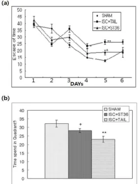
Fig. 1a shows the mean group latencies recorded to reach the hidden platform, in the MWM, for all groups over 6d. An ANOVA (3 x 6, treatment x time) performed on the swimming time during the acquisition trials revealed significant group difference effects (F2,14=63.151, p<0.001), day effects (F5,70=78.125, p<0.001) and day * by group effects (F10,70=4.232, p<0.001). A significant delayed latency was observed in the tail-acupuncture group (ISC + TAIL) compared to the SHAM group from the third day of the trials to the last day. However, repeated treatment with acupuncture at ST36 markedly reduced the time spent in searching for the platform compared to the SHAM group on the fifth day (p<0.05).
To examine the spatial memory, analysis of the performance on the probe trial was compared to the time spent swimming to the platform; this is illustrated in Fig. 1b.
All animals were examined by a retention test involving the removal of the platform from the pool on the seventh trial
day. The time spent in the quadrant was statistically different among groups (F2,14=6.749, p<0.001). Retention performances of the ST36 acupuncture group (ISC + ST36) was not different from the SHAM group; however, the ST36 acupuncture group (ISC + ST36)(28.13 ± 1.24) a significant increase in the time spent in the quadrant compared to the tail-acupuncture group (ISC + TAIL)(23.01 ± 1.82).
Fig. 1. The effecf of stimulation at ST36 on Morris water maze. (a) Time to escape on the platform during acquisition trials of the MWM test. Four trials per day over 6 days were performed for the acquisition test. Mongolian gerbils were treated with acupuncture at tail (ISC + TAIL group, n=7) and acupuncture at ST36 (ISC + ST36 group, n=7) for 2 weeks after induction of cerebral ischemia. The sham-operated control group (SHAM, n=7) was not given any treatments for 2 weeks after induction of ischemia. Significance with Turkey’s test following a repeated ANOVA is indicated as **p<0.01 (SHAM vs. ISC + TAIL), or + p<0.05 (ISC + TAIL vs. ISC + ST36). Vertical lines indicate SEM. (b) Time spent around the platform on the water maze test. The task was performed with four daily trials on the 7th day without the platform for the retention test.
Mongolian gerbils were treated with acupuncture at tail (ISC + TAIL group, n=7) and acupuncture at ST36 (ISC + ST36 group, n=7) for 2 weeks after induction of cerebral ischemia. The sham-operated control group (SHAM, n=7) was not given any treatments for 2 weeks after induction of ischemia. Significance with Turkey’s test following a one-way ANOVA is indicated as **p<0.01 (SHAM vs. ISC + TAIL), or +p<0.05 (ISC + TAIL vs. ISC + ST36). Vertical lines indicate SEM.
2. Effect of acupuncture treatment on cholineacetyltransferase changes in ischemic gerbils
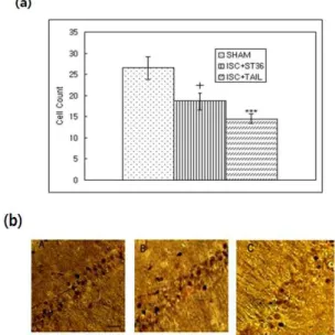
The results of the ChAT immunoreactive analysis of CA1 are shown in Fig. 2. The number of ChAT immunoreactive cells in the hippocampal CA1 was decreased after the induction of transient global ischemia. The number of ChAT immunoreactive cells was statistically different among groups (F2,34=11.210, p<0.001). The number of ChAT immunoreactive cells in the ST36 acupuncture group (ISC + ST36) was not
different from the SHAM group; however, the results of the ST36 acupuncture group (ISC + ST36)(29.82 ± 2.02) were significantly increased compared to the tail-acupuncture group (ISC + TAIL)(23.11 ± 2.32). The number of ChAT positive neurons was significantly increased to 129.0% of the tail-acupuncture group (ISC + TAIL) in the ST36 acupuncture group (ISC + ST36)(p<0.05). This result demonstrated that stimulation of the ST36 acupuncture point was associated with improved memory function.
Fig. 2. The effecf of stimulation at ST36 on expression of ChAT. (a) The number (mean ± SEM) of choline acetyltransferase (ChAT) immunostained nuclei in hippocampal CA1 area of the experimental groups after the water maze learning task. Mongolian gerbils were treated with acupuncture at tail (ISC + TAIL group, n=7) and acupuncture at ST36 (ISC + ST36 group, n=7) for 2 weeks after induction of cerebral ischemia. The sham-operated control group (SHAM, n=7) was not given any treatments for 2 weeks after induction of ischemia. Significance with Turkey’s test following a one-way ANOVA is indicated as ***p<0.001 (SHAM vs. ISC + TAIL), or +p<0.05 (ISC + TAIL vs. ISC + ST36). Vertical lines indicate SEM. (b) Representative microphotographs of coronal section showing ChAT expression in the hippocampal CA1 of (A)SHAM, and (B)ISC+ST36, (C)ISC+TAIL group.
Calibration bars represents 100 ㎛ (located in A)in A-C.
Discussion
In this study, we investigated whether treatment with acupuncture could improve memory retention functions on the MWM and measured the ChAT immunoreactive cells in the hippocampal CA1 region, in a transient cerebral ischemia gerbil model. We found that acupuncture at ST36 improved the function of memory retention on the MWM and increased ChAT immunoreactivity in the hippocampal CA1 area in the cerebral ischemia gerbil model.
Acupuncture is a widely used clinical treatment used for
various diseases in oriental medicine. Treatment with acupuncture promotes homeostasis, improves brain circulation, pain control, and neuromodulatory functions in the central nervous system7,10-24). Treatment with acupuncture is particularly effective for symptomatic improvement of cerebrovascular accidents including ischemia10,12,13). Consistent with previous studies, our results showed that the tail-acupuncture group (ISC + TAIL) had deficits in learning and memory on the MWM test. However, the ST36 acupuncture group (ISC + ST36) had significantly improved spatial learning and memory compared to the tail-acupuncture group (ISC + TAIL) group.
One logical consequence of neuronal loss, observed after ischemia, is cholinergic deficits in affected brain areas25). The hippocampus has been associated with memory processes and is densely innervated by cholinergic fibers26). The degeneration of the cholinergic innervations, from the basal forebrain to the hippocampal formation in the temporal lobes, is thought to be one of the factors determining the progression of memory decay, both during normal aging and in AD11). The best available marker for cholinergic neurons, in the basal forebrain, is ChAT activity. ChAT synthesizes the neurotransmitter acetylcholine, in the basal forebrain, cortex, hippocampus, and amygdala. A significant reduction in ChAT activity in the postmortem brains of mentally impaired patients has been reported. In addition, Ischemic insult decreases the acetylcholine concentration in affected regions of the brain27). There is a 20-50% decrease in ChAT activity in the hippocampus after cerebral ischemia in the gerbil model.
However, treatment with acupuncture at ST36 modulates the function of these neurons and appears to play a role in their maintenance by preventing the ischemia-induced decrease in ChAT activity2). The results of this study show that stimulation with acupuncture at ST36 exerts beneficial effects on cholinergic neurotransmission in the brain by increasing ChAT activity in the hippocampus. The findings suggest that treatment with acupuncture at ST36 helps to recover cholinergic systems following ischemic insults, which may be beneficial to the plastic responses of cholinergic nerves after ischemic injury.
In this study, treatment with acupuncture suppressed the ischemia-induced decrement in the number of ChAT positive cells in the hippocampal CA1 region. The most potent inhibitory effect was observed at the ST36 acupoint and acupuncture at a non-acupoint showed no significant effects.
Acupuncture exerted no significant effect on the number of ChAT positive cells under normal conditions. One of the most impressive effects of acupuncture is the rapid recovery from
complications associated with a stroke26,27). Acupuncture provides neuroprotection against cerebral ischemia in the monkey10) and gerbil1) regionresults of the present study show that acupuncture, especially at the ST36 acupoint, ameliorates ischemia-induced deficits of learning and memory, suggesting that acupuncture might aid in recovery following ischemic cerebral injury.
Acknowledgments
This work was supported by the Korea Science and Engineering Foundation (KOSEF) grant funded by the Korea government (MEST) (No. R11-2005-014).
References
1. Kirini T. Delayed neuronal death in the gerbil hippocampus following ischemia: Brain Research, 239:
57-69, 1982.
2. Pulsinelli, W.A., Brierley, J.B., Plum, F. Temporal profile of neuronal damage in a model of transient forebrain ischemia: Annals of Neurology, 11: 491-498, 1982.
3. Amano, M., Hasegawa, M., Hasegawa, T., Nabeshima, T.
Characteristics of transient cerebral ischemia-induced deficits on various learning and memory task in male Mongolian gerbils: Japanese Journal of Pharmacology, 63:
469-477, 1993.
4. Davis, H.P., Baranowski, J.R., Pulsinelli, W.A., Volpe, B.T.
Retention of reference memory following ischemic hippomcapal damage: Physiology and Behavior, 39:
783-786, 1987.
5. Davis, H.P., Tribuna, J., Pulsinelli, W.A., Volpe, B.T.
Reference and working memory of rats following hippocampal damage induced by transient forebrain ischemia: Physiology and Behavior, 37: 387-392, 1986.
6. Ohno, M., Yamamoto, T., Ueki, S. Effect of the k-receptor agonist, U-50, 488H, on cerebral ischemia-induced impairment of working memory assessed in rats by a three-panel runway task: European Journal of Pharmaocology, 193: 357-361, 1991.
7. Cho, Z.H., Chung, S.C., Jones, J.P., Park, J.B., Park, H.J., Lee, H.J., Wong, E.K., Min, B.I. New findings of the correlation between acupoints and corresponding brain cortices using functional MRI: Proceedings of the National Academy of Sciences of the United States of America, 95:
2670-2673, 1998.
8. Lee, B., Choi, E.J., Lee, E.J., Han, S.M., Hahm, D.H., Lee, H.J., Shim, I. The neuroprotective efect of methanol extract
of gagamjungjihwan and fructus euodiae on ischemia-induced neuronal and cognitive impairment in the rat:Evidence Based Complementary Alternative Medicine, 2009.
9. Liu, J.X., Liu, Y., Chen, X.L., Zhao, J.J., Song, T.S., Qian, Y.H. Breviscapine improves functions of spatial learning and memory of focal cerebral ischemia rats. Zhong Yao Cai. 32: 548-556, 2009.
10. Kim, E.H., Kim, Y.J., Lee, H.J., Huh, Y., Chung, J.H., Seo, J.C., Kang, J.E., Lee, H.J., Yim, S.V., Kim, C.J. Acupuncture increases cell proliferation in dentate gyrus after transient global ischemia in gerbils: Neuroscience Letters, 297: 21-24, 2001.
11. Wu, X., Glinn, M.A., Ostrowski, N.L., Su, Y., Ni, B., Cole, H.W., Bryant, H.U., Paul, S.M. Raloxifene and estradiol benzonate both fully restore hippocampal choline acetyltransferase activity in ovariectomized rats: Brain Resesrch, 847: 98-104, 1999.
12. Gao, H., Guo, J., Zhao, P., Cheng, J. The neuroprotective effects of electroacupuncture on focal cerebral ischemia in monkey: Acupuncture & electro-therapeutics research, 27:
45-57, 2002.
13. Ying, S.X., Cheng, J.S. Effects of electro-acupuncture on c-fos expression in gerbil hippocampus during transient global ischemia: Acupuncture & electro-therapeutics research, 19: 207-213, 1994.
14. Pang, J., Itano, T., Sumitani, K., Negi, T., Miyamoto, O.
Electroacupuncture attenuates both glutamate release and hyperemia after transient ischemia in gerbils: American Journal of Chinese Medicine, 31: 295-303, 2003.
15. Han, Z., Jiang, Y.H., Wan, Y., Chang, J.K., Han, J.S.
Endomorphin-1 mediates 2Hz but not 100Hz electroacupuncture analgesia in the rat: Neuroscice letters, 274: 75-78, 1999.
16. Kim, E.H., Chung, J.H., Kim, C.J. Auricular acupuncture increases cell proliferation in the dentate gyrus of sprague-dawley rats. Acupunture electrotherpy research, 26: 187-194, 2001.
17. Paxinos, G., Watson, C., Pennisi, M. Bregma, lamda and the interaural midpoint in stereotaxic surgery with rats of different sex: Strain and weight, 13: 139-143, 1985.
18. Puurunen, K., Sirviö, J., Koistinaho, J., Miettinen, R., Haapalinna, A., Riekkinen, P Sr., Sivenius, J. Studies on the influence of enriched-environment housing combined with systemic administration of an alpha2-adrenergic antagonist on spatial learning and hyperactivity after global ischemia in rats: Stroke, 28: 623-631, 1997.
19. Kiprianova, J., Sandhkuhler, S., Schwab, S., Hoyer, S.,
Spranger, M. Brain derived neurotrophic factor improves long term potentiation and cognitive functions after transient forebrain ischemia in the rat: Experimental Neurology, 159: 511-519, 1999.
20. Okada, M., Tamura, A., Urae, A., Nakagomi, T., Kirino, T., Mine, K., Fujiwara, M. Long term spatial cognitive impairment following middle cerebral artery occlusion in rats: A behavioral study: Journal of cerebral blood flow and metabolism, 15: 505-512, 1995.
21. Seren, M.S., Lazzaro, A., Yang, C.L., Canella, R., Bassan, M., Zanoni, R., Manev, H. Orally administered glycolipid derivative LIGA20 reduced infarct volume and behavioral impairment after focal ischemia: The Journal of pharmacology and experimental therapeutics, 268: 460-465, 1994.
22. Jang, M.H., Shin, M.C., Lee, T.H., Lim, B.V., Shin, M.S., Min, B.I., Kim, H., Cho, S., Kim, E.H., Kim, C.J.
Acupuncture suppresses ischemia-induced increase in c-fos expression and apoptosis in the hippocampal CA1 region in gerbils: Neuroscience Letters, 347: 5-8, 2003.
23. Sato, A., Sato, Y., Suzuki, A., Uchida, S. Neuronal mechanisms of the reflex inhibition and excitation of gastric motility elicited by acupuncture-like stimulation in anesthetized rats: Neuroscience Letters, 18: 53-62, 1993.
24. Uchida, S., Kagitani, F., Suzuki, A., Aikawa, A. Effect of
acupuncture-like stimulation on cortical cerebral blood flow in anesthetized rats: The Japanese journal of physiology, 50: 495-507, 2000.
25. Ott, E.O., Abraham, J., Meyer, J.S., Achari, A.N., Chee, A.N., Mathew, N.T. Disordered cholinergic neurotransmission and dysautoregulation after acute cerebral infarction:
Stroke, 6: 172-180, 1975.
26. Huang, Y.L., Onodera, H., Takeda, A., Itoyama, Y., Kogure, K. The effect of long-term post-ischemic bifemelane hydrochloride treatment on cholinergic systems in the gerbil hippocampus: Brain Research, 722: 195-199, 1996.
27. Schetinger, M.R., Bonan, C.D., Frassetto, S.S., Wyse, A.T., Schierholt, R.C., Webber, A., Dias, R.D., Sarkis, J.J., Netto, C.A. Pre-conditioning to global cerebral ischemia changes hippocampal acetylcholinesterase in the rat: Biochemistry and molecular biology international, 47: 473-478, 1999.
28. Inoune, I., Chen, L., Zhou, L., Zeng, X., Wang, H.
Reproduction of scalp acupuncture therapy on strokes in the model rats, spontaneous hypertensive rats-stroke prone (SHR-SP): Neuroscience Letters, 333: 191-194, 2002.
29. Chung, J.H., Lee, E.Y., Jang, M.H., Kim, C.J., Kim, J., Ha, E., Park, H.K., Choi, S., Lee, H., Park, S.H., Leem, K.H., Kim, E.H. Acupuncture decreases ischemia-induced apoptosis and cell proliferation in dentate gyrus of gerbils:
Neurological Research, 29 suppl 1, S23-S27, 2007.