Molecular identification and expression analysis of bactericidal permeability-increasing protein/ LPS-binding protein (BPI/LBP) from
Black rockfish Sebastes schlegeli
Mun Gyeong Kwon*, Ju Won Kim, Myoung Ae Park*, Jee Youn Hwang*, Hyung Jun Park, Gun Wook Baeck and Chan Il Park†6)
Department of Marine Biology & Aquaculture, Institute of Marine Industry, College of Marine Science, Gyeongsang National University, 455, Tongyeong, 650-160, Korea
*Pathology Division, National Fisheries Research and Development Institute, Busan 619-900, SouthKorea Bactericidal/permeability-increasing protein (BPI) and lipopolysaccharide-binding protein (LBP) are important components of the mammalian innate defence system against Gram-negative infections. The BPI/LBP cDNA was identified from the black rockfish ConA/PMA or LPS stimulated leukocyte cDNA library. The full-length BR-BPI/LBP cDNA was 2118 bp long and contained an open reading frame (ORF) of 1422 bp that encoded 473 amino-acid residues. The 5’ UTR had a length of 57 bp, and the 3’ UTR 639 bp. The molecular weight and theoretical isoelectric point (pI) values were calculated 51.4 kDa and 9.72, respectively. Compared with other known BPI or BPI/LBP peptide sequences, the most conserved regions of the black rockfish BPI/LBP peptide were found to be the BPI1 N-terminal, BPI2 C-terminal domains and a LPS binding domain. Phylogenetic analysis based on the deduced amino acid sequence revealed a homologous relationship between the BPI/LBP sequence of black rockfish and that of other teleosts. The black rockfish BPI/LBP gene was predominantly expressed in the PBLs, head kidney, trunk kidney and spleen. The expression of the black rockfish BPI/LBP molecule was induced in the peripheral blood leukocytes (PBLs) from 1 to 24 h following LPS stimulation, with a peak at 12 h post-stimulation.
Key words :Black rockfish, BPI/LBP, LPS stimulation, Real-time PCR
There are two types of immunity, innate immunity and adaptive immunity, which have different roles against the infection of pathogens (Dempsey et al., 2003). The innate immune system has the potential for quick, powerful, and non-specific responses to a wide range of pathogens (Xu et al., 2005). Moreover, innate immune system is a varied and complex group of phenomena and has a role as a first line of defense
†Corresponding Author: Chan-Il Park E-mail: [email protected] TEL: 82-55-640-3103,
FAX: 82-55-642-4509
against infections (Kono and Sakai, 2003). The host innate immune system recognises invading pathogens by their conserved pathogen associated molecular patterns (PAMPs) using pattern recognition receptors (PRRs) (Medzhitov and Janeway, 2002). Lipopolysaccharide (LPS), a complex glycolipid and one of the major components of the outer surface membrane of Gram-negative bacteria, is one of the PAMPs. LPS plays an important role in the activation of innate immunity (Alexander and Rietschel, 2001).
Two of the proteins crucial to the mediation of signals for LPS are bactericidal permeability-increasing protein (BPI) and LPS-binding protein (LBP), which share several characteristics. Both belong to a family of lipid transfer/LPS-binding proteins (LT/LBP), which also include the mammalian phospholipid transfer protein (PLTP) and cholesteryl ester transfer protein (CETP) (Bingle and Craven, 2004). They are composed of an N-terminal domain that binds to LPS and a C-terminal domain that mediates the delivery of LPS to other host molecules involved in the response to LPS (Schumann et al., 1990; Iovine et al., 1997; Beamer et al., 1998).
Even though these two proteins have similar structures, they are considered to have antagonistic functions. LBP binds LPS and enhances cellular responses to LPS, while BPI binds LPS but reduces cellular responses (Tobias et al., 1988; Tobias et al., 1997).
The complete cDNA sequences of BPI and LBP have been cloned from human (Gray et al., 1989), bovine (Leong and Camerato, 1990), rabbit (Zarember et al., 1997; Schumann et al., 1990), mouse (Gallay et al., 1993; Lengacher et al., 1995) and rat (Su et al., 1994).
The BPI or LBP homologues of teleosts have not been classified as either a BPI or a LBP because the functional properties of these proteins have not been characterized.
Therefore, in teleosts, they have been referred to as BPI/LBP (Nam et al., 2010). The BPI/LBP genes have been isolated and characterized in some fish species.
In particular, the BPI/LBP gene was sequenced from rainbow trout (Inagawa et al., 2002), Atlantic cod (Stenvik et al., 2004), common carp (Kono and Sakai, 2003), channel catfish (Xu et al., 2005), olive flounder (Nam et al., 2010), and ayu (Suzuki et al., 2009).
Furthermore, a cDNA sequence with homologies to members of the LBP/BPI family was identified in the oyster Crassostrea gigas, and this invertebrate LBP/BPI family is the first to have been characterized by functional analysis using a BPI recombinant protein (Gonzalez et al., 2007). The presence of the BPI/LBP gene in black rockfish, however, has not been reported to date.
Black rockfish (Sebastes schlegeli) is one of the more significant fish species in Korea due to the human interest in aquaculture and fisheries, but diseases, especially infectious ones, have occurred frequently in them and have limited the production efficiencies of this economic fish. To improve the black rockfish production efficiency, it is necessary to understand the cellular and molecular processes involved in the disease resistance to this species. In this paper, the cloning, characterization, and expression analysis of the BPI/LBP gene from black rockfish (Sebastes schlegeli) are reported.
Materials and Methods
Identification of Black rockfish BPI/LBP cDNA Black rockfish BPI/LBP full-length cDNA was identified via the EST analysis of the ConA/PMA or LPS stimulated PBLs library (Baeck et al., 2008).
5’-termini base sequencing of the selected cDNA clones in phagemid form was performed using an ABI 3100 automatic DNA sequencer (PE Applied Biosystems, CA, USA) and an ABI Prism Big Dye Terminator Cycle Sequencing Ready Reaction kit (PE Applied Biosystems, USA). Bioinformatic analysis was conducted to determine the gene identities, using the Genetyx ver.
8.0 software (SDC Software, Japan). Briefly, the vector
sequences were removed, and the database search was limited to the ESTs that are >400 bp in length.
Multiple alignment and phylogenetic analysis The sequence of BR-BPI/LBP cDNA was compared with the sequences in the peptide sequence databases at the National Center for Biotechnology Information (NCBI), using the BLAST network service. Nucleotide sequence comparisons were carried out using the BLASTX program (Gish and David, 1993).
The multiple alignment of the amino acid sequence of the BR-BPI/LBP proteins was performed using the Clustal W program (Thompson et al., 1994). The BPI or BPI/LBP sequences of that were used for the comparison and their GenBank accession numbers were as follows: Cattle BPI (NP_776320), Channel catfish BPI (AAX20011), Dog BPI (XP_534417), House mouse BPI (NP_808518), Human BPI (ABD66755), Norway rat BPI (AAH79318), Pig BPI (ABO34136), Atlantic cod BPI/LBP-A (AAM52335), Atlantic cod BPI/LBP-B (AAM52336), Ayu BPI/LBP (BAG49475), Common carp BPI/LBP (BAC56095), Olive flounder LBP/BPI (ACV74252), Rainbow trout LBP/BPI-1 (NP_001118057), and Rainbow trout LBP/BPI-2 (NP_001117670). The primary structure of the deduced amino acid sequence of BR-BPI/LBP was analyzed by ProtParam (http ://cn.expasy.org/tolls/protparam). The positions of signal peptide and BPI domains was predicated with the Simple Modular Architecture Tool (SMART) (http ://smart.
embl-heidelberg.de/).
The phylogenetic relationships were inferred using the Mega 4 program and distance analysis through the neighbor-joining method. The accession numbers of BPI
or BPI/LBP for the creation of phylogenetic tree used are described above. In addition to accession number of LBP amino sequences are as follows: Cattle LBP (NP_001033763), Dog LBP (XP_542993), House mouse LBP (NP_032515), Human LBP (AAB31143), Norway rat LBP (NP_058904), Pig LBP (NP_001121907). The values supporting each node were derived from 2,000 re-samplings.
RT-PCR analysis of the BR-BPI/LBP transcripts in different tissues
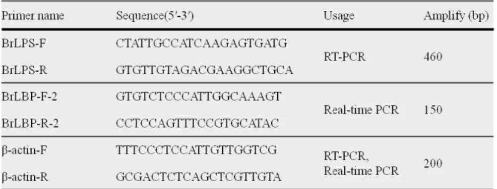
Eight tissues (PBLs, head kidney, trunk kidney, spleen, liver, intestine, gill, and muscle) were isolated from approximately 350 g healthy black rockfish. The total RNA from each tissue was extracted using TRIzol reagent (Invitrogen, USA), according to the manufacturer’s instructions. First-strand cDNA synthesis was carried out using a first-strand cDNA synthesis kit (Takara, Japan), according to the manufacturer’s instructions. The first-strand cDNAs were used as a PCR amplification template, with the specific primers. The oligonucleotide primers used to amplify the BR-BPI/LBP cDNA fragment were BrLPS-F and BrLPS-R. Thermal cycling was performed using TaKaRa PCR Thermal Cycler (Takara, Japan) with ExPrime Taq premix (GENET BIO, Korea) as follows:
predenaturation at 94℃ for 5 min, 25 cycles of denaturation at 94℃ for 30 s, annealing at 55℃ for 1 min, extension at 72℃ for 1 min, and final extension at 72℃ for 7 min. The expression of β-actin was used as the internal control. The PCR amplification products were observed on 1.5% agarose gel electrophoresis with a 100-bp DNA ladder (Takara, Japan). The primer
Table 1. Primers used for all the experiments
sequences used in this study are listed in Table 1.
Real-time PCR analysis
Black rockfish (300-350 g) was purchased from the Fisheries market in Tong-yeong, Korea. Peripheral blood samples were collected in a heparinized (100 Ul) syringe from the caudal blood vein. Peripheral blood leukocytes (PBLs) isolation and PBLs stimulation with LPS (50
㎍/ml) were carried out as a previously reported method (Beack et al., 2008). The total RNA was extracted, and first-strand cDNA synthesis was performed as described above. Real-time PCR was performed on a Thermal cycler Dice real time system (Takara, Japan) with SYBR Green I (Takara, Japan). The threshold cycle (Ct) values were automatically calculated as follows: the cycle when the fluorescence of the sample exceeded a threshold level corresponding to 10 standard deviations of the mean of the baseline fluorescence. Amplification was performed 3 step PCR methods as follows: 1 cycle at 95℃ for 10 s and 45 cycles at 95℃ for 30 s, 62℃
for 30 s and 72℃ for 30 s, with a dissociation step at 95℃ for 15 s, 62℃ for 30 s and 95℃ for 15 s.
β-actin was detected as an internal control. All the samples were analyzed in triplicate and the expression
of target genes was calculated as relative folds of the expression of β-actin according to the 2−ΔΔCT methods.
Results and discussion
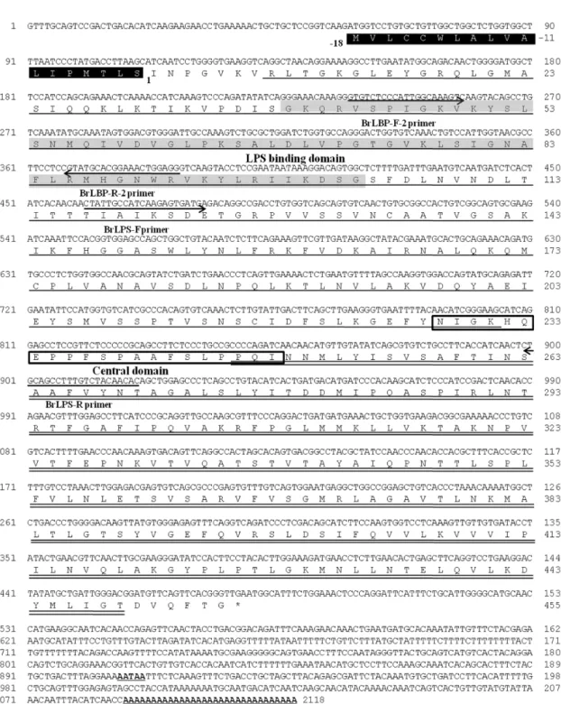
In the present study, we report for the cloning and characteristics of the LBP/BPI gene from the black rockfish, Sebastes schlegeli. The full-length BPI/LBP cDNA was 2118 bp long and contained an open reading frame (ORF) of 1422 bp that encoded 473 amino acid residues. The molecular mass were calculated 51.4 kDa and theoretical isoelectric point (pI) values were calculated 9.72. The 5’ UTR was 57 bp in length, and the 3’ UTR 639 bp, and they contain a polyadenylation signal (AATAA) and a polyadenylation site (accession number: AB548677) (Fig. 1). These sequences had a putative 18 AA signal peptide, followed by 455 AA residues namely identified as putative mature proteins.
Mammalian BPI or LBP family proteins have signal peptide. This suggests that these proteins could be secreted in the mature form after cleavage.
The Black rockfish BPI/LBP has a BPI/LB P/CETP-specific sequence motifs BPI1 (BPI/LBP/CETP N-terminal domain) between amino acid residues 8 and
Fig. 1. Nucleotide and deduced amino acid sequences of black rockfish BPI/LBP. The oligonucleotide primers that were used in the study are indicated with arrows. The putative signal peptide is indicated black box. Central domain and LPS binding domain is indicated box and gray. N-terminal domain and C-terminal domain was indicated underline and double underline, respectively. The polyadenylation signal (AAUAA) and poly A tails is indicated in boldface and underline.
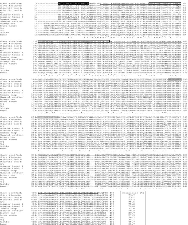
Fig. 2. Comparison between the black rockfish BPI/LBP amino acid sequence and other known BPI or BPI/LBP sequences.
The amino acids identical to the black rockfish sequence are indicated by an asterisk (*), and the absent amino acids are indicated by dashes (–).The accession numbers of BPI or BPI/LBP for the creation of multiple alignment used are described Fig. 4. The putative signal peptide is indicated black box. Identities of between black rockfish BPI/LBP and other BPI/LBP or BPI is indicated box. Central domain and N-terminal domain and C-terminal domain is indicated gray and underline, respectively.
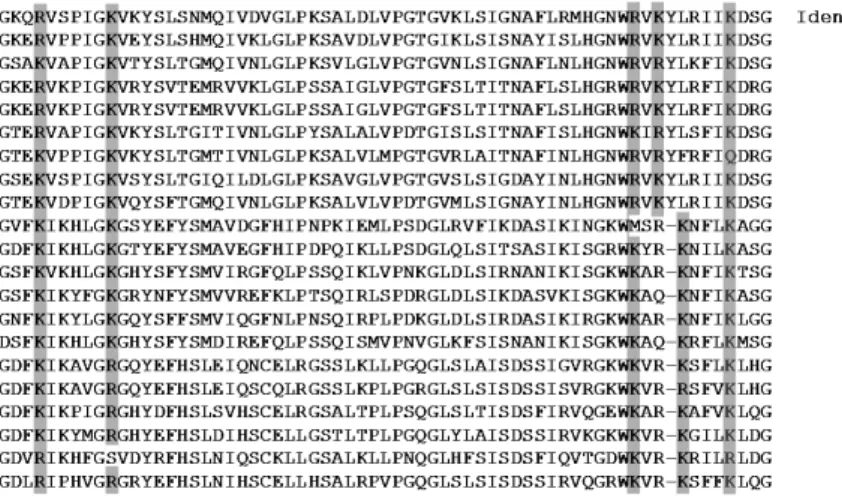
Fig. 3. Alignment of the predicted LPS-binding domain of the black rockfish BPI/LBP with that of other species. Identical amino acid residues are indicated by asterisks (*). Dots (·) indicate the black rockfish BPI/LBP. The positively charged amino acid residues (K or R) conserved in BPI/LBP are indicated by shaded regions. The number at the end of each sequence represents amino acid identities (%) to that of the black rockfish LBP/BPI.
231 and BPI2 (BPI/LBP/CETP C-terminal domain) between amino acid residues 246 and 449 were identified by the SMART program (Fig. 1). The mature Black rockfish BPI/LBP was predicted by comparing the amino acid sequence with that of other mammalian LBP and BPI. The mature Black rockfish BPI/LBP was also separated into the N-terminal domain (residues 8-231) and C-terminal domain (residues 246-449). The proline-rich central domain (resi dues 228-248) was located between the N-terminal and C-terminal domains (Fig. 1). The two domains form the three structural elements, the N-terminal barrel (barrel-N), a central β-sheet, and the C-terminal barrel (barrel-C) (Beamer et al., 1998).
The deduced amino acid sequence of black rockfish BPI/LBP had a 79.7% identity with that of Olive flounder LBP/BPI, 75.5% with Ayu BPI/LBP, 75.2% with Rainbow trout LBP/BPI-1, 74.8% with Rainbow trout LBP/BPI-2, 72% with Common carp BPI/LBP, 68.2%
with Channel catfish BPI, 63.2% with Atlantic cod BPI/LBP-A and B, 38.7% with Dog BPI, 35.7% with
Cattle BPI and Human, 34.1% with Pig BPI, 29.7%
Norway rat BPI, and 27.5% with House mouse BPI. (Fig.
2). Furthermore, multiple sequence alignment revealed that the LPS binding domain was well conserved.
Moreover, the LPS-binding domain included in barrel-N was aligned with those of olive flounder LBP/BPI, ayu BPI/LBP, Atlantic cod BPI/LBP-a and -b, rainbow trout BPI/LBP-1 and -2, channel catfish BPI, common carp BPI/LBP, house mouse BPI and LBP, Norway rat BPI and LBP, dog BPI and LBP, pig BPI and LBP, cattle BPI and LBP, and human BPI and LBP (Fig. 3). When LPS-binding domain of black rockfish BPI/LBP was compared with that of other teleosts, that of black rockfish BPI/LBP showed the highest identity (80.0%) to that of olive flounder LBP/BPI. The identities to that of human BPI and LBP were 33.8 and 35.7%, to that of black rockfish BPI/LBP, respectively (Fig. 3).
LBP and BPI are distinguished from PLTP and CETP because they have the function of binding LPS with high affinity (Inagawa et al., 2002). Recent analyses
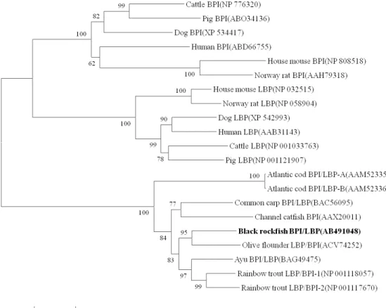
Fig. 4. Neighbor-joining tree of BPI/LBP constructed with Mega4. The bootstrap confidence values shown at the nodes of the tree are based on 2000 bootstrap 2000 replications (DDBJ/EMBL/GenBank Accession numbers:
Black rockfish BPI/LBP (AB491048), Olive flounder LBP/BPI (ACV74252), Ayu BPI/LBP (BAG49475), Rainbow trout LBP/BPI-1 (NP 001118057), Rainbow trout LBP/BPI-2 (NP 001117670), Channel catfish BPI (AAX20011), Common carp BPI/LBP ( BAC56095), Atlantic cod BPI/LBP-A(AAM52335), Atlantic cod BPI/LBP-B (AAM52336), Cattle BPI (NP 776320), Pig BPI (ABO34136), Dog BPI (XP 534417), Human BPI (ABD66755), House mouse BPI (NP 808518), Norway rat BPI (AAH79318), House mouse LBP (NP 032515), Norway rat LBP(NP 058904), Dog LBP (XP 542993), Human LBP (AAB31143), Cattle LBP (NP 001033763), Pig LBP(NP 001121907)).
suggest that conserved positive charge amino acids in the N-terminal barrel of mammal LBP and BPI (lysine 42, 48, 92, 95, and 99 in human BPI) were involved in binding the anionic portion of lipid A (Beame et al., 1998). As these residues are clustered at the tip of the NH2-terminal domain, they may cause electrostatic interactions with negatively charged groups of LPS (Beame et al., 1998; Lamping et al., 1996). As shown
in Fig. 3, basic amino acid residues in black rockfish BPI/LBP (lysine or arginine 42, 48, 92, 94, and 100 in BR-BPI/LBP) were well conserved at a similar position (Fig. 3). These data suggest that black rockfish BPI/LBP proteins might bind LPS, and thus may have a pivotal role in the innate immune system in fish.
The phylogenetic analysis indicated that the BPI and/or LBP sequences from mammals and teleosts
Fig. 6. Quantitative real-time PCR analysis of the BPI/LBP expression in black rockfish leukocytes stimulated with LPS (50㎍/ml) at 0, 1, 3, 6, 12, and 24 h.
Fig. 5. Detection of the black rockfish BPI/LBP mRNA levels and β-actin from various tissues of healthy black rockfish via RT-PCR. The marker indicates a 100-bp ladder marker.
segregated into two separate clusters and is closest to that of olive flounder LBP/BPI among the examined species. Moreover, all the mammalian BPI and LBP sequences clustered into distinct clades, This grouping was well-supported by bootstrapping (Fig. 4). The majority of fish BPI/LBP sequences clustered according to the species of origin. This result indicates that BPI and LBP in mammals are molecules of common origin,
and duplication of BPI and LBP genes in mammals occurred after divergence from teleosts.
The expression of the BPI/LBP gene in the black rockfish tissues was detected via RT-PCR. The BPI/LBP gene was expressed in all the tissues of the black rockfish.
Especially, the expression was found to be predominantly in the PBLs, and less dominantly in the head kidney, trunk kidney, spleen, and least in the liver, gill, intestine,
muscle after 25 cycles of PCR (Fig. 5). Some kind of leukocytes are probably the main source of BPI/LBP mRNA in blood as well. It is thus possible that leukocytes may be the main source of BPI/LBP in the spleen, heart, gills, and liver, as these organs are blood filled. This pattern was also similar to that found in Atlantic cod (Stenvik et al., 2004) and large yellow croaker (Huang et al., 2008), which inhabit seawater. However common carp (Kono and Sakai, 2003) and channel catfish (Xu et al., 2005), which inhabit freshwater, was a contrast to black rockfish BPI/LBP expression pattern. In the latter BPI/LBP mRNA expression was high in the gill, intestine, and skin. Further work is necessary to reveal the reason why the different fish species have distinct gene expression patterns of BPI/LBP.
Black rockfish peripheral blood leukocytes (PBLs) were treated with LPS and the expression level of BR-BPI/LBP was examined. The LPS treated PBLs showed a marked increase in BR-BPI/LBP expression.
Especially, the BPI/LBP levels seemed to reach a peak in expression at 12 h post-LPS stimulation, and decreased at 24 h (Fig. 6). Stenvik et al. (2004) repoted that the possible lack of the main components of the mammalian LPS-recognition system, including LBP, CD14, and TLR4, was speculated to explain the low LPS sensitivity of fish. Moreover, the CD14 and TLR4 orthologs have not been identified in black rockfish and the other teleosts. Even though, the black rockfish BPI/LBP mRNA expression level seemed like to lower tendency, these results indicate that the increased BR-BPI/LBP mRNA level is an early and short-lived response to stimulation by LPS. Through further investigation, we substantiate either the possibility of
LPS-recognition system in black rockfish, or the presence of CD14 and TLR4 orthologs.
The study of innate immunity of fish has been hampered by a lack of molecular tools like gene probes and specific antibodies. Thus, BPI/LBP could become useful as a molecular tool for the study of innate immune mechanisms of fish. This information could help explain the immune system in fish, and might also illuminate the role of the innate immune system in other species.
Acknowledgement
This Research was supported by National Fisheries Research and Development Institute (RP-2010-AQ-038) grant.
References
Alexander, C., Rietschel, E.T.: Bacterial lipopolysaccharides and innate immunity. J Endotoxin Res., 7:167-202, 2001.
Baeck, G.W., Kim, J.W., Park, C.I.: Identification and expression analysis of an interferon stimulated gene 15 (ISG15) from black rockfish, Sebastes schlegeli.
Fish Shellfish Immunol., 25:679-81, 2008.
Beamer, L.J., Carrol, S.E., Eisenberg, D.: The BPI/LBP family of proteins: a structural analysis of conserved regions. Prot. Sci., 7: 906-914, 1998.
Bingle, C.D., Craven, C.J.: Meet the relatives: a family of BPI- and LBP-related proteins. Trends Immunol., 25:53-55, 2004.
Dempsey, P.W., Vaidya, S.A., Cheng, G.: The art of war:
innate and adaptive immune responses. Cell Mol Life Sci., 60:2604-2621, 2003.
Gallay, P., Carrel, S., Glauser, M.P., Barras, C., Ulevitch, R.J., Tobias, P.S.: Purification and characterization of murine lipopolysaccharidebinding protein. Infect Immun., 61:378-383, 1993.
Gish, W., David, J.S.: Identification of protein coding regions by database similarity search. Nature Genetics, 3:266-272, 1993.
Gonzalez, M., Gueguen, Y., Destoumieux-Garzón, D., Romestand, B., Fievet, J., Pugnière, M., Roquet, F., Escoubas, J.M., Vandenbulche, F., Levy, O., Sauné, L., Bulet, P., Bachère, E.: Evidence of a bactericidal permeability increasing protein in an invertebrate, the Crassostrea gigas Cg-BPI. Proc.
Natl. Acad. Sci. U.S.A., 104:17759-17764, 2007.
Gray, P.W., Flaggs, G., Leong, S.R., Gumina, R.J., Weiss, J., Ooi, C.E.: Cloning of the cDNA of a human neutrophil bactericidal protein. Structural and functional correlations. J Biol Chem., 264:9505-9509, 1989.
Huang, Y., Lou, H., Wu, X., Chen, Y.: Characterization of the BPI-like gene from a subtracted cDNA library of large yellow croaker (Pseudosciaena crocea) and induced expression by formalin-inactivated Vibrio alginolyticus and Nocardia seriolae vaccine challenges. Fish Shellfish Immunol., 25:740-750, 2008.
Inagawa, H., Honda, T., Kohchi, C., Nishzawa, T., Yoshiura, Y., Nakanishi, T., Yokomizo, Y., Soma, G.: Cloning and characterization of the homolog of mammalian lipopolysaccharide binding protein and bactericidal permeability-increasing protein in rainbow trout Oncorhynchus mykiss. J. Immunol., 168:5638-5644, 2002.
Iovine, N., Elsbach, P., Weiss, J.: An opsonic function of
the neutrophil bactericidal/permeability-increasing protein depends on both its N- and C-terminal domains. Proc. Natl. Acad. Sci. U.S.A., 94:10973- 10978, 1997.
Kono, T., Sakai, M.: Molecular cloning of a novel bactericidal permeability-increasing protein/lip opolysacc har ide-binding protein (BPI/LBP) from comment carp Cyprinus carpio L. and its expression. Mol.
Immunol., 40:269-278, 2003.
Lamping, N., Hoess, A., Yu, B., Park, T.C., Kirschning, C.J., Pfeil, D., Reuter, D., Wright, S.D., Herrmann, F., Schumann, R.R.: Effects of site-directed mutagenesis of basic residues (Arg 94, Lys 95, Lys 99) of lipopolysaccharide (LPS)-binding protein on binding and transfer of LPS and subsequent immune cell activation. J Immunol., 157(10):4648-4656, 1996.
Lengacher, S., Jongeneel, C.V., LeRoy, D., Lee, J.D., Kravchenko, V., Ulevitch, R.J., Glauser, M.P., Heumann, D.: Reactivity of murine and human recombinant LPS-binding protein (LBP) within LPS and gram negative bacteria. J Inflamm., 47(4):165-172, 1995.
Leong, S.R., Camerato, T.: Nucleotide sequence of the bovine bactericidal permeability increasing protein (BPI).
Nucleic Acids Res., 18:3052, 1990.
Medzhitov, R., Janeway, Jr C.A.: Decoding the patterns of self and nonself by the innate immune system.
Science 296:298-300, 2002.
Nam, B.H., Ahn, K.J., Kim, Y.O., Kong, H.J., Kim, W.J., Kim, H.S., Lee, S.J., Kim, K.K.: Molecular cloning and characterization of LPS-binding protein/
bactericidal permeability-increasing protein (LBP/BPI) from olive flounder, Paralichthys olivaceus. Vet.
Immunol. Immunopathol., 133:256-263, 2010.
Schumann, R.R., Leong, S.R., Flaggs, G.W., Gray, P.W., Wright, S.D., Mathison, J.C.: Structure and function of lipopolysaccharide binding protein. Science, 249:1429-1431, 1990.
Stenvik, J., Soltrad, T., Strand, C., Leiros, I., Jørgensen, T.: Cloning and analyses of BPI/LBP cDNA of the Atlantic cod (Gadus morhua L.). Dev. Comp.
Immunol., 28:307-323, 2004.
Su, G.L., Freeswick, P.D., Geller, D.A., Wang, Q., Shapiro, R.A., Wan, Y.H., Billiar, T.R., Tweardy, D.J., Simmons, R.L., Wang, S.C.: Molecular cloning, characterization, and tissue distribution of rat lipopolysaccharide binding protein. Evidence for extrahepatic expression. J Immunol, 153(2):743-752, 1994.
Suzuki, K., Izumi, S., Tanaka, H., Katagiri, T.: Molecular cloning and expression analysis of the BPI/LBP cDNA and its gene from ayu Plecoglossus altivelis altivelis. J. Fish. Sci., 75:673-681, 2009.
Thompson, J.D., Higgis, D.G., Gibson, T.J.: CLUSTAL W: improving the sensitivity of progressive multiple sequence alignment through sequence weighting, position specific gap penalties and weight matrix choice. Nucleic Acids Res., 22: 4673-4680, 1994.
Tobias, P.S., Mathison, J.C., Ulevitch, R.J.: A family of lipopolysaccharide binding proteins involved in responses to gramnegative sepsis. J Biol Chem., 263:13479-13481, 1988.
Tobias, P.S., Soldau, K., Iovine, N.M., Elsbach, P., Weiss, J.: Lipopolysaccharide (LPS)-binding proteins BPI and LBP form different types of complexes with LPS. J Biol Chem., 272:18682- 18685, 1997.
Xu, P., Bao, B., He, Q., Peatman, E., He, C., Liu, Z.:
Characterization and expression analysis of bactericidal permeability-increasing protein (BPI) antimicrobial peptide gene from channel catfish Ictalurus punctatus. Dev. Comp. Immunol., 29:865-878, 2005.
Zarember, K., Elsbach, P., Shin-Kim, K.,Weiss, J.: p15s (15-kD antimicrobial proteins) are stored in the secondary granules of rabbit granulocytes: implications for antibacterial synergy with the bactericidal/perm eability-increasing protein in inflammatory fluids.
Blood, 89:672-679, 1997.
Manuscript Received : August 26, 2010 Revised : November 15, 2010 Accepted : November 27, 2010