Introduction
Lateral compartment osteoarthritis in a young patient repre
sents a challenge for the orthopedic surgeon. Realignment oste
otomy, such as distal femoral varus osteotomy (DFO), is an estab
lished alternative to arthroplasty for the treatment of degenerative or traumatic valgus arthritis of the knee joint
1). This procedure aims to reduce pain, slow the rate of progression of arthritis by correcting deformity, offloading the affected compartment, and potentially allowing a return to heavy functional loading
24).
Open wedge distal femoral varus osteotomy (OWDFO) and closed wedge distal femoral varus osteotomy (CWDFO) are two main surgical options. It is known that the OWDFO is a good choice for medium or large corrections and is particularly easy to perform and precise. Height restoration is one of the advan
tages of the procedure; however, the opening gap may necessitate bone grafting and increase the risk of opposite site hinge fracture,
Distal Femoral Varus Osteotomy for Valgus Arthritis of the Knees: Systematic Review of Open versus Closed Wedge Osteotomy
Young Chan Kim, MD
1,*, JaeHyuk Yang, MD
2,*, Hyun Jung Kim, MD
3, Tulyapruek Tawonsawatruk, MD
4, Yong Suk Chang, MD
1, Jong Seong Lee, MD
5, Nikhil N Bhandare, MD
6, Ki Seong Kim, MD
7,
Giorgio D.G. Delgado, MD
8, and Kyung Wook Nha, MD
11Department of Orthopaedic Surgery, Inje University Ilsan Paik Hospital, Goyang; 2Department of Orthopedic Surgery, Hanyang University Guri Hospital, Guri;
3Department of Preventive Medicine, College of Medicine, Korea University, Seoul, Korea; 4Department of Orthopaedic Surgery, Mahidol University, Ramathibodi Hospital, Bangkok, Thailand; 5Department of Orthopaedic Surgery, KS Hospital, Ansan, Korea; 6Department of Orthopaedic Surgery, Bhandare Hospital, Goa, India;
7Department of Orthopaedic Surgery, Cheongju St. Mary’s Hosptial, Cheongju, Korea; 8Department of Orthopaedic Surgery, University of the Philippines, Philippine General Hospital, Manila, Philippines
Purpose: The purpose of this review is to compare the clinical and radiological outcomes between open and closed wedge distal femoral varus osteotomy (DFO).
Methods: A literature search of online databases (MEDLINE, EMBASE, and Cochrane Library database) was made in addition to manual search of major orthopedic journals. Data were searched from the time period of January 1990 to October 2016. A modified Coleman Methodology Score system was used to assess the methodologic quality of the included studies. A total of 20 studies were included in the review. All studies were level IV evidence.
Results: Comparative analysis of open and closed wedge DFO did not demonstrate clinical and radiological differences. The survival rates were also similar. Five studies (56%) on open wedge DFO mentioned the need for either bone grafting or substitute for osteotomy gap filling and reported higher incidences of reoperation for plate removal than the closed wedge DFO studies.
Conclusions: The present systematic review showed similar performance between open and closed wedge DFO. Outcomes including survival rates were not statistically significantly different. However, additional bone grafting or substitutes were often needed to prevent delayed union or nonunion for open wedge techniques. Additional operations for plate removal were commonly required due to plate irritation in both techniques.
Keywords: Knee, Femur, Valgus, Osteotomy pISSN 2234-0726 · eISSN 2234-2451
Knee Surgery & Related Research
Received October 25, 2016; Revised December 31, 2016;
Accepted January 12, 2017
Correspondence to: KyungWook Nha, MD
Department of Orthopaedic Surgery, Inje University Ilsan Paik Hospital, 170 Juhwaro, Ilsanseogu, Goyang 10380, Korea
Tel: +82319107312, Fax: +82319107319 Email: kwnhamj@hotmail.com
*The first two authors contributed equally to this study.
3
This is an Open Access article distributed under the terms of the Creative Commons Attribution NonCommercial License (http://creativecommons.org/licenses/bync/4.0/) which permits unrestricted noncommercial use, distribution, and reproduction in any medium, provided the original work is properly cited.
Copyright © 2018 KOREAN KNEE SOCIETY
www.jksrr.org
which may eventually result in collapse of the osteotomy site
5,6). CWDFO heals the osteotomy site faster with a shorter rehabilita
tion time and there are lower risks of opposite hinge fracture
6). However, CWDFO carries technical difficulties
7,8). Various results of CWDFO
7,913)and OWDFO
6,12,1420)have been described in the literature.
The survivorship of DFO may depend on 1) the open vs closed osteotomy, 2) fixation method (staple vs blade plate vs anatomi
cal plate; locking vs nonlocking), and 3) the use of bone graft materials etc.
16,19,21). To the authors’ knowledge, this is the first review written in English comparing the results of OWDFO and CWDFO. The purpose of this study was to compare the clinical and radiological outcomes including the survivorship and com
plications between OWDFO and CWDFO. The hypothesis was
that CWDFO would have fewer complications with better clini
cal outcome than OWDFO.
Methods
1. Eligibility Criteria
Published studies meeting the selection criteria listed in Table 1 were included in the systematic review.
2. Search Strategy
A literature search of online databases (MEDLINE, EMBASE, and Cochrane Library database) was performed. The following keywords were used for search strategy (which was modified for each database): osteoarthritis, knee, femur, genu valgum, joint
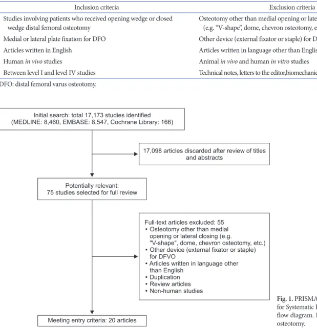
Table 1. Inclusion and Exclusion Criteria
Inclusion criteria Exclusion criteria
Studies involving patients who received opening wedge or closed
wedge distal femoral osteotomy Osteotomy other than medial opening or lateral closing (e.g. “Vshape”, dome, chevron osteotomy, etc.) Medial or lateral plate fixation for DFO Other device (external fixator or staple) for DFO
Articles written in English Articles written in language other than English
Human in vivo studies Animal in vivo and human in vitro studies
Between level I and level IV studies Technical notes, letters to the editor,biomechanical reports, or review articles DFO: distal femoral varus osteotomy.
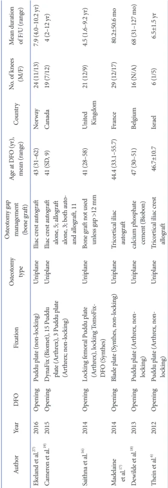
Initial search: total 17,173 studies identified (MEDLINE: 8,460, EMBASE: 8,547, Cochrane Library: 166)
17,098 articles discarded after review of titles and abstracts
Potentially relevant:
75 studies selected for full review
Meeting entry criteria: 20 articles
Full-text articles excluded: 55 Osteotomy other than medial opening or lateral closing (e.g.
"V-shape", dome, chevron osteotomy, etc.) Other device (external fixator or
for DFVO
Articles written in language other than English
Duplication Review articles Non-human studies
staple)
Fig. 1. PRISMA (Preferred Reporting Items for Systematic Reviews and Metaanalyses) flow diagram. DFVO: distal femoral varus osteotomy.
deformities, and DFO. The search was performed from January 1990 to October 2016. Next, the references from the included studies were screened, and experts in the field were contacted for help in identifying additional studies. Two independent reviewers selected citations based on the titles and the abstracts. The eligi
bility of the full papers of those citations for study inclusion was then assessed. In cases where a consensus could not be reached, a third reviewer was consulted.
3. Data Abstraction
Each of the selected studies was evaluated by the two indepen
dent reviewers for methodological quality. The following data were extracted from each article: study type, level of evidence, demographic information, prostheses used, surgical details, outcome measures, clinical and radiographic findings, compli
cations, and survival rates. The extracted data were then cross
checked for accuracy. Any disagreements were settled by the third reviewer.
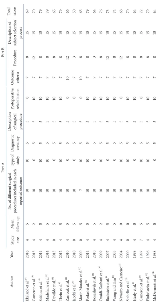
4. Quality Assessment
The methodological quality of the included studies was assessed by the two reviewers using the 10 critical appraisal criteria of the Coleman Methodology Score (CMS). The final scores ranged from 0 to 100, a perfect score (100) indicating a study design that completely avoids the influences of chance, various biases, and confounding factors
22).
Results
1. Included Studies
Following the fulltext review, 16 studies on DFO were ulti
mately included in the systematic review. There were 8 studies on OWDFO and 8 studies on CWDFO. A flowchart illustrating the study selection process is provided in Fig. 1. The characteristics of included studies are listed in Table 2. The number of knees included in each study ranged from 6 to 49. The preoperative diagnosis for DFO was lateral osteoarthritis with genu valgum deformity in all studies. All of the included studies except one study
23)that did not mention the followup period had a mini
mum followup of 2 years. All studies considered conversion to total knee arthroplasty (TKA) as an endpoint for cumulative sur
vival analysis.
2. Quality Assessment
The mean CMS for all included studies was 71 (range, 50 to 79).
Each score for each CMS criterion is shown in Table 3.
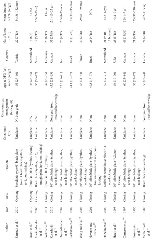
Table 2. Characteristics of Included Studies AuthorYearDFOFixationOsteotomy type Osteotomy gap management (bone graft)
Age at DFO (yr), mean (range)CountryNo. of knees (M/F)Mean duration of F/U (range) Ekeland et al.27) 2016OpeningPuddu plate (nonlocking)UniplaneIliac crest autograft43 (31–62)Norway24 (11/13)7.9 (4.0–10.2 yr) Cameron et al.19) 2015OpeningDynaFix (Biomet), 15 Puddu plate (Arthrex), 3 Puddu plate (Arthrex; nonlocking)
UniplaneIliac crest autograft alone, 5; allograft alone, 3; both auto and allograft, 11
41 (SD, 9)Canada19 (7/12)4 (2–12 yr) Saithna et al.16) 2014OpeningLocking femoral Puddu plate (Arthrex), locking TomoFix DFO (Synthes)
UniplaneBone graft not used unless gap >12 mm41 (28–58)United Kingdom21 (12/9)4.5 (1.6–9.2 yr) Madelaine et al.17)2014OpeningBlade plate (Synthes, nonlocking)UniplaneTricortical iliac autograft44.4 (33.1–55.7)France29 (12/17)80.2±50.6 mo Dewilde et al.18) 2013OpeningPuddu plate (Arthrex, non locking)Uniplanecalcium phosphate cement (Biobon)47 (30–51)Belgium16 (N/A)68 (31–127 mo) Thein et al.6) 2012OpeningPuddu plate (Arthrex, non locking)UniplaneTricortical iliac crest allograft46.7±10.7Israel6 (1/5)6.5±1.5 yr
Table 2. Continued AuthorYearDFOFixationOsteotomy type Osteotomy gap management (bone graft)
Age at DFO (yr), mean (range)CountryNo. of knees (M/F)Mean duration of F/U (range) Zarrouk et al.14) 2010OpeningSterelitziatype 95° blade plate (n=21), blade plate (Synthes; n=1, nonlocking)
UniplaneNo bone graft53 (27–66)Tunisia22 (7/13)54 (36–132 mo) Jacobi et al.20) 2010OpeningTomoFixDFO (Synthes, locking) UniplaneN/A46 (28–63)Switzerland14 (8/6)45 mo Marin Morales et al.12)2000OpeningBlade plate (Synthes, n=13), straight plate (n=4, nonlocking)UniplaneN/A55 (50–72)Spain19 (5/12)6.5 (2–15 yr) Forkel et al.21) 2014ClosingTomoFixDFO (Synthes)UniplaneNone47 (25–55)Germany23 (6/17)13.6 yr Kosashivili et al.11)2010Closing90° offset blade plate (Synthes, nonlocking)UniplaneBone graft from resected bone wedge45.5 (24–63)Canada33 (23/8)15.1 (10–25 yr) OmidiKashani et al.26)2009Closing90° offset blade plate (Synthes, nonlocking)UniplaneNone23.3 (17–41)Iran23 (4/12)16.3 (8–25 mo) Backstein et al.9) 2007Closing90° offset blade plate (Synthes, nonlocking)UniplaneNone44.1 (10–67)Israel38 (10/28)123 (39–245 mo) Wang and Hsu13) 2005Closing90° offset blade plate (Synthes, nonlocking)UniplaneNone53 (31–64)Taiwan30 (2/28)99 (61–169 mo) Navarro and Carneiro23)2004Closing90° offset blade plate (Synthes), fixation from lateral side (non locking) UniplaneNone49.5 (17–77)Brazil26 (4/18)N/A Stahelin et al.25) 2000ClosingMalleable semitubular plate (AO, nonlocking)UniplaneNone57 (39–71)Switzerland
21 (9/10, 2 b
ilateral)5 (2–12 yr) Healy et al.4) 1998Closing90° offset blade plate (AO, non locking)UniplaneNone56 (19–70)USA23 (5/18)4 (2–9 yr) Cameron et al.29) 1997Closing90° offset blade plate (Synthes, nonlocking)UniplaneNone60 (23–84)Canada49 (15/34)3.5 (1–7 yr) Finkelstein et al.10)1996Closing90° offset blade plate (Synthes, nonlocking)UniplaneNone56 (27–77)Canada21 (6/15)133 (97–240 mo) McDermott et al.24)1988ClosingBlade plate (nonlocking)UniplaneBone graft from resected bone wedge53 (22–74)Canada24 (4/20)4 (2–11.5 yr) DFO: distal femoral osteotomy, F/U: followup, SD: standard deviation, N/A: not applicable, AO: arbeitsgemeinschaft für osteosynthesefragen.
Table 3. Coleman Scores for Each Selected Article AuthorYear Part APart B Total scoreStudy sizeMean followup
No. of different surgical procedures included in each reported outcome Type of studyDiagnostic certainty Description of surgical procedure
Postoperative rehabilitationOutcome criteriaProcedureDescription of subject selection process Ekeland et al.27) 2016451010550781569 Cameron et al.19) 20154210055107121570 Saithna et al.16) 20144510105510781579 Madelaine et al.17) 20144510105510781579 Dewilde et al.18) 2013051005510781565 Thein et al.6) 20124510105510781579 Zarrouk et al.14) 20104510055010121566 Jacobi et al.20) 201005100550731550 Marin Morales et al.12) 2000057105501081565 Forkel et al.21) 20144510105510781579 Kosashivili et al.11) 2010451005510731564 OmidiKashani et al.26) 20094210105510781576 Backstein et al.9) 20074510055107121573 Wang and Hsu13) 2005451005510781574 Navarro and Carneiro23) 20044010105507121568 Stahelin et al.25) 20000010105510781570 Healy et al.4) 1998401005510781564 Cameron et al.29) 1997751010550781572 Finkelstein et al.10) 19964510105510781579 McDermott et al.24) 1988401005510781564
3. Surgical Intervention and Rehabilitation
Most of the cases in the included studies used either a locking plate or a blade plate to provide strong stability after osteotomy (Table 2). The plate was fixed on the medial side in cases of CWDFO and lateral side in cases of OWDFO. In Navarro and Carneiro
23)series, medial CWDFO was performed using the anterior approach and plate fixation on the lateral side. For the gap filling material after OWDFO, a majority of the studies used autologous bone graft while allografts
6,19)or calcium phosphates were used in the rest
18). Saithna et al.
16)mentioned that the gap was filled only if the gap size was over 12 mm in their OWDFO series. Bone grafting was not done in one study
14). In CWDFO series, most studies did not use additional bone graft material.
Two studies mentioned the use of morcellized bone grafting which was obtained from the resected bone wedge
11,24). The post
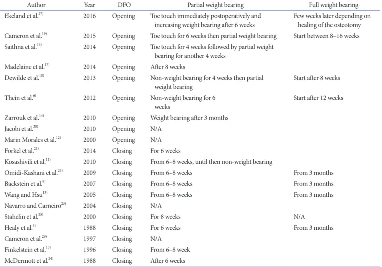
operative weight bearing permit time is demonstrated in Table 4.
Generally, weight bearing was delayed for OWDFO by 2–4 weeks compared to CWDFO.
4. Clinical Outcomes
The clinical outcomes are provided in Table 5. All but one study
23)reported clinical outcome. Various knee scoring systems were used for clinical assessment including Knee Society score (KSS, the French version), modified KSS, Hospital of Special Surgery score, Oxford knee score, Knee Injury and Osteoarthritis Outcome score (pain, symptoms, and function in daily living, kneerelated quality of life, and function in sport and recreation), International Knee Documentation Committee score, Lysholm, Tegner, and Short Form 36. All series showed improvement in clinical scores after DFO.
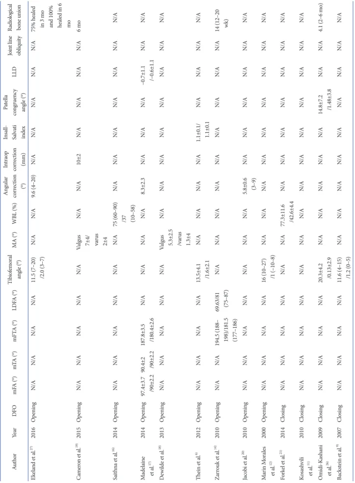
5. Radiological Outcomes
The radiological outcomes are provided in Table 6. Seventeen of the 20 studies reported radiological outcome. Kosashivili et al.
11)did not report the radiological results. Navarro and Car
neiro
23)only reported the joint line obliquity value, and Stahelin et al.
25)reported the mean angular correction after CWDFO. The
Table 4. Rehabilitation (Weight Bearing Period)
Author Year DFO Partial weight bearing Full weight bearing
Ekeland et al.27) 2016 Opening Toe touch immediately postoperatively and
increasing weight bearing after 6 weeks Few weeks later depending on healing of the osteotomy Cameron et al.19) 2015 Opening Toe touch for 6 weeks then partial weight bearing Start between 8–16 weeks Saithna et al.16) 2014 Opening Toe touch for 4 weeks followed by partial weight
bearing for another 4 weeks Madelaine et al.17) 2014 Opening After 8 weeks
Dewilde et al.18) 2013 Opening Nonweight bearing for 4 weeks then partial
weight bearing Start after 8 weeks
Thein et al.6) 2012 Opening Nonweight bearing for 6
weeks Start after 12 weeks
Zarrouk et al.14) 2010 Opening Weight bearing after 3 months
Jacobi et al.20) 2010 Opening N/A
Marin Morales et al.12) 2000 Opening N/A
Forkel et al.21) 2014 Closing For 6 weeks
Kosashivili et al.11) 2010 Closing From 6–8 weeks, until then nonweight bearing
OmidiKashani et al.26) 2009 Closing From 6–8 weeks From 3 months
Backstein et al.9) 2007 Closing From 6–8 weeks From 3 months
Wang and Hsu13) 2005 Closing From 6–8 weeks From 3 months
Navarro and Carneiro23) 2004 Closing N/A
Stahelin et al.25) 2000 Closing For 8 weeks N/A
Healy et al.4) 1988 Closing For 6 weeks From 3 months
Cameron et al.29) 1997 Closing N/A
Finkelstein et al.10) 1996 Closing From 6–8 week McDermott et al.24) 1988 Closing After 6 weeks DFO: distal femoral osteotomy, N/A: not applicable.
Table 5. Clinical Outcome AuthorYearDFOKnee Society score HSS soreOxford knee sore
KOOS IKDCIKSLysholm Tegner Short Form 36French versionModifiedPainSexADLQOLSport Ekeland et al.27)2016OpeningN/AN/AN/AN/A53/7251/6267/7929/5819/42N/AN/AN/AN/AN/A Cameron et al.19)2015OpeningN/AN/AN/AN/AN/AN/AN/AN/AN/A47 (SD, 15)/ 67 (SD, 10)
N/AN/AN/AN/A Saithna et al.16)2014OpeningN/AN/AN/AN/A49.1 (14–97) /70.3 (39–94)45.3 (11–100) /57.5 (32–86)62.7 (21–99) /73.9 (43–100)33.5 (13–100) /42 (6–75)23.7 (0–90) /32.7 (0–95)36.4 (10.3– 51.7) /52.6 (20.7– 94.3)
N/A48.5 (18–100) /54 (31–92)2.8 (1–6) /3.2 (1–9)79.8 (61.1– 107.2) /88.7 (65–105.9) Madelaine et al.17)2014Opening80.5±19 /65.8±21.3 Functional score 50.4±14.6 /68.5±27.6
N/AN/AN/AN/AN/AN/AN/AN/AN/AN/AN/AN/AN/A Dewilde et al.18)2013OpeningN/A43±8/78±23N/AN/AN/AN/AN/AN/AN/AN/AN/AN/AN/AN/A Thein et al.6)2012OpeningN/AN/AN/A13.1±8.6 /26±12.5N/AN/AN/AN/AN/AN/AN/AN/AN/AN/A Zarrouk et al.14)2010OpeningN/AN/AN/AN/AN/AN/AN/AN/AN/AN/A49.28 (14–70) /74.23 (41–92) Functional score 50.68 (30–80)/ 72.85 (40–90)
N/AN/AN/A Jacobi et al.20)2010OpeningN/AN/AN/AN/A36±20 /76±2446±28 /79±1851±26 /84±1712±9 /51±2812±10 /55±30N/AN/AN/AN/AN/A Marin Morales et al.12)
2000OpeningN/AN/A47.5 (36–67) /83.3 (57–97)N/AN/AN/AN/AN/AN/AN/AN/AN/AN/AN/A Forkel et al.21)2014ClosingN/AN/AN/AN/A55±34 /90±1054±10 /88±954±29 /90±939±39 /83±1049±39 /80±19N/AN/AN/A3.5±1.1 /4.2±1.1N/A Kosashvili et al.11)2010ClosingN/A36.8/77.5N/AN/AN/AN/AN/AN/AN/AN/AN/AN/AN/AN/A
Table 5. Continued AuthorYearDFOKnee Society score HSS soreOxford knee sore
KOOS IKDCIKSLysholm Tegner Short Form 36French versionModifiedPainSexADLQOLSport OmidiKashani et al.26)2009ClosingN/AN/A90.7 (77–96) /98.13 (93–100)N/AN/AN/AN/AN/AN/AN/AN/AN/AN/AN/A Backstein et al.9)2007ClosingN/A18 (0–74) /87.2 (50–100) Functional score 54 (0–100)/ 85.6 (40–100)
N/AN/AN/AN/AN/AN/AN/AN/AN/AN/AN/AN/A Wang and Hsu13)2005ClosingN/AN/A46 (20–63) /88 (65–99)N/AN/AN/AN/AN/AN/AN/AN/AN/AN/AN/A Navarro and Carneiro23)2004ClosingN/AN/AN/AN/AN/AN/AN/AN/AN/AN/AN/AN/AN/AN/A Stahelin et al.25)2000ClosingN/AN/A65 (56–70) /84 (61–100)N/AN/AN/AN/AN/AN/AN/AN/AN/AN/AN/A Healy et al.4)1998ClosingN/AN/A65 (42–100) /86 (36–100)N/AN/AN/AN/AN/AN/AN/AN/AN/AN/AN/A Cameron et al.29)1997ClosingN/APreop score, not recorded; postop score, 84.8±18.5 (functional 64.5±21.5)
N/AN/AN/AN/AN/AN/AN/AN/AN/AN/AN/AN/A Finkelstein et al.10)1996ClosingN/AN/AN/AN/AN/AN/AN/AN/AN/AN/AN/AN/AN/AN/A McDermott et al.24)1988ClosingN/AN/AN/AN/AN/AN/AN/AN/AN/AN/AN/AN/AN/AN/A Values are presented as mean±standard deviation and preoperative/postoperative score (range). DFO: distal femoral osteotomy, HSS: Hospital for Special Surgery, KOOS: Knee Injury and Osteoarthritis Outcome score (pain, symptoms, function in daily living, kneerelated quality of life, function in sport and recreation), IKDC: International Knee Documentation Committee, IKS: International Knee Score, ADL: activites of daily living, QOL: kneerelated quality of life, N/A: not applicable, SD: standard deviation, Preop: preoperative, Postop: postoperative.
Table 6. Radiological Results AuthorYearDFOmFA (°)mTA (°)mFTA (°)LDFA (°)Tibiofemoral angle (°)MA (°)WBL (%)Angular correction (°) Intraop correction (mm)
Insall Salvati index
Patella congruency angle (°)LLDJoint line obliquityRadiological bone union Ekeland et al.27)2016OpeningN/AN/AN/AN/A11.5 (7–20) /2.0 (3–7)N/AN/A9.6 (4–20)N/AN/AN/AN/AN/A75% healed in 3 mo and 100% healed in 6 mo Cameron et al.19) 2015OpeningN/AN/AN/AN/AN/AValgus 7±4/ varus 2±4
N/AN/A10±2N/AN/AN/AN/A6 mo Saithna et al.16)2014OpeningN/AN/AN/AN/AN/AN/A75 (60–90) /37 (10–58)
N/AN/AN/AN/AN/AN/AN/A Madelaine et al.17)2014Opening97.4±3.7 /90±2.290.4±2 /90±2.2187.8±3.5 /180.4±2.6N/AN/AN/AN/A8.3±2.3N/AN/AN/A–0.7±1.1 /–0.6±1.1N/AN/A Dewilde et al.18)2013OpeningN/AN/AN/AN/AN/AValgus 5.3±2.5 /varus 1.3±4
N/AN/AN/AN/AN/AN/AN/AN/A Thein et al.6) 2012OpeningN/AN/AN/AN/A13.5±4.1 /1.6±2.1N/AN/AN/AN/A1.1±0.1/ 1.1±0.1N/AN/AN/AN/A Zarrouk et al.14)2010OpeningN/AN/A194.5 (188– 198)/181.5 (177–186)
69.63/81 (75–87)N/AN/AN/AN/AN/AN/AN/AN/AN/A14 (12–20 wk) Jacobi et al.20) 2010OpeningN/AN/AN/AN/AN/AN/AN/A5.8±0.6 (3–9)N/AN/AN/AN/AN/AN/A Marin Morales et al.12)2000OpeningN/AN/AN/AN/A16 (10–27) /1 (–10–8)N/AN/AN/AN/AN/AN/AN/AN/AN/A Forkel et al.21) 2014ClosingN/AN/AN/AN/AN/AN/A77.3±11.6 /42.6±4.4N/AN/AN/AN/AN/AN/AN/A Kosashvili et al.11)2010ClosingN/AN/AN/AN/AN/AN/AN/AN/AN/AN/AN/AN/AN/AN/A OmidiKashani et al.26)2009ClosingN/AN/AN/AN/A20.3±4.2 /0.13±2.9N/AN/AN/AN/AN/A14.8±7.2 /1.48±3.8N/AN/A4.1 (2–6 mo) Backstein et al.9)2007ClosingN/AN/AN/AN/A11.6 (4–15) /1.2 (0–5)N/AN/AN/AN/AN/AN/AN/AN/AN/A
reported parameters were mechanical femoral axis, mechanical tibia axis, mechanical femorotibial axis, lateral distal femoral angle, tibiofemoral angle, mechanical axis, weight bearing line, leg length discrepancy (LLD), and patellarelated parameters.
All studies demonstrated improvement toward neutraltovarus alignment after DFO. According to few studies
6,26), there were minimal impact on the patella. The LLD was not a significant factor after OWDFO
17). The mean radiological bone union time was between 3–6 months for OWDFO
14,19)and around 4 months for CWDFO
13,26).
6. Complications and Survivorship
Complications of both procedures are shown in Table 7. Among various complications, plate irritation needing a removal pro
cedure was reported in up to 12/14 cases (86%) in an OWDFO study
20). On the contrary, the incidence of plate removal was low in CWDFO series beside one study reporting 16/23 cases (70%)
21). One study
27)reported 3/13 cases (13%) of delayed union in their OWDFO series. The incidence of other complications, such as loss of correction, nonunion, infection, and fractures, did not differ between OWDFO and CWDFO. The survivorship (TKA as endpoint) was reported in some studies (Table 7). There was no notable difference in survivorship between OWDFO and CWDFO.
Discussion
The important finding of this systematic review is that the clini
cal and radiological outcome including the survival rate did not significantly differ between OWDFO and CWDFO contrary to our initial hypothesis.
It has been known that OWDFO is effective for medium or large corrections and particularly easy to perform and allows for precise control of the correction amount
5,6). By contrary, CWDFO is known to be technically more difficult than OWDFO because the surgeon is very reliant on the accuracy of preoperative plan
ning and bony resection
16). However, differences in the improve
ment of postoperative radiological alignment between OWDFO and CWDFO series were not demonstrated in this study. The reason may be multifactorial and include improvement of surgi
cal techniques for CWDFO. Compared to CWDFO techniques, however, the main concern for OWDFO techniques is the infe
rior mechanical stability
28)at the osteotomy site as well as the lon
ger healing time of the defect. In a previous biomechanical study on axial and torsional stability after supracondylar osteotomies, the least amount of motion and highest stiffness were measured
Table 6. Continued AuthorYearDFOmFA (°)mTA (°)mFTA (°)LDFA (°)Tibiofemoral angle (°)MA (°)WBL (%)Angular correction (°)Intraop correction (mm)
Insall Salvati index
Patella congruency angle (°)LLDJoint line obliquityRadiological bone union Wang and Hsu13) 2005ClosingN/AN/AN/AN/A18.2 (12–27) /1.2 (–6–10)N/AN/AN/AN/AN/AN/AN/AN/A4.7 (3–9 mo) Navarro and Carneiro23)2004ClosingN/AN/AN/AN/AN/AN/AN/A5N/AN/AN/AN/A+3.1/ –2N/A Stahelin et al.25)2000ClosingN/AN/AN/AN/AN/AN/AN/A1.7 (0–4)N/AN/AN/AN/AN/AN/A Cameron et al.29) 1997ClosingN/AN/AN/AN/A13 (7–23) /1.0 (8–10)N/AN/A11.8±4N/AN/AN/AN/AN/AN/A Finkelstein et al.10) 1996ClosingN/AN/AN/AN/A1.7 (0–3)/10N/AN/AN/AN/AN/AN/AN/AN/AN/A McDermott et al.24)1988ClosingN/AN/AN/AN/A0 degree, 18; 2–8 varus degree, 4; 6 valgus degree, 2
N/AN/AN/AN/AN/AN/AN/AN/AN/A Values are presented as mean±standard deviation and preoperative/postoperative (range). DFO: distal femoral osteotomy, mFA: mechanical femoral axis, mTA: mechanical tibial axis, mFTA: mechanical femorotibial axis, LDFA: lateral distal femoral angle, MA: mechanical axis, WBL: weight bearing line, Intraop: intraoperative, LLD: leg length discrepancy, +: medial inclincation, –: lateral inclincation, N/A: not applicable.
Table 7. Complications and Survivorship AuthorYearNo.of casesF/U period (range)Plate irritation (removal) Loss of correction Non union Delayed union Infection Fracture OthersConversion to TKA
Survivorship (TKA as endpoint) Ekeland et al.27) 2016247.9 (4.0–10.2 yr)3 (13)02One patient with antecurvation after fall injury and 1 patient of arthroscopic adhesiolysis for reduced flexion
6 (25)88% at 5 yr and 74% at 10 yr Cameron et al.19)2015194 (2–12 yr)3 (16)01 (5)00Four additional arthroscopic surgeries for persistent symptoms5 (26)74% at 5 yr Saithna et al.16) 2014214.5 (1.6–9.2 yr)10 (48)2 (10)1 (5)1 (5)Two additional arthroscopic surgeries for persistent symptoms4 (19)79% at 5 yr Madelaine et al.17)20142980.2±50.6 mo23 (79)2 (7)1 (3)One case of Judet's arthromyolysis for stiffness5 (17)91.4% at 5 yr Dewilde et al.18) 20131668 (31–127 mo)4 (25)1 (6)2 (13)82% at 7 yr Thein et al.6) 201266.5±1.5 yr000000N/A Zarrouk et al.14) 20102254 (36–132 mo)N/A Jacobi et al.20) 20101445 mo12 (86)1 (7)N/A Marin Morales et al.12)2000196.5 (2–15 yr)2 (11)01 (5)N/A Forkel et al.21) 20142313.6 yr16 (70)1 (4)00N/A Kosashvili et al.11)20103315.1 (10–25 yr)1 (3)15 (45)N/A OmidiKashani et al.26)20092316.3 yr (8–25 mo)1 (4)One patient with plate revision after fall injury N/A Backstein et al.9)200738123 (39–245 mo)12 (32)82% at 10 yr and 45% at 15 yr Wang and Hsu13)20053099 (61–169 mo)1 (3)1 (3)1 (3)3 (10)87% at 10 yr Navarro and Carneiro23)200426N/AN/A Stahelin et al.25) 2000195 (2–12 yr)11Two hematoma and one popliteal vein thrombosis (conservative treatment) Healy et al.4) 1998234 (2–9 yr)321One patient with manipulation, two additional arthroscopic surgeries for persistent symptoms at lateral joint
2N/A
in medial oblique CW osteotomy fixated with an angled blade plate. The lateral OW techniques resulted in less stability and lower stiffness than the medial CW osteotomy
28). Both of these factors are considered to work in favor when direct bonetobone apposition is obtained as in a CW technique. To overcome the concern, addition of bone substitute in the osteotomy gap or iliac corticocancellous bone graft has been performed in a majority of OWDFO series
6,18,19,29).
The cumulative survival of DFO series should be noted. Saithna et al.
16)reported 79% at 5 years and Madelaine et al.
17)reported an even higher rate of 91.4% at 5 years in their OWDFO series.
Likewise, Backstein et al.
9)reported 82% at 10 years and 45% at 15 years in their recent CWDFO series. Finkelstein et al.
10)previ
ously reported 83% at 4 years and 64% cumulative survival at 10 years. Although heterogeneity between studies may prevent fur
ther statistical analyses, the survivorship figures were favorable for both OWDFO and CWDFO series with similar performance.
On the surgical aspect of the procedures, the OWDFO tech
nique allows finetuning of deformity correction with application of an opening device such as a laminar spreader until the desired angle is achieved. By contrast, in a CW osteotomy, the surgeon is very reliant on the preoperative plan for accuracy of bony resec
tion; however, precise resection of a wedge is technically difficult although not demonstrated in this study.
The choice of implant is an important consideration. Edgerton et al.
7)reported 17/24 patients (70%) complications including 7 cases of delayed union or nonunion by using staples for fixa
tion. Stahelin et al.
25)used a malleable semitubular plate. They modified the conventional tubular plate into a fixed angle blade plate to improve the mechanics of fixation. They suggested that the strong fixation device is one of critical factors for successful outcome. Although the studies using the Puddu plate (Arthrex, Naples, FL, USA)
6,18)did not demonstrate inferior results com
pared to the studies using the blade plate (Synthes, Oberdorf, Switzerland) or the locking TomoFix plate (Synthes), it has been recommended to use these devices with greater axial and torsion
al stability
30). In contrast to the tibial bone, the femur has a longer lever arm with more rotational force applied requiring more stable plate configuration than the previously used or currently used implants for high tibial osteotomy. Improving plate stability will also facilitate rapid rehabilitation shortening the nonweight bearing or partial weight bearing period.
Evaluation of the type of graft (i.e., autograft vs. allograft or syn
thetic materials) among the OW osteotomy studies was limited due to the heterogeneity of graft choice. Given the wide variabil
ity, no conclusions can be drawn on the optimal graft choice for
Table 7. Continued AuthorYearNo.of casesF/U period (range)Plate irritation (removal)Loss of correction Non union Delayed union Infection Fracture OthersConversion to TKA
Survivorship (TKA as endpoint) Cameron et al.29)1997493.5 (1–7 yr)1 (2)6 (12)0One patient with derotational osteotomy (15 degrees of external rotational deformity developed after DFO) and 14 additional arthroscopic surgeries for persistent symptoms
5 (10)87% at 7 yr Finkelstein et al.10) 199621133 (97–240 mo)1 (5)1 (5)One patient with stiffness underwent manipulation under anesthesia7 (33)64% at 10 yr McDermott et al.24) 1988244 (2–11.5 yr)1One patient with plate revision after fixation failureN/AN/A Values are presented as mean±standard deviation or number (%). F/U: followup, TKA: total knee arthroplasty, N/A: not applicable, DFO: distal femoral osteotomy.