As the general population ages, the number of patients with degenerative knees also continues to increase1). In addition, more patients of advanced age are choosing surgical treatment to im
prove their quality of life. Total knee arthroplasty (TKA) is the most costeffective and safest procedure for restoring function to an arthritic knee joint2). However, despite its safety, TKA may have unsatisfactory outcome associated with immediate or short
term complications after surgery in a population with other concomitant diseases. Perioperative complications include blood loss, infection, early hemorrhage and wound breakdown, intra
operative fractures, and anesthetic problems as well as cardiovas
cular, respiratory, renal, electrolyte, and other medical problems3). Acute arterial occlusion is rarely associated with TKA. A rare but serious complication is occlusion of the artery, the incidence of which is stated to be 0.033%4) in large patient populations. Here, we describe a case of acute arterial occlusion after TKA associ
ated with atrial fibrillation (AF). The case presented in this report
is different from that in previous studies in that thrombotic oc
clusion occurred even after adequate prophylaxis following knee arthroplasty in a patient who had AF.
Case Report
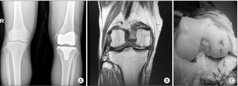
An 83yearold male patient had osteoarthritis for 7 years in the right knee, with no flexion contracture or highly varus and valgus deformity. Because of extreme pain, the patient underwent magnetic resonance imaging, which showed lesions in the medial femoral condyle cartilage in the weightbearing region; the same findings were confirmed during the TKA operation (Fig. 1). He had previously undergone left primary TKA for incapacitating knee pain recalcitrant to conservative therapy at our hospital 25 months ago. We considered salvage procedures such as cartilage restoration procedure or high tibial osteotomy considering the size of the knee joint lesion and combined medical disease with age. However, we planned TKA since the patient complained of severe pain, and satisfaction was high on the contralateral side after TKA. He had been taking warfarin for paroxysmal AF. He had a smoking history of 1.5 packs per day for 32 years and cho
lecystectomy 2 years ago, but he had no specific medical history or history of peripheral vascular disease, being bedridden for a long period, or glucocorticoid usage. The patient had normal femoral, popliteal, and ankle pulses bilaterally on palpation.
The ankle brachial index (ABI; right, 1.06; left, 1.04) and echo
cardiography showed no specific clinical manifestation. After 3 Acute arterial occlusion is a rare complication following total knee arthroplasty (TKA). This is a report of a case of acute femoral artery occlusion and its sequelae following TKA in a patient with a history of atrial fibrillation. Arterial circulation of the lower limb could not be restored by thrombectomy treatments, and aboveknee amputation had to be carried out.
Keywords: Knee, Arthroplasty, Artery, Occlusion, Atrial fibrillation
Received April 17, 2017; Revised (1st) June 8, 2017; (2nd) July 5, 2017;
Accepted July 10, 2017
Correspondence to: HongMan Cho, MD
Department of Orthopedic Surgery, Gwangju Veterans Hospital, 99 Cheomdanwolbongro, Gwangsangu, Gwangju 62284, Korea Tel: +82626026162, Fax: +82626026936
Email: chm1228@hanmail.net
84
This is an Open Access article distributed under the terms of the Creative Commons Attribution NonCommercial License (http://creativecommons.org/licenses/bync/4.0/) which permits unrestricted noncommercial use, distribution, and reproduction in any medium, provided the original work is properly cited.
Copyright © 2018 KOREAN KNEE SOCIETY www.jksrr.org
days of warfarin hold for perioperative bridging anticoagulation therapy, the patient’s international normalized ratio (INR) was 1.72. Enoxaparine sodium (40 mg Cnoxane; YooYoung Pharm co., Seoul, Korea) was administered to the patient. He underwent TKA under spinal anesthesia. TKA was initiated with a tourni
quet inflated to a pressure of 300 mmHg, and the anterior parapa
tellar approach was used. The total blood loss for the procedure was 350 mL, which was measured by weighing the gauze plus the suction drain amount without the irrigating solution. The tourni
quet time lasted for 96 minutes. No intraoperative complications were noted during the surgical procedure. Compressive dressing was applied on the lower limb after the operation. After surgery, the patient was transferred to the recovery room. The patient’s motor and sensory functions were not checked in the recovery room due to spinal anesthesia, but the patient’s lower limb was warm and the pulsation of the dorsalis pedis artery was intact.
After about 20 minutes in the recovery room, the patient’s mental state declined and he became drowsy, his respiration decreased, and his blood pressure decreased to 80/40 mmHg; thus, he was transferred to the intensive care unit. Computed tomography (CT) images showed no specific lesions in the brain and lungs.
Supportive therapy was administered, and the patient’s vital signs recovered to normal range on postoperative day 1. However, on postoperative day 1, after waking up, the patient showed an un
stable mental status without awareness of places or persons and signs of delirium; he was trembling and tried to get out of bed.
He was given a sedative, which did not provide much improve
ment, and restraint bands were used to limit the patient’s hand and foot movements. On postoperative day 2, the drain tube was
removed because the Hemovac drain amount decreased to 60 mL. To remove the drain, the compressive dressing was removed for the first time after surgery. However, the pulse and tempera
ture of the lower limb were not checked, which should have also been checked on postoperative day 1. We should have classified him as a high risk patient for thrombotic events, but which we did not. If we had examined more carefully, arterial occlusion could have been found earlier. On postoperative day 2, Cnoxane 40 mg and warfarin were administered because the patient’s INR increased to 1.83. An air pressure device was used for mechanical prophylaxis to prevent deep vein thrombosis after the removal of the compressive dressing. On postoperative day 3, the dosage of warfarin was the same as that used before the surgery. On the afternoon of postoperative day 2, when the compressive dressing was removed for dressing change, there was notable necrosis of the foot on gross examination. No pulsation of the dorsalis pedis artery was noted during palpation or capillary filling of the toe
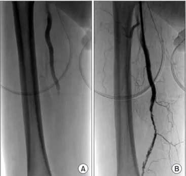
nail. Emergency lowerextremity CT angiography showed total occlusion of the right femoral artery (Fig. 2). After emergency consultation with the cardiovascular department, angiography was performed, which showed total embolic occlusion of the right femoral artery. Fogarty thrombectomy was attempted via the right inguinal site; however, the reperfusion of the popliteal and anterior and posterior tibial arteries was not satisfactory (Fig.
3). No back bleeding was noted, which means that there is no bleeding through the catheter where it is approached below the occluded artery. Open thrombectomy was performed when the occlusion site was not completely resolved, but flow maintenance was observed on completion angiography, thus precluding the
A B C
Fig. 1. (A) Radiograph showing proximal tibial vara in the right knee and previous total knee arthroplasty status of the left knee. (B) T1weighted coronal magnetic resonance imaging scan showing degeneration of the medial meniscus, loss of articular cartilage in the medial femoral condyle, and tiny marginal osteophytes in the right knee joint. (C) Removal of the right knee joint capsule with cartilage loss in the medial femur.
need for additional open thrombectomy. However, upon evalu
ation, we were dissatisfied because the flow was insufficient. On postoperative day 4, sepsis combined with rhabdomyolysis was suspected. The patient was drowsy, with decreased blood pres
sure (70/50 mmHg), and the necrosis below the knee deteriorat
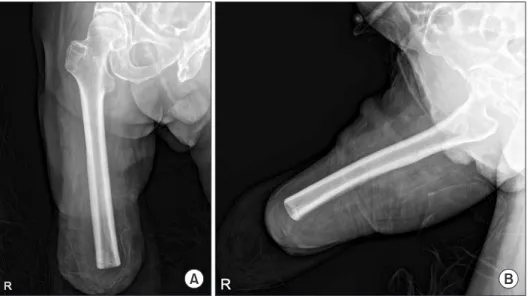
ed rapidly (Fig. 4). On postoperative day 17, aboveknee amputa
tion was performed (Fig. 5). After amputation, the vital signs of the patient stabilized. He was discharged, using a wheelchair for ambulation. The patient has been monitored for 6 months after amputation.
Discussion
Arterial complications after TKA are relatively uncommon.
Although infrequent, the arterial complications after TKA are heterogeneous; the most frequently reported ones are arterial thrombosis, arterial transection, arteriovenous fistula, and aneu
rysm formation5). The reported incidence of complications after TKA including arterial occlusion, arteriovenous fistula, arterial aneurysm, and arterial severance ranged from 0.03% to 0.17%6). Nonetheless, the infrequency of these complications may make the diagnosis and treatment extremely challenging to surgeons, and limbthreatening ischemia may subsequently occur7). Two possible causes of necrosis were considered in this case. The first is the patient’s AF. AF is a very common type of arrhythmia, and its prevalence increases significantly with age. This can lead to an increase in blood loss and thrombotic events such as cerebrovas
cular accident during the perioperative surgical period. AF pa
tients need longterm (usually lifelong) anticoagulation therapy to decrease the risk of a thrombotic event8). We presented a case of arterial occlusion due to thrombosis despite the use of periop
erative bridging anticoagulation therapy (warfarin and enoxapa
rine).
Fig. 2. Computed tomography angiography showing superficial femoral artery occlusion in the right limb.
A B
Fig. 3. (A) Arteriogram showing right superficial femoral artery occlu
sion. (B) Arteriogram showing the reperfusion of the superficial femoral, popliteal, and anterior and posterior tibial arteries after Fogarty throm
bectomy.
Fig. 4. Photograph showing necrosis of the lower leg on postoperative day 7.
A higher risk of cardiocerebral events, including acute myocar
dial infarction and cerebral stroke, has been reported in AF. In addition, orthopedic surgery can significantly increase this risk on account of the hypercoagulability status, elevated sympathetic activation, and higher level of catecholamine9), and patients with a history of AF were more likely to develop thrombotic events after orthopedic surgery10). To the best of our knowledge, only a few studies have assessed the impact of AF on the outcomes after TKA10). In addition to the complication experienced in this present case, which is very rare, given the implementation of appropriate perioperative bridging anticoagulation therapy, the lower limb arterial condition of the patient was overlooked. Close observation of blood circulation in the lower extremity should be done regularly in patients with high thrombotic risk such as AF. Although the screening tests (palpation and ABI test) of the lower extremity arterial circulations showed no preoperative ab
normality, a more precise examination of the lower limb artery of the patient was not performed before the operation. We sug
gest that Doppler ultrasound or CT angiography may be useful in preventing such fatal complications in patients at high risk for thrombotic events after orthopedic surgery. Furthermore, using a tourniquet in TKA is controversial, as plaque disruption has been reported after tourniquet use in patients with calcification of the femoral or popliteal artery11). It has been suggested that the mechanical pressure from the tourniquet may traumatize athero
matous vessels, causing fractures and dislodgment of the plaque, especially in old patients11). The vessels of old patients may have calcified and lost their elasticity, and intimal tear and plaque em
bolization could result from the fixation of the femoral artery by the tourniquet and the stretching of the distal artery during in
traoperative manipulation12). Therefore, if calcified blood vessels
are noted in preoperative plain radiographs, it is necessary to pay more attention to the use of customary tourniquet.
Acute arterial occlusions are serious and can lead to amputa
tion if inadequately treated. Treatment comprises anticoagula
tion or surgical intervention, which includes thrombectomy and bypass grafting. Therefore, surgeons should take care to prevent the occurrence of this serious complication and avoid the worst scenario, i.e., amputation. Patients who have acute limb occlusion with severe symptoms such as sensory loss in the toe, rest pain, and moderate motor deficit require emergent surgical revascular
ization13). However, in this case, we considered the patient’s dis
comfort after surgery due to delirium and missed the appropriate period of treatment by not performing a basic physical examina
tion of the lower extremities on postoperative day 2. Therefore, we suggest careful checking of motor, sensory, and circulation of patients’ lower extremity, which is a basic but highly overlooked procedure in patients at high risk for thrombotic events after orthopedic surgery. If acute occlusion of the lower limb is identi
fied, bypass grafting and revascularization6) or thrombectomy using an aspiration catheter12) including thrombolytic therapy is necessary. Thrombectomy is the most frequently described treat
ment, which can lead to complete recovery; however, in patients with extreme necrosis, as in this case, thrombectomy might be futile and ultimately need amputation. In our case, the patient presented with foot coldness, decreased sensation, and paresthe
sias, thus prompting immediate thrombectomy with a Fogarty catheter for revascularization of acute arterial occlusion, which, however, failed. Calligaro et al.6) reported the largest singlecenter experience with management of acute ischemic complications as
sociated with TKA. They claimed that this complication was best managed by an aggressive protocol including arterial bypass and
A B
Fig. 5. Radiographs of the right femur after aboveknee amputation: anteroposterior (A) and lateral (B).
patient’s arterial condition should be considered to avoid acute arterial occlusive disease. Moreover, the fundamental process of examination of blood circulation in the lower extremity per
formed periodically and thoroughly after surgery is very impor
tant in patients at high risk for thrombotic events.
Conflict of Interest
No potential conflict of interest relevant to this article was re
ported.
References
1. Zhang Y, Jordan JM. Epidemiology of osteoarthritis. Clin Geriatr Med. 2010;26:35569.
2. Jones CA, Beaupre LA, Johnston DW, SuarezAlmazor ME.
Total joint arthroplasties: current concepts of patient out
comes after surgery. Clin Geriatr Med. 2005;21:52741.
3. Menendez ME, Memtsoudis SG, Opperer M, Boettner F, Gonzalez Della Valle A. A nationwide analysis of risk factors for inhospital myocardial infarction after total joint arthro
plasty. Int Orthop. 2015;39:77786.
8. Schulman S, Hwang HG, Eikelboom JW, Kearon C, Pai M, Delaney J. Loading dose vs. maintenance dose of warfarin for reinitiation after invasive procedures: a randomized trial.
J Thromb Haemost. 2014;12:12549.
9. Kurtz S, Ong K, Lau E, Mowat F, Halpern M. Projections of primary and revision hip and knee arthroplasty in the Unit
ed States from 2005 to 2030. J Bone Joint Surg Am. 2007;89:
7805.
10. Aggarwal VK, Tischler EH, Post ZD, Kane I, Orozco FR, Ong A. Patients with atrial fibrillation undergoing total joint arthroplasty increase hospital burden. J Bone Joint Surg Am.
2013;95:160611.
11. Smith DE, McGraw RW, Taylor DC, Masri BA. Arterial complications and total knee arthroplasty. J Am Acad Or
thop Surg. 2001;9:2537.
12. Berger C, Anzbock W, Lange A, Winkler H, Klein G, Engel A. Arterial occlusion after total knee arthroplasty: successful management of an uncommon complication by percutane
ous thrombus aspiration. J Arthroplasty. 2002;17:2279.
13. Sedghi Y, Collins TJ, White CJ. Endovascular management of acute limb ischemia. Vasc Med. 2013;18:30713.