Received: 11 November, 2015 Revised: 18 November, 2015 Accepted: 19 November, 2015 Corresponding author: Su-Young Lee
Department of Physical Therapy, Division of Health Science, Baekseok University, 76 Munam-ro, Dongnam-gu, Cheonan 31065, Republic of Korea Tel: 82-41-550-2546 Fax: 82-41-550-2829 E-mail: [email protected]
This is an Open-Access article distributed under the terms of the Creative Commons Attribution Non-Commercial License (http://creativecommons.org/licens es/by-nc/4.0) which permits unrestricted non-commercial use, distribution, and reproduction in any medium, provided the original work is properly cited.
Copyright © 2015 Korean Academy of Physical Therapy Rehabilitation Science
http://dx.doi.org/10.14474/ptrs.2015.4.2.108 pISSN 2287-7576
eISSN 2287-7584
Phys Ther Rehabil Sci 2015, 4 (2), 108-114 www.jptrs.org
A comparison of vital capacity values and respiratory muscles activities on pelvic tilt position
Seo-Young Janga, Su-Young Leeb
aPhysical Therapy Room, Department of Rehabilitation Medicine, Seoul Medical Center, Seoul, Republic of Korea
bDepartment of Physical Therapy, Division of Health Science, Baekseok University, Cheonan, Republic of Korea
Objective: The purpose of this study was to examine the effect on vital capacity (VC) and inspiratory muscle activation according to the anterior and posterior pelvic tilt positions.
Design: One group pretest-posttest design.
Methods: Twenty-six healthy adult men and women, age 19 to 27 years, volunteered to participate in this study. Forced vital ca- pacity (FVC), and forced expiratory volume in 1 second (FEV1) were measured by desktop spirometer in the pelvic positions dur- ing respiration, and muscle activation was recorded from sternocleidomastoid, upper trapezius, external intercostal, rectus abdom- inis, and external oblique muscles by surface electromyography (EMG) at the same time. EMG values were normalized by max- imum muscle contractions (% maximum voluntary isometric contraction). Subjects were to breathe in as much air as possible and then exhale as quickly as possible in both anterior and posterior pelvic tilt positions. To measure lung capacity, inspiration was measured for 5 seconds and expiration was measured for 7 seconds with data collection taken place during the middle three seconds. Lung capacities were measured in each position three times.
Results: For the results of this study, there was a significant increase in both FVC and FEV1 values during the anterior pelvic tilt- ing compared to the posterior pelvic tilting posture (p<0.05). The sternocleidomastoid, upper trapezius muscle, rectus abdominus and external oblique muscle activation was significantly increased during anterior pelvic tilt compared to the posterior pelvic tilt position (p<0.05).
Conclusions: These findings suggest that pelvic anterior tilt position could be more effective for vital capacity and respiratory muscles activation during respiration.
Key Words: Electromyography, Posture, Respiratory muscles, Vital capacity
Introduction
Respiration involves the process of oxygen from the out- side being drawn into the body and delivered and consumed into the cells and therefore undergo metabolic consumption, which leads to the production of carbon dioxide, which then is then discharged outside of the body [1]. The breathing mechanism is largely divided into two parts. The two parts are inspiration and expiration, where inspiration is accom- panied by the active movement of the diaphragm, and ex- piration involves the inspiratory muscles being relaxed and
the passive process of the ribcage returning back to its origi- nal position [1].
Lung capacity is the amount of air forced out during ex- halation, the largest possible change in lung volume, which indicates a person’s maximum inspiration capacity. In gen- eral, the pulmonary functions tests include lung capacity, ti- dal volume, inspiratory capacity, expiratory reserve volume, forced vital capacity (FVC), forced expiratory volume in 1 second (FEV1), which all measures the breathing index and assess the general breathing ability [2]. Specifically, in de- termining the total lung volume, lung capacity is considered
a very important indicator [3]. With lung capacity use during maximum inspiration, the diaphragm and external inter- costal muscles contract more forcefully, and the accessory inspiration muscles expand the chest cavity. Of the inspira- tion muscle, the sternocleidomastoid (SCM) is one of the most important accessory muscles, and together with the up- per trapezius, they work together to raise the sternum up and play a role in pulling the ribs up [4]. Campbell and Green [5]
(1955) has reported that lung during capacity measurements on abdominal muscles during voluntary contraction, the thoracic cavity and lung volume, decreases, in which the muscle activity must increase. However, this activity is not apparent during the inspiratory phase.
Park et al. [6] (1993) reported that lung capacity will be decreased due to impairments related to breathing center dysfunction, such as ribcage size reduction, weakness and paralysis of the expiratory and inspiratory muscles. Just as the human body responds or changes to any type of stimulus, so does lung function. The general functions of the lungs are affected by low body weight, height, gender, and age [7], and it is also stated that there is a proportional relationship between physical build and lung function in mammals [8].
Knudson et al. [9] (1983) described three different stages of age growth, maturity and decline of each lung function, spe- cifically in growth in children less than ages 11 to 12 years, the maturity is usually seen in women up to 20 years old, men up to 25 years old, and after that, decline in physique and lung ventilation are all reduced. There is a high correla- tion between lung volume and height [10], and although in normal, healthy persons, the lung capacity is used as a pre- dictive index, and the subject’s height is most commonly used as the standard [11]. Although it has been stated that of all the racial backgrounds, it has been observed that colored people distinctively have reduced lung capacities [12], there is no standardized normal value between racial groups [13].
In addition, body posture influences respiration [14]. Just like how the head and neck are positioned according to the location of the thoracic vertebrae, the thoracic vertebrae is influenced by the location of the pelvis and hip bones. When the pelvic and hip bones are at an ideal condition or state, an ideal position of the thoracic vertebrae can be observed.
Pelvic tilting and the position of the spine, lordosis and ky- phosis in the sagittal plane all have a close correlation [15].
If the pelvis is tilted forward, this will increase the lordosis on the lumbar vertebrae, and the whole spinal alignment will be optimally arranged in a line [16]. In contrast, if the pelvis is posteriorly tilted, the length of the abdominal muscles will
be decreased, the curves of the thoracic vertebrae will in- crease, causing a backward bend [17]. Forces causing an in- crease in kyphosis of the spine are the inspiratory muscles, and the built in force will lead to difficulty breathing due to decreased pressure on the diaphragm [18]. Being related to an increase in the kyphosis of thoracic vertebrae, a weak- ened and sagging chest position leads to a decreased vital ca- pacity and thoracic cavity [19].
Thus, recently, studies on factors that influence lung ca- pacity, the comparison on lung capacity in accordance to posture [20], and changes in abdominal and respiratory mus- cle activity according to position are in progress [21,22].
However, studies comparing lung capacity and muscle ac- tivity, and studies examining the position of the pelvis and changes in the form of the spine and the influence of the lung capacity, and simultaneously looking at the influences of lung capacity and muscle activity are lacking. Accordingly, the purpose of this study was to compare the lung capacity and respiratory muscle activation during anterior and poste- rior pelvic tilt in healthy subjects.
Methods
SubjectsA total of 26 healthy students (14 males, 12 females) at- tending Baekseok University in Cheonan-si of Chungnam have participated in this study. Subjects read and signed the consent of agreement after being informed of the research purpose and procedure.
Subjects without a history of cardiac or respiratory dis- ease, who have not experienced any acute stomach or related diseases, without neurological diseases or spinal arthritis or any diseases that might affect lung function, without any dis- eases that might affect the lateral spinal flexion and anterior and posterior pelvic tilting process, those with full joint range of motion allowing anterior and posterior pelvic tilt, and who could physically and cognitively cooperate during the examination were included in the study. The average age was 22.2 years, average height was 170.5 cm, and average weight was 62.9 kg (Table 1).
Equipment
In order to measure lung capacity, the Pony Fx desktop spirometer (COSMED, Rome, Italy) meeting American Thoracic Society criteria for human testing for FVC, FEV1, and peak flow was used: This precision was the average range 0.03 L (0.80%) in FVC , 0.01 L (0.49%) in FEV1, and
Table 1. General characteristics of subjects (N=26) Variable Male (n=14) Female (n=12) Total Age (y) 22.6 (2.5) 21.8 (1.7) 22.2 (2.1) Height (cm) 176.2 (4.5) 164 (5.4) 170.5 (7.9) Weight (kg) 67.9 (10.7) 56.9 (9.9) 62.9 (11.6) Values are presented as mean (SD).
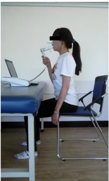
Figure 1. Anterior pelvic tilt position.
−0.016 L (−0.21%) in peak flow. During anterior and pos- terior pelvic tilt, inspiration was measured for 5 seconds, and expiration was measured for 7 seconds. Lung capacity was measured according to FVC, FEV1, FEV6, peak expiratory flow rate, peak inspiratory flow rate, and basic measure- ments of lung capacity and FEV1 was measured.
To measure the respiratory muscle activity, the Trigno surface electromyography (EMG; Delsys Inc., MA, USA) and a common mode rejection ratio 80 dB was used. After measuring with the Trigno sensors, with the Trigno base sta- tion, the signals from the wireless EMG were analyzed using the EMGworks 3.7 (Delsys Inc., Natick, MA, USA). A sam- pling rate of 2,000 Hz was used and was bandpass filtered at 400 to 500 Hz.
Measurements
Lung capacity measurements were carried out after a lapse of 2 hours after eating, and in order to exclude the ef- fects of smoking on lung capacity, subjects were not allowed to smoke for two hours before taking the measurements [23].
Prior to taking measurements, subjects were instructed on how to use the lung capacity measuring equipment and pel- vic tilt positions. The measuring process included the sub- jects making a tight seal with their mouths over the mouth- piece and attaching a clip onto their nose. One respiration was considered as the subject breathing as they normally would 2 to 3 times, and then after breathing in as much air as possible, the subjects were to exhale as quickly as possible.
After observing maximal suction of air, the subjects were in- structed to “quickly exhale, more, more, more, more!” so that they could exhale all the air out, and tried not to have them stop exhaling too soon (Figures 1, 2) [24]. Prior to the course of measuring the subjects’ lung capacities, the sub- jects were shown from a computer monitor a graph of their lung capacities to provide motivation, and inspiration was measured for 5 seconds while expiration was measured for 7 seconds. Lung capacities were measured in each position three times, and the highest scores were selected [25]. After each trial, there was a 1-minute rest period. Also, there was a 5-minute rest period between each position [23].
To assess the pelvic tilt positions, a chair without wheels was used to provide a safe and stable support, and the sub- ject’s bottom and knee joint was at 90 degrees, and the feet were at shoulder width apart. After marking the subject’s greater trochanter, the outer end of the acromion, and the spi- nous process of the lumbar vertebrae, the subjects were in- structed on how to perform an anterior and posterior pelvic
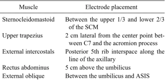
tilt so that there was a difference of greater than 20 degrees (Figures 1, 2) [26]. EMG signal process and collection was taken from one side of the external intercostal muscles, SCM, upper trapezius, rectus abdominis, and external obli- que, and in order to measure muscle activity, the electrode placements for each muscle was marked with a pen (Table 2) [27]. In order to standardize each muscle’s root mean square (RMS) value, the maximal voluntary isometric contraction
Figure 2. Posterior pelvic tilt position.
Table 2. Electrode placement of the respiratory muscles (N=26)
Muscle Electrode placement
Sternocleidomastoid Between the upper 1/3 and lower 2/3 of the SCM
Upper trapezius 2 cm lateral from the center point bet- ween C7 and the acromion process External intercostals Posterior 5th rib interspace along the
line of the axillary Rectus abdominus 5 cm above the umbilicus External oblique Between the umbilicus and ASIS SCM: ternocleidomastoid, ASIS: anterior superior iliac spine.
Table 3. Comparison of lung capacity on pelvic posture (N=26) Variable Anterior
pelvic tilt
Posterior
pelvic tilt t p
FVC (L) 4.4 (1.1) 4.2 (1.1) 2.723 0.012 FEV1 (L) 3.3 (1.1) 3.1 (1.2) 2.147 0.042 Values are presented as mean (SD).
FVC: forced vital capacity, FEV1: forced expiratory volume in 1 second.
(%) was normalized as a percentage. The hand positions for assessing maximal voluntary isometric contractions were performed according to Kendall et al. [28] (2005). Maximal voluntary isometric contractions were performed for 5 sec- onds, three times, with data being measured for 3 seconds except for the first and last second, and the average was quantified using the RMS. Together with measuring for lung capacity, inspiration was measured for 5 seconds with data collection taken place during the middle three seconds, and expiration was measured for 7 seconds, with data collection taken place during the middle three seconds. To prevent muscle fatigue, there was a rest period of 1 minute after each session and a 5-minute break between each posture.
Statistical analysis
In order to obtain the mean and standard deviation values
of the subjects’ age, height, and weight, descriptive statistics was used. To compare the pelvic posture and muscles, a two-way ANOVA was used. In order to compare the pelvic position and respiratory muscles (inspiratory and expira- tory), a one-way ANOVA was used. In order to analyze lung capacity and respiratory muscle activities during the pelvic postures, the paired t-test was used. Data analysis was done using SPSS ver. 12.0 (SPSS Inc., Chicago, IL, USA), and for statistical validation, a significance value of p<0.05 was used.
Results
A comparison of lung capacity on pelvic posture During anterior pelvic tilting posture, the FVC and FEV1
values were significantly increased compared to the posteri- or pelvic tilting posture (p<0.05) (Table 3).
Other respiratory muscle activities of the pelvic posture There was a statistically significant difference between the pelvic position and respiratory (inspiratory and expira- tory) muscle activity (p<0.05). Of the inspiratory muscles, the SCM and upper trapezius muscle activation was sig- nificantly increased during anterior pelvic tilt compared to posterior pelvic tilt. However, there was no significant dif- ference in the external intercostal muscles. Of the expiratory muscles, the rectus abdominus and external oblique muscle activation was significantly increased during anterior pelvic
Table 4. Comparison of muscle activation on respiratory muscles (N=26)
Muscle Anterior
pelvic tilt
Posterior
pelvic tilt t p Inspirators (%MVIC)
Sternocleidomastoid 24.1 (16.0) 14.9 (9.8) 3.856 0.001 Upper trapezius 13.4 (11.5) 8.9 (7.9) 2.815 0.009 External intercostals 22.6 (21.9) 19.8 (15.1) 0.919 0.367 Expirators (%MVIC)
Rectus abdominus 12 (8.8) 8.2 (5.5) 2.576 0.016 External oblique 27.8 (14.1) 22.1 (12.9) 2.466 0.021 Values are presented as mean (SD).
%MVIC: % maximum voluntary isometric contraction.
tilt compared to posterior pelvic tilt (p<0.05) (Table 4).
Discussion
The purpose of this study was to investigate whether hav- ing the pelvic in an anterior or posterior tilt posture had an ef- fect on lung capacity and respiratory muscle activities. Body position has an effect on lung and expiratory volume. The posterior bend of the thoracic vertebrae and the anterior bend of the lumbar vertebrae, the curve of the spine is related to body posture, which in turn, changes or has an effect on lung capacity [29]. In the standard standing position, the pel- vis is in neutral position. When the pelvis is lying in the neu- tral position, the lumbar vertebrae inclining toward the front can be observed [28]. In other words, increased lung ca- pacity occurs due to strengthening of the respiratory mus- cles, that is, having an optimal alignment of the spine in- creases overall lung capacity even further compared to the natural or kyphotic position. Therefore, previous studies warrant the examination of the effect of respiratory exer- cises on various pelvic postures. Previous studies also state that there is a statistically significant increase in lung ca- pacity and expiratory volume in the standing position [7,30].
Like previous studies, in this study anterior pelvic tilt posi- tion was higher than posterior pelvic tilt position in lung capacity. This study in order to analyze lung capacity ac- cording to posture, the FVC and FEV1 were assessed. FVC and FEV1 are used to facilitate the judgment of restrictions or obstructive ventilation failure, and are indicators that are commonly used within the clinic [31], and compared to oth- er indicators, they are considered to have the least amount of variance and so they are most commonly used [32]. In this study, the FVC and FEV1 values were higher during anterior
pelvic tilt compared to posterior pelvic tilt (p<0.05). Similar to a lordotic curve, an anterior pelvic tilt created greater lung capacity, which supports and provides a backboard for the results of other related research.
In this study, the muscle activation of all respiratory and expiratory muscles were more increased during anterior pel- vic tilt compared to posterior pelvic tilt. As for the inspi- ratory muscles, the SCM muscle activation was 24.1% with an anterior pelvic tilt, with a change value of 9.2% higher than with a posterior pelvic tilt, and it showed the highest amount of muscle activity out of all the inspiratory muscles.
In terms of expiratory muscles, the external oblique muscles produced a muscle activity of 27.8%, which was higher than the rectus abdominus muscle activity of 12%. In Kera and Maruyama’s [33] (2005) study on the effect of posture on respiratory activity of the abdominal muscle, subjects were placed in the supine, standing, sitting, and sitting-with-elbow- on-the-knee (SEK). And the electromyographic activity of the abdominal muscle and mouth pressure were measured during breathing. The external oblique abdominis muscle was higher during breathing in the SEK position than in any other position and the internal oblique. The rectus abdomi- nis muscle activity did not change with changes in posture during both inspiration and expiration. This results was dif- ferent with ours that expiratory muscles activities were high- er in anterior pelvic tilt position. On Increase in the external oblique abdominis activity in the SEK position, this re- searchers commented it was due to stretching of the abdomi- nal wall by the viscera.
In O'Sullivan et al.’s [26] (2002) study on the effect of dif- ferent standing and sitting postures on trunk muscle activity in 20 healthy adults, surface EMG was used to measure ac- tivity in the lumbar mulifidus, internal oblique, rectus ab- dominis, external oblique, and thoracic erector spinae mus- cles for standardized standing and sitting postures. Internal oblique, multifidus, erector spinae muscles showed a sig- nificant decrease in activity during sway standing and slump sitting, as compared with erect postures. Rectus abdominis activity increased significantly in sway standing, as com- pared with erect standing. This results was correspond with ours that expiratory muscles activities were higher in ante- rior pelvic tilt position. This researchers showed that lumbo- pelvic stabilizing musculature was active in maintaining op- timally aligned, erect postures, and that these muscles are less active during the adoption of passive postures.
Optimal lung capacity is possible with optimal muscle alignment. The erector spinae and upper trapezius muscles
play a role in straightening the upper back, however, if these muscles are weakened, this roles will be disabled. The mus- cles also play a role in bringing up the chest and facilitating the most optimal lung capacity. Specifically, the role of the upper trapezius muscle is to pull up and aid in the expanding the rib cage [34]. Postural dysfunction is related to kyphosis, scoliosis, osteoporosis, and funnel breast [28]. Just like the lung capacity results seen in this study, the SCM especially plays a role in hyperventilation, however, the muscle is not active during expiration. In contrast, there are many con- troversies as to whether the intercostal muscles play an exact role in expiration [28].
The more aggressive the respiratory muscle exercises, the more quickly the increase in need for air. Activation of the accessory muscles supports the process of aggressive brea- thing. The greater the abdominal muscle strength, the more forceful contraction of the abdominal muscles, and the more enhanced the expiratory pressure [28]. Similarly, in order to increase the respiratory muscle strength and lung capacity, lumbar and pelvic posture needs to be slightly in an anterior direction in order to create an optimal spinal alignment and to be more effective, rather than in a comfortable or mala- ligned posture. Further studies will need to include the effect of respiratory exercises on spinal alignment.
This study had several limitations. First, the number of subjects was small and all subjects were 20’s healthy adults.
Thus, this study was difficulty in generalizing to all ages and unhealthy people. Second, it was that the muscle activity of the diaphragm was not assessed using sEMG in spite of one of primary respiratory muscles. Because surface EMG used in the measurement of the respiratory muscles activities has non-invasive trait, sEMG electrodes couldn’t be placed on the subjects’ diaphragm. Third, the measurement and pos- ture maintenance of the anterior and posterior pelvic tilt po- sition wasn’t specific. Thus, subjects was difficult in main- taining same pelvic tilting angle.
This results showed that when the pelvis was tilted ante- riorly, a greater lung capacity and respiratory muscle activa- tion was observed more than when the pelvis was tilted posteriorly. Therefore, the study results suggest that the most effective method of respiration and performing respiratory exercises, there needs to be a proper alignment of the spine and the pelvis tilted anteriorly.
Conflict of Interest
The authors declared no potential conflicts of interest
with respect to the authorship and/or publication of this article.
References
1. Lee HK, Koo HM, Kim CY, Baek HS, Sim JM, Jung HK.
Pathology. Paju: Soomoonsa; 2010.
2. Park CI, Park ES, Kim C, Shin JS. Pulmonary function test in spi- nal cord injury. Ann Rehabil Med 1990;14:19-26.
3. Haas A, Lowman EW, Bergofsky EH. Impairment of respiration after spinal cord injury. Arch Phys Med Rehabil 1965;46:399- 405.
4. Nam HC, Jang JH, Im KI. Influence of sling stability exercise to have on pain lumbar stability in patients with back pain. Sports Sci Phys Ther 2009;5:37-48.
5. Campbell EJ, Green JH. The behaviour of the abdominal mus- cles and the intra-abdominal pressure during quiet breathing and increased pulmonary ventilation; a study in man. J Physiol 1955;
127:423-6.
6. Park JM, Rah UW, Lee JH. Phonation time and pulmonary func- tion in spinal cord injured patients. J Korean Acad Rehabil Med 1993;17:436-43.
7. Cotes JE. Lung function: assessment and application in medi- cine. 5th ed. Oxford: Blackwell Scientific; 1993. p. 74.
8. Stahl WR. Scaling of respiratory variables in mammals. J Appl Physiol 1967;22:453-60.
9. Knudson RJ, Lebowitz MD, Holberg CJ, Burrows B. Changes in the normal maximal expiratory flow-volume curve with growth and aging. Am Rev Respir Dis 1983;127:725-34.
10. Cook CD, Hamann JF. Relation of lung volumes to height in healthy persons between the ages of 5 and 38 years. J Pediatr 1961;59:710-4.
11. Doershuk CF, Lough MD, Stern RC. Pediatric respiratory therapy. 3rd ed. Chicago: Year Book MP Inc; 1985. p. 226-89.
12. Wilson MG, Edwards DJ. Diagnostic value of lungs of children.
JAMA 1922;78:1107-10.
13. Binder RE, Mitchell CA, Schoenberg JB, Bouhuys A. Lung function among black and white children. Am Rev Respir Dis 1976;114:955-9.
14. Bracco P, Deregibus A, Piscetta R. Effects of different jaw rela- tions on postural stability in human subjects. Neurosci Lett 2004;
356:228-30.
15. Legaye J, Duval-Beaupère G. Sagittal plane alignment of the spine and gravity: a radiological and clinical evaluation. Acta Orthop Belg 2005;71:213-20.
16. Neumann DA. Kinesiology of the musculoskeletal system. 2nd ed. St. Louis: Mosby; 2010.
17. Toppenberg RM, Bullock MI. The interrelation of spinal curves, pelvic tilt and muscle lengths in the adolescent female. Aust J Physiother 1986;32:6-12.
18. Lindseth RE, Stelzer L Jr. Vertebral excision for kyphosis in chil- dren with myelomeningocele. J Bone Joint Surg Am 1979;61:
699-704.
19. Harrison DE, Cailliet R, Harrison DD, Janik TJ. How do ante- rior/posterior translations of the thoracic cage affect the sagittal lumbar spine, pelvic tilt, and thoracic kyphosis? Eur Spine J 2002;11:287-93.
20. Nielsen KG, Holte K, Kehlet H. Effects of posture on post- operative pulmonary function. Acta Anaesthesiol Scand 2003;
47:1270-5.
21. De Troyer A, Kirkwood PA, Wilson TA. Respiratory action of the intercostal muscles. Physiol Rev 2005;85:717-56.
22. Drysdale CL, Earl JE, Hertel J. Surface electromyographic activ- ity of the abdominal muscles during pelvic-tilt and abdomi- nal-hollowing exercises. J Athl Train 2004;39:32-6.
23. Song JY, Sim HV, Current ME, Lee YR. A comparison of vital capacity values with healthy subjects in standing and head-down positions. KAUPT 1996;3:40-7.
24. Lee KH. Use of office spirometry in primary: care clinics. J Korean Med Assoc 2006;49:612-22.
25. Irwin S, Tecklin JS. Cardiopulmonary physical therapy. 2nd ed.
St. Louis: Mosby; 1990. p. 524.
26. O'Sullivan PB, Grahamslaw KM, Kendell M, Lapenskie SC, Möller NE, Richards KV. The effect of different standing and sit- ting postures on trunk muscle activity in a pain-free population.
Spine (Phila Pa 1976) 2002;27:1238-44.
27. Hansson GA, Nordander C, Asterland P, Ohlsson K, Strömberg U, Skerfving S, et al. Sensitivity of trapezius electromyography to differences between work tasks: influence of gap definition and normalisation methods. J Electromyogr Kinesiol 2000;10:
103-15.
28. Kendall FP, McCreary EK, Provance PG, Rodgers MM, Romani WA. Muscles: testing and function with posture and pain. 5th ed.
Oxford: Lippincott Williams and Wilkins; 2005.
29. Lin F, Parthasarathy S, Taylor SJ, Pucci D, Hendrix RW, Makhsous M. Effect of different sitting postures on lung ca- pacity, expiratory flow, and lumbar lordosis. Arch Phys Med Rehabil 2006;87:504-9.
30. Allen SM, Hunt B, Green M. Fall in vital capacity with posture.
Br J Dis Chest 1985;79:267-71.
31. Kreitzer SM, Saunders NA, Tyler HR, Ingram RH Jr. Respiratory muscle function in amyotrophic lateral sclerosis. Am Rev Respir Dis 1978;117:437-47.
32. Harber P, SooHoo K, Tashkin DP. Is the MVV:FEV1 ratio useful for assessing spirometry validity? Chest 1985;88:52-7.
33. Kera T, Maruyama H. The effect of posture on respiratory activ- ity of the abdominal muscles. J Physiol Anthropol Appl Human Sci 2005;24:259-65.
34. McCreary CR, Chilibeck PD, Marsh GD, Paterson DH, Cunningham DA, Thompson RT. Kinetics of pulmonary oxygen uptake and muscle phosphates during moderate-intensity calf exercise. J Appl Physiol (1985) 1996;81:1331-8.