Ⅰ. 서 론
. VivoMetrics
‘Life shirt’[1] 1999 ,
34%
, CPOD NASA .
. , .
, ,
, ,
, .
박세형
1, 이종실
2, 김인영
1, 김선일
21 2
Development of Neuromuscular Stimulus System using Wearable Ultra-miniature Lighting Modules
and its Verification of Clinical Effectiveness
Se Hyeong Park
1, Jong Shill Lee
2, In Young Kim
1, Sun I. Kim
21
Department of Biomedical Engineering, Hanyang University School of Medicine
2
Department of Biomedical Engineering, Graduate School of Hanyang University (Received August 4, 2008. Accepted January 19, 2009)
It can be used easily to reduce rehabilitation and treatment time if diagnostic and therapeutic devices are attached to cloth or body. Therefore we developed neuromuscular wearable ultra-miniature lighting modules which can improve the neuromuscular function and verified its clinical effectiveness. The system is based on the ultra-miniature lighting treatment module and there are two types of systems. One of them is designed as an attached type and the other type is combined with clothing. The wearable ultra-miniature lighting module is composed of controller (battery, MCU, bidirectional transmitter and receiver), cable, treatment medium generating device and other peripheral devices.
To verify the clinical effectiveness of this device, we observed the difference of the strength of a muscle before and after 650nm and 25mW laser irradiation on the reflex point for 1 to 4 seconds. Among 48 patients having the degenerative osteoarthritis, the muscle strength before and after irradiation of laser was 21.8±7.99 and 27.3±8.43. According to the result, the muscle strength after treatment was significantly increased (p<0.01). To whom having difficulty in visiting to OPD(Out-Patient Department), doctors medically examine the patients and find the therapeutic point, attachment of this wearable ultra-miniature lighting module can function as self treatment (treating instrument) and treatment assist at home. If doctor can remotely control the patient and take part in treatment, the therapeutic device could contribute to prevention and care device.
Wearable device, Muscle testing, Laser irradiation, Neuromuscular Stimulus
Corresponding Author :
김인영서울시 성동구 행당동
17
번지 한양대학교 제2
의학관 의공학교실Tel : +82-2-2291-1713 / Fax : +82-2-2220-4949 E-mail : [email protected]
본 연구는 대구
technopark
진단과 치료가 가능한 의복 사업화 사업의 연구비 지원으로 수행되었습니다.
,
[2].
, [2], 4000~5000Hz
[2]. cm , 1.5~
2.5W/cm
2,
[2].
27.12MHz ,
, 915MHz 2456MHz
[2]. -
[2]. , ,
.
. ,
.
. [3-10]
48
. .
(sartorius),
(gracillis) [11].
. .
[2].
.
, [2].
, ,
.
[2].
CDMA .
.
. .
.
,
[3-8]
[9]
[10]
.
Ⅱ. 재료 및 방법
A. 연구대상 연구대상 연구대상 연구대상
2007 1 2007 2
48 .
(chief complaint) ,
. B. 초소형 초소형 초소형 초소형 발광모듈의 발광모듈의 발광모듈의 발광모듈의 설계 설계 설계 설계 및 및 및 구현 및 구현 구현 구현
.
,
.
, , ,
. .
, ,
,
,
, contractile element Ia
α γmotor neuron ,
. . LED
650nm ,
, 1 .
, , ,
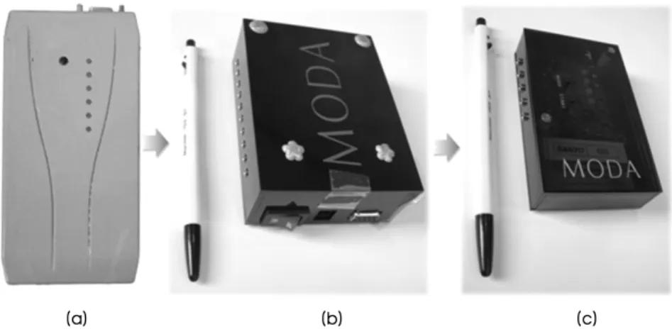
. 1-(a) ,
, (b) , .
1)
Hitachi chip ,
, 32 mode ( 2).
2006 RF
, ( 4). 12V/
2Ah Pb-Acid 10 , 3 .
( 2) ATMEL ATMEGA 16/
8 chip , 5V/2.8Ah SANYO
( 20 , 10 ) /lithium ion battery ( 15 , 8
) .
( 2)
probe 32 , W10.5xH3.5xD8cm ,
probe 20 ( 2-(a))
, probe 10
( 2-(b),(c)).
650nm InGaAlP(Indium-Gallium-Aluminium-
Phosphide) 25mW
.
,
. ,
(a)
(b)
그림 1. 초소형 발광모듈 시스템 (a) 유선 시스템 , (b) 무선 시스템 .
Fig. 1. Ultra-miniature lighting module system (a) Wire System, (b) wireless system.
, LED , . LED
650nm .
,
. 1 probe .
. controller
. 25mW
.
,
2cm 66microwatt/cm
26.6×10
211 1cm
2.
30 2.2×10
20.
25mW , 2mm( 0.0314
cm
2) 25mW 0.796W/ cm
2.
2mm
0.796W/ cm
2. LED 150mW ,
4cm( 12.56 cm
2) 150mW
0.0119W/ cm
2.
. 25mW 25mW ,
25mW .
, probe 796mW/ cm
2. LED
11.9 mW/ cm
2.
2) a.
2006 Controller
, , CDMA
kit ( 4), 2007 zigbee
( 3), 2008 OOK( 5)
controller 1/30 .
Biotemp BioNIBP
그림 3. 무선 시스템 . Fig. 3. Wireless system.
그림 4. 진단과 치료가 가능한 무선 레이저 의료기 . Fig. 4. Wireless Laser medical instrument for diagnosis and treatment.
(a) (b) (c)
그림 2. 유선 시스템 (a) 초기 시스템 , (b) MD_LE rev.1, (c) MD_LE rev.2.
Fig. 2. Wire System (a) The prototype, (b) MD_LE rev.1 (c) MD_LE rev.2.
. b.
, ,
RS-232 JACK
, ,
. cable
, flexible 2 3 fiber-optic cable
. ,
, .
3)
LASER 2mm, LED 4cm .
1 .
LASER 0.796 J/cm
21
. LED
30mA 5V 150mW 650nm . LASER 1
4 0.796 J/cm
2. LED 0.476J/ cm
2. LASER
0.199W/ cm
2. LED 0.0119W/ cm
2. ,
. D=P x t/A [J/cm
2]
. D: , P : (W), t: (sec), A
: (cm
2) . 25mW(0.025W) , 1 4
, 2mm(0.0314 cm
2) , 0.796 J/cm
2. LED 150mW , 1 4 , 4cm(12.56 cm
2) . 1 cm
2GaAlAs
0.1J .
1-4J J/cm
2.
1cm .
.
. spot 2mm .
2cm . ,
, 1cm
.
1 1 4
. 20
. .
4)
( 3) 4 pattern
, alarm
. 1 240 , Device-
Programmer . PC
program . ( 4)
, CDMA
.
( 2) (20 ),
( ), 1 (20
), 1 (20
) 4 . OOK
그림 5. OOK 무선 시제품 . Fig. 5. OOK Wireless medical instrument.
그림 6. 진단과 치료가 가능한 의복 및 장치 시뮬레이션 .
Fig. 6. Simulation of clothing and apparatus for diagnosis and treatment.
( 5) On-off , 20 , .
( 6).
C. 방법 방법 방법 방법
( )
, ,
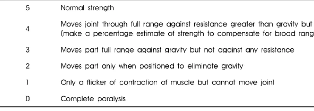
. British Medical Research Council Numeric
Scale [12].
1 4 ‘
(MSC SERIES, Ametek, FL, USA)’
[5]. Digital Strength Comparator ozf, gf, lbf, kgf, N
kgf .
0~5 numeric scale , 2
.
, , BELLY, MOTOR POINT, . , NOCICEPTION MECHANO-
RECEPTOR IA, IB, II FIBER
THALAMUS
GAMMA MOTOR NEURON
.
. ,
(preMuscle).
. (postMuscle)
British Medical Research Council Numeric Scale
. (preMSC)
, 1 4
(postMSC) . ,
,
.
SPSS version 15.0 for windows .
, ,
paired T-test .
Ⅲ. 실험 및 결과
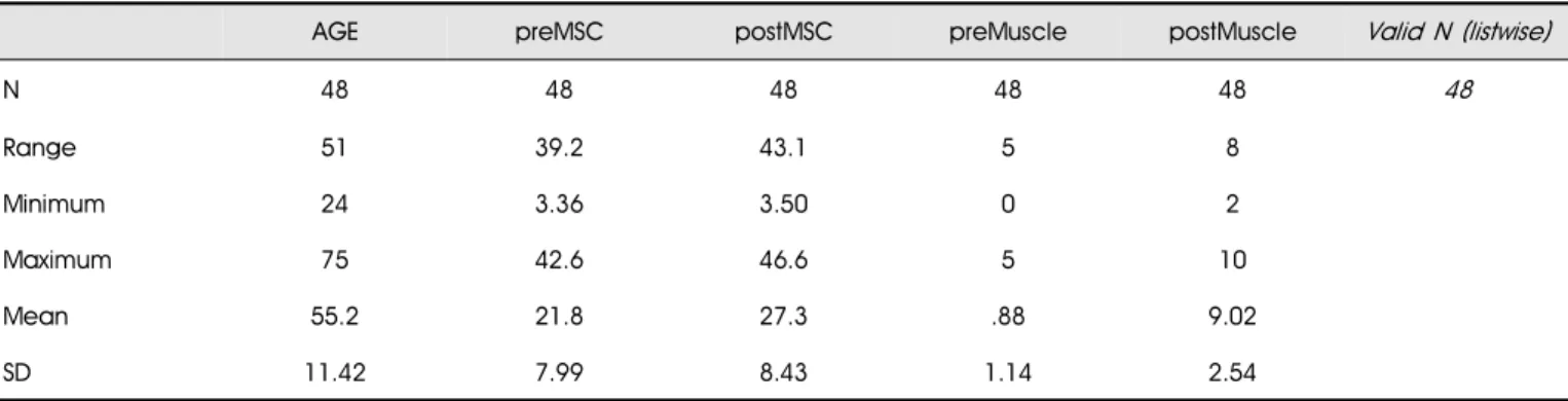
A. 연구 연구 연구 연구 대상자들의 대상자들의 대상자들의 일반적인 대상자들의 일반적인 일반적인 일반적인 특징 특징 특징 특징
48 7 , 41 55.2±
11.42 ( 2). ( 3)
. 50~60
( ) .
48 75 24 51 .
55.2 11.42 1.65
.
42.6 3.4 39.2
. 21.8 7.99 .
46.6 3.5 43.1 . 27.3
8.43 .
0 5 5 .
0.9 1.14 .
2 10 8
9.0 2.54 .
gastrocnemius(9 ), sartorius (23 ), deltoid (1 ), rhomboid(1 ), piriformis(1 ), biceps(3 ), infraspinatus
5 Normal strength
4 Moves joint through full range against resistance greater than gravity but examiner can overcome the action (make a percentage estimate of strength to compensate for broad range of this number)
3 Moves part full range against gravity but not against any resistance 2 Moves part only when positioned to eliminate gravity
1 Only a flicker of contraction of muscle but cannot move joint 0 Complete paralysis
표 1. British Medical Research Council 의 근력 검사의 척도
Table 1. Numeric Scale of British Medical Research Council
(2 ), rotator cuff muscle (2 ), paraspinal muscle(1 ), rectus femoris(1 ), flexor digitorum(1 ) .
11 , 34 ,
33 .
(38 ) spinal stenosis(1 ), bicipital calcific tend- onitis(2 ), thoracic spine fascitis(1 ), intervertebral disc
herniation(2 ), rotator cuff tear(1 ), rheumatoid arthritis(1 ), AVN of femur(1 ), gastrocnemius tendonitis(9 )
.
(42 ), (7 ), (4 ), (2
) .
B. 치료적 치료적 치료적 치료적 접촉 접촉 접촉 접촉 검사 검사 검사 검사 전 전 전 전 후 후 후 후 및 및 및 및 레이저 레이저 레이저 레이저 치료 치료 치료 전 치료 전 전 전 후 후 후 후 근력 근력 근력 근력 비교 비교 비교 비교 British Medical
Council 5 2 ,
kgf .
100%
,
. ‘
’ 21.8±7.99 27.3±8.43 (P<0.01), ‘
’ 0.9±1.14 9.0±2.54 (P<0.01). ‘
’ ‘
’ .
Age (years old) 21-40.9 41-60.9
≥61
55.2±11.42(mean±SD) 4( 8.3) 30(62.4) 14(29.3) Sex male
female
7(14.6) 41(85.4) Disease(number)
Degenerative OA Spinal stenosis Bicipital tendonitis Fascitis
Intervertebral disc herniation Roator cuff tearing
Rheumatoid arthritis AVN of femur
Gastrocnemius tendonitis
38 1 2 1 2 1 1 1 1 Muscles(number)
Sartorius Gastrocnemius Biceps Infraspinatus Rotator cuff muscle Deltoideus
Rhomboideus Piriformis Paraspinalis Rectus femoris Flexor digitorum
23 9 3 2 2 1 1 1 1 1 1
Data are Number(%), except mean age표 2. 연구 대상자들의 나이 , 성별 , 질병 , 검사한 근육 분포
Table 2. Age, sex, disease, tested muscle distribution of study subjects (N=48)
LASER Manual
Before 21.8±7.99 0.9±1.14
After(LBF) 27.3±8.43 9.0±2.54
P-value* 0.000 0.001
Data are mean ± SD, *P- value by paired t-test
표 4. 치료적 접촉검사 혹은 레이저 치료 이전과 이후의 근력 검사 수치의 비교
Table 4. Comparisons of muscle strength before and after manual or LASER therapy(N=48)
AGE preMSC postMSC preMuscle postMuscle Valid N (listwise)
N 48 48 48 48 48 48
Range 51 39.2 43.1 5 8
Minimum 24 3.36 3.50 0 2
Maximum 75 42.6 46.6 5 10
Mean 55.2 21.8 27.3 .88 9.02
SD 11.42 7.99 8.43 1.14 2.54
표 3. 기술통계량
Table 3. Descriptive statistics.
Ⅳ. 결론 및 고찰
, .
. , , , . Georgia
Institute of Technology 1996
, GTWM-C,M,P
2006 ,
,
, 2006
.
, ,
. Shafton[3]
tetrodotoxin nicardipine , Ridding
[4] ‘2 ’ ‘
(first dorsal interossei, FDI)’ short-interval intrac-
ortical inhibition(SICI) ‘
(abductor digiti minimi, ADM)’ , ‘5
’ ‘ ’ SICI
‘ ’ ‘ ’
. Fallon [5]
motor synaptic coupling . Nicholas [6]
cybex II 17%
8%
. Tokimura [7] transcranial magnetic stimulation
,
. Zimlichman [8]
‘Medex(Medex Screen Ltd.,)’
, , , 70%
.
[9]. George J. Goodheart Jr2 Pubmed, CINAHL (Cumulative Index to Nursing and Allied Health Literature)
‘manual muscle testing or manual muscle test(MMT)’
(peer reviewed studies, commmentaries,
reviews) , MMT
inter- and intra examiner reliability studies, (construct) (content), (convergent) (discriminant), con- current and predictive validity studies
. 10 0.55~0.99 .
.
. “ ?”
“ ?” .
8
0.05~0.001 .
.
. 14
, post-polio syndrome . , (ileocecal valve, ICV)
. Concurrent vali- dity
. MMT (hand held dynamometer,
HHD) (EMG)
0.55~0.99 0.001 .
MRI , EMG ,
. Hsieh and Philips
(Intratester reliability)
0.55,0.75,0.76 , MMT
0.96,0.99, 0.97 . (Intertester
reliability) 0.77 0.59 0.95
0.96 .
.
. Michener [10]
(HHD) .
4
, .
40 ,
, , 3 HHD
kg . 4
. 24 72
. 3 .
Intraclass 0.89~0.96 . 4
‘ (Construct) ’ , . 1~3
. Construct validity
, . construct validity
. 2008 12
Cochrane 1
, Review article
10 ‘
( )’ 3 ,
8 ,
, , 14
3 , ,
, 103 ,
19 .
.
[13]
.
[14] , Livens[15], Amaral
[16], Weiss[17] Bibikova[18]
,
.
. Schwartz
[19] HeNe ‘ ’
. ,
, .
, Ohno
[20] ‘ ,
C-fiber ,’ Kasai[21]
‘
A-δ ’
, Ia, Ib
. van Breugel[22], Mulligan[23], Wollman[24], Anders[25]
. Jimbo[26]
‘ process bradykinin
’ C- fiber
.
,
. Khullar [27-32] ‘
, ,
, ,
’
. Miloro[33], Yamada[34], Paolini[35], Brugnera [36], Bernal[37], Murakami[38]
, ,
. ,
1 ,
. Roc- hkind[39] ‘780nm
, ,
’ ,
, . Byrnes[40] ‘810nm 1589J/cm
2’ ,
.
650nm , 0.796 J/cm
2.
, .
. ,
. Sensory Evoked Potential Motor Evo- ked Potential
.
, , , B-scan ,
, , , ,
.
.
‘ ’
.
참고문헌