Core Stabilization With the Lumbar Extension Exercise in Low Back Pain
Dong-koog Noh1, PhD, PT, Young-joo Cha2, BHSc, PT, Dae-hun Kim3, PhD, PT, Joshua (Sung) H. You2, PhD, PT
1Department of Physical Medicine and Rehabilitation, Seoul Hyu Hospital, Gyeonggi-do, Republic of Korea
2Sports·Movement·Artificial Robotics·Techology (SMART) Institute, Department of Physical Therapy, Yonsei University
3Department of Physical Therapy, School of Medical & Public Health, Kyungdong University, Kangwon-do, Republic of Korea
Abstract1)
Background: We developed a novel integrative lumbar stabilization technique that combines lumbar extension (LE) exercise with abdominal drawing-in maneuver (ADIM) to ameliorate low back pain (LBP) associated with neuromuscular imbalance and instability, based on the collective evidence of contemporary spinal rehabilitation.
Objects: The specific aim of the present study was to investigate the effects of LE exercise with and without ADIM on core muscle strength, lumbar spinal instability, and pain, as well as functional characteristics in individuals with LBP using advanced radiographic imaging techniques.
Methods: patients with mechanical LBP (N = 40, 6 males; 35.1±7.6 years) were recruited and randomly assigned either to the combined LE and ADIM (experimental group) or the LE alone (control group).
Outcome measures included the visual analog scale, the modified Oswestry Disability Index, muscle strength imbalance (MSI), and radiographic imaging. The lumbar intervertebral displacement (LID), intervertebral (IV) and total lumbar extension (TLE) angles were calculated to evaluate the lumbar segmental instability.
Results: The experimental group showed significant differences in the L3-L4, L5-S1 LIDs, L4-L5 and L5-S1 IV angles, and TLE angle as compared to the controls (p<.05). Immediate pain reduction and muscle strength imbalance ratio were significantly different between the groups (p<.05).
Conclusion: These results suggest that the addition of ADIM significantly increased lumbar spinal stabilization in individuals with LBP, thereby reducing pain associated with functional lumbar flexion during daily activities.
Key Words: Abdominal drawing-in maneuver; Low back pain; Lumbar extension exercise.
Introduction
Posterior derangement syndrome (PDS) is a com- mon type of mechanical low back pain (LBP) that presents as mechanical obstruction of affected lumbar joint movement and pain (Mckenzie and May, 2003).
Pathomechanically, PDS may involve mechanical ob-
struction of the affected joint structure that com- presses the intervertebral disc and nerve root, result- ing in either centralized or peripheralized pain during repetitive lumbar flexion (Harris-Hayes et al, 2005;
Shirazi-Adl et al, 2005). The lumbar extension (LE) exercise is a commonly used and effective technique to mechanically centralize peripheralized pain and re-
Corresponding author: Joshua (Sung) H. You [email protected]
This research was in part supported by a Brain Korea 21 PLUS Project grant of Korean Research Foundation awarded to the Department of Physical Therapy of the Graduate School, Yonsei University.
Characteristic Experimental group (n1=20) Control group (n2=20) p
Gender (male/female) 2/18 4/16
Age (years) 35.4±7.3a 34.9±8 .719
Height (㎝) 162.4±6.2 193.1±6.1 .841
Weight (㎏) 56.4±8.8 57.4±9.5 .387
amean±standard deviation.
Table 1. Demographic and clinical characteristics of patients (N=40) store lumbar spinal movement in individuals with
PDS (Browder et al, 2007; Donelson et al, 1997;
Mckenzie and May, 2003).
Accumulating clinical evidence suggests that lum- bar core stabilization exercises can reduce low back pain and associated radiating pain during lumbar ex- tension in individuals with mechanical low back (Akuthota and Nadler, 2004) or lumbar radiculopathy (Hibbs et al, 2008). Muscle strength imbalances (MSI) between deep core muscles (weak or under- active) and superficial (strong or overactive rectus abdominal and external oblique) or antagonist (strong or overactive erector spinae) muscles is a potentially important pathomarker of lumbar spine instability.
Such lumbar core instability contributes to inter- vertebral (IV) angle and total lumbar extension (TLE) angle (Dupuis et al, 1985; Kong et al, 2009), which may be linked to PDS (Fredericson et al, 2001). As such, clinical improvements in low back pain and spinal movement suggest that the combina- tion of core stabilization with LE exercise produces an augmented or additive effect in individuals with PDS and associated core instability (Hosseinifar et al, 2013; Miller et al, 2005).
We developed a novel integrative lumbar stabiliza- tion technique that combines LE exercise with ab- dominal drawing-in maneuver (ADIM) to ameliorate LBP associated with neuromuscular imbalance and instability, based on the collective evidence of con- temporary spinal rehabilitation (Teyhen et al, 2008;
Vasseljen et al, 2012). ADIM is a lumbar stabiliza- tion exercise that focuses on the activation of the deep core muscles such Transverse abdominis (TrA) and internal oblique (IO) in coordination with super- ficial abdominal muscles. ADIM is effective for im-
proving lumbopelvic stability and reducing pain in individuals with LBP (Hodges and Richardson, 1999;
Richardson et al, 2002). This core stabilization ex- ercise helps restore neuromuscular control in im- paired active and neural subsystems, thereby in- creasing lumbopelvic core stability or stiffness and reducing concurrent pain in individuals with LBP (Cresswell et al, 1992; Hodges and Richardson, 1999).
Our combined exercise would be effective not only mechanical pain but also lumbopelvic stability. The specific aim of the present study was to investigate the effects of LE exercise with and without ADIM on core muscle strength, lumbar spinal instability, and pain, as well as functional characteristics in in- dividuals with LBP using advanced radiographic imaging techniques.
Methods
Subjects
The PDS was evaluated by a physical therapist that was specialized in the McKenzie method. The experimental protocol was approved by the Ministry of Health and Welfare Institutional Review Board, and informed consent was obtained from all patients.
The sample size was estimated based on a power of 80% with large differences (.8) in effect size. patients were recruited from Korean spine rehabilitation hospitals. Inclusion criteria were as follows: (1) con- sistently diagnosed with mechanical LBP by physia- trists, (2) chronic low back pain (>3 months), and (3) LBP at L4-L5 and lumbosacral segments asso- ciated with PDS with or without radiating pain to- ward the buttock/lower leg. The PDS was classified
based on the directional preference for lumbar ex- tension, not requiring any lateral compartment proce- dures (Clare et al, 2007; Mckenzie and May, 2003).
Exclusion criteria were a history of low back sur- gery, spondylolisthesis, increased peripheral pain with repeated lumbar extension, and neurological or mus- culoskeletal impairments that could affect the ex- perimental tests (Van et al, 2003). patients were ran- domly assigned to either the experimental group (n1=20) or the control group (n2=20). The ex- perimental group received a combination of ADIM and LE, whereas the control group received LE alone. Demographic and clinical characteristics of pa- tients are presented in Table 1.
Study design
This study was based on a longitudinal sin- gle-blind randomized controlled study. visual analog scale, the modified Oswestry disability index were used to measure the pain and function. Trunk mus- cle strength, and radiographic imaging, lumbar inter- vertebral displacement (LID), IV and TLE angles were calculated to evaluate the lumbar segmental in- stability in patients with mechanical LBP.
Procedure
This study is a single-blind randomized controlled design in which the two investigators who performed the radiographic examinations and the patients were blinded to group allocation and the intervention provided. All experimental procedures were im- plemented by the same investigator. A randomization sequence was created with Microsoft Excel (Microsoft corp., Roselle, IL, USA) to assign patients randomly to either the experimental group or the control group for a 2-week course of treatment. To standardize tests and interventions, the certified and experienced physical therapists were trained in the standardized clinical tests, ADIM training and/or lumbar extension technique (Clare et al, 2007; Mckenzie and May, 2003).
Clinical radiographic imaging was performed with AccuRay-525R (Dong Kang Medical Systems Co.,
Ltd., Seoul, Korea) to determine lumbar spine move- ment characteristics. Each participant was instructed to lie prone with the pelvis stabilized and to extend his or her lumbar spine within pain-free maximal extension for 5 seconds. The risks are minimal; but the examiner be requested to be present if there is serious concern about the risks involved. The radio- graphic images were acquired at the end of the ex- piration phase. The baseline or pretest radiographic imaging was obtained under the LE condition for both groups. However, to evaluate intervention-re- lated changes in lumbar spine kinematics, post-test imaging was performed under the LE condition for the control group as well as under the ADIM + LE condition for the experimental group. Ultrasound imaging and a pressure biofeedback unit (PBU) were used to monitor core stabilization performance during ADIM to ensure consistency in the implementation of experimental procedures.
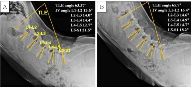
Clinical radiographic measures included the TLE angle (the angle between the inferior endplate of T12 and the superior endplate of S1) (Powers et al, 2008), IV angles (Hodges and Richardson, 1999) [the angles between the inferior and superior endplates of con- tiguous vertebrae from L1 to S1 (Figure 1)], and the LID [the vertical distance from the inferior-posterior edge of the superior vertebral body to the posterior surface of the inferior vertebral body (Figure 2)]
(Dupuis et al, 1985; Kong et al, 2009). LID is recog- nized as an important indicator of lumbar spinal in- stability (Kong et al, 2009). Interspinal instability was determined by computing the LID and IV angles. Excessive or abnormal spinal instability was defined as >2-3 ㎜ of LID and a >10˚ IV angle (Berlemann et al, 1999).
A visual analog scale (VAS) was used to de- termine the severity of LBP (Shaffer et al, 1990). The VAS ranged from 0 (“no pain”) to 10 (“worst pain”).
The modified Oswestry disability index (MODI) was used to determine the pain and physical and social disabilities related to LBP (Marshall and Murphy, 2010). The MODI consists of 10 items (pain intensity,
A B
Figure 1. The X-ray images of the LE assessment; the total LE angle and the intervertebral angles were measured in lumbar extension position (A : LE position without ADIM, B : LE position with ADIM) (LE : lumbar extension, ADIM : abdominal drawing-in maneuver).
personal care-washing and dressing, lifting, walking, sitting, standing, sleeping, sex life-if applicable, social life, and traveling) on a scoring scale ranging from 0 (“no pain”) to 5 (“worst pain”). The reliability and validity of the VAS and MODI measures ranged from r=.60 to r=.92, suggesting good to excellent correla- tions (Boonstra et al, 2008; Kim et al, 2005).
MSI ratio was determined by measuring maximal voluntary isometric muscle contraction (MVIC) for the abdominal and erector spinae muscles using hand-held dynamometry (HHD) (JTech Medical, Salt Lake City, USA) measurements. The MSI ratio was expressed as erector spinae MVIC/abdominal MVIC.
To assess the abdominal muscles, each participant was positioned supine with the hip and knee at 90˚
of flexion and asked to raise his or her trunk from the table (off of the inferior scapula border) and push against a hand-held dynamometer applied at the ster- num (Kendall et al, 1973). For the erector spinae muscle test, the participant was asked to lie prone with hands resting on the buttocks while the pelvis was stabilized with a strap and then asked to extend the trunk (off of the umbilicus) and push against a dynamometer applied at the thoracic spine (T6-8). All patients performed 3 consecutive trials, with each test lasting for 5 seconds and a resting interval of 3 mi-
nutes between tests. The reliability and validity of the strength test used in this study is considered good to excellent (Abizanda et al, 2012).
Intervention
The experimental group underwent ADIM ex- ercises during LE intervention while the control group underwent LE intervention alone. The inter- ventions were consistently provided 30 minutes per day, 3 times per week, over a 2-week period.
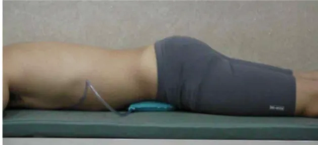
For the ADIM exercise, the participant was asked to lie in the prone position, and a PBU was placed under the anterior superior iliac spine (ASIS) and inflated to 70 ㎜Hg. The participant was then asked to inhale and stabilize the lumbar spine by coordi- nated and balanced co-activation of deep and super- ficial core muscles while maintaining pressure within the target pressure range of 4-10 ㎜Hg (Figure 3).
The coordinated co-activation of deep and superficial core muscles and lumbar core stability were con- currently monitored by real-time ultrasound imaging and PBU, respectively. Specifically, ultrasound imag- ing was used to guide accurate activation of the deep core muscles in coordination with overactive external oblique muscle or rectus abdominus to im- prove muscle balance between the overactive super-
Figure 2. Measurement of relative lumbar spinal intersegmental displacement.
Figure3. Core stability test using a pressure biofeedback unit.
ficial and underactive deep core muscles (Frank et al, 2013; Janda et al, 1996). A linear transducer was transversely placed 2.5 ㎝ anteromedial to the point midway between the iliac crest and the 12th rib (Whittaker, 2008).
For the LE method, once core stabilization was ach- ieved from the ADIM training, LE exercise was im- plemented according to the specific procedure: 1. The participant was asked to lie prone with hands palms down and aligned under the shoulders while maintain- ing the core stabilization. 2. The participant then raised the upper body using the arms, but the pelvis and lower legs remained stable. 3. The participant maintained this position for one to two seconds and then returned to the neutral prone position. 4. The participant rhythmically repeated this movement to move further towards the end range of motion with each repetition. 5. The movement was repeated up to 10 times. The use of the “force progressions” concept in the LE method was applied via dynamic pa- tient-generated force progression to improve the cen- tralization of the symptoms and lumbar extension movement. After the participant successfully performed repeated LE, patients progressed to end-range with patient overpressure, which involved the patient lock- ing the elbows straight and exhaling while allowing the pelvis to sag. Applications of force progressions and force alternatives were conducted according to the clinical reasoning and attentive interpretation of symp- tomatic and mechanical responses described in the LE method (Clare et al, 2007; Mckenzie and May, 2003).
Statistical analysis
Descriptive and standard statistical analyses in- cluded mean, standard deviation, and computations of the independent t-test was used to determine group difference of age, height, and weight, χ2 test was used to determine whether there were between group differences of gender. A independent t-test was used to compare intervention-related changes in VAS, MODI, trunk strength, LID, IV, and TLE angles within and between groups. A paired t-test was used to investigate pre-posttest within group. The level of significance was set at p<.05. All statistical analyses were performed with SPSS ver. 18.0 (IBM corp., Armonk, NY, USA).
Results
Demographic and clinical data: Independent t-tests showed no significant differences in baseline demo- graphic or clinical variables between groups, indicat- ing that the groups were homogenous (Table 1).
Pain and function data
Independent t-tests revealed a significant with- in-group differences (p<.001) but not between-group differences in the VAS (p=.17) or MODI (p=.16), al- though some results approached statistical sig- nificance (Table 2).
Muscle strength imbalance ratio
The muscle strength imbalance ratio at pretest was not significantly different between groups
Variable Experimental group (n1=20) Control group (n2=20)
Pretest Posttest Changes Pretest Posttest Changes
VASa 5.7±1.7b 2.2±1.4 -3.5±1.4 5.5±1.3 2.6±1.5* -2.9±1.3
MODIc 11.2±6.2 6.3±6.7* -4.8±3.6 11.6±4.3 8.1±4.1* -3.5±2.1
Strength measure
(N)
Abdominal
muscle 82.5±13.2 86.9±13.2 4.4±12.4 83.5±29.2 84.5±21.3 1.0±15.3 Erector
spinae muscle 80.7±16.2 100.8±17.5*† 20.0±11.0 80.1±19.7 83.2±10.6 3.1±17.3
Ed/Ae Ratio .9±.1 1.1±.1 .1±.1† .9±.1 1.0±.2 .0±.1
avisual analog scale, bmean±standard deviation, cthe modified Oswestry disability Index, derector spinae muscle,
eabdominal muscle, *significant difference between pretest and posttest for each group (p<.05), †significant difference in the intervention-related changes of variables between the two group (p<.05).
Table 2. Clinical outcome data in pain, lumbar function, and trunk muscle strength
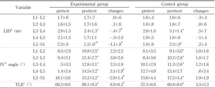
Variable Experimental group Control group
pretest posttest changes pretest posttest changes
LIDa (㎜)
L1-L2 1.7±.6 1.7±.7 .0±.6 1.6±.4 1.6±.6 -.3±.4
L2-L3 1.6±1.5 1.7±1.6 .1±.6 1.8±.8 1.8±.7 .0±.6
L3-L4 2.8±1.3 2.4±1.3* -.4±.7† 2.6±1.0 3.1±1.4* .5±.7
L4-L5 2.1±1.3 1.7±1.1 -.3±1.0 1.9±.5 1.8±.6 -.1±.4
L5-S1 2.3±.9 1.2±.6*† -1.1±.8† 1.9±.9 2.2±.9* .2±.4
IVb angle (˚)
L1-L2 8.5±2.9 10.8±2.5* 2.2±2.2 8.1±3.5 9.1±3.0* 1.0±1.6
L2-L3 9.4±2.1 12.4±2.7* 3.0±2.0 8.4±3.6 10.2±2.8* 1.8±1.7
L3-L4 .5±3.3 12.8±3.1* 2.3±1.9 10.1±2.9 11.3±2.6* 1.2±1.8
L4-L5 1.4±3.4 14.5±3.2* 3.1±1.9† 12.7±4.0 13.4±2.7 .6±3.4
L5-S1 18.1±3.6 15.2±3.2* -2.9±1.4† 15.6±4.4 17.5±4.4* 1.9±1.9 TLEc (˚) 60.2±8.6 69.1±9.3* 8.9±6.3† 57.3±8.9 60.8±8.0* 3.5±5.3
alumbar intervertebral displacement, bintervertebral, ctotal lumbar extension, *significant within-group difference (p<.05), †significant between-group difference (p<.05).
Table 3. Comparative radiological imagining data in the lumbar intersegmental displacements, intervertebral, and total lumbar extension angles between groups
(p=.948), but the ratio was significantly increased in the experimental group following the intervention at the posttest (p=.041) (Table 2).
Clinical radiographic data
There were significant differences between groups in LIDs (posttest-pretest) for the L3-L4 (p<.001) and L5-S1 (p<.001) segments, indicating greater spinal stability after the combination of LE and ADIM than after LE intervention alone. Similarly, the combined intervention group showed greater improvement in
the TLE angle (p=.006) and IV angles at L4-L5 (p=.008) and L5-S1 (p<.001) than did the control group. Both groups showed significant improvements in IVs at L1-S1, with the exception of the IV angle at L4-L5 in the control group (Table 3).
Test-retest reliability of the radiographic measure- ments: Test-retest reliability of the radiographic measurements for the TLE angle, IV angles, and LID was determined by intraclass correlation co- efficients (ICC) [3, 1] analysis. The correlations were .91 (.53 to .98) for the TLE angle, .96 (.83 to .99) for
the IV angles, and .86 (.41 to .97) for the LID, sug- gesting good to excellent consistency.
Discussion
This is the first study to demonstrate the effects of lumbar stabilization with an ADIM technique dur- ing LE in individuals with chronic LBP. As hypothe- sized, the experimental group, which received a com- bination of LE and ADIM, showed greater reduction in LIDs (L3-L4 and L5-S1) and IV angles of the lumbosacral segment (L5-S1) than did patients in the control group who received only LE exercise. These results suggest that the addition of ADIM sig- nificantly increased lumbar spinal stabilization in in- dividuals with LBP, thereby reducing pain associated with functional lumbar flexion during daily activities.
Importantly, radiological imaging revealed that the baseline LID (2.1 ㎜) and IV angle (10.9˚) for L1-5 instability in the present study were comparable to those identified as indicating abnormal spinal in- stability in previous studies (LID: >2-3 ㎜ and IV angle: >10˚) (Dupis et al, 1985; Berlemann U et al, 1999). This lumbar instability was noticeably de- creased in the L3-4 and L5-S1 LID lumbar segments in the experimental group (L3-4: 14.2%, L5-S1:
47.8%, within group changes) but increased in the control group (L3-4: -19.2%, L5-S1: -15.7%, within group changes). Similarly, the L5-S1 segment IV an- gles were substantially more decreased in the ex- perimental group (16.0%, within group changes) than in the control group (-12.1%, within group changes), suggesting that the combined method was more ef- fective for lumbosacral stabilization. The total lumbar extension angle was greater in the experimental group (15%, within group changes) than the control (6%, within group changes) group, indicating that a superior stabilization effect was achieved with the combined ADIM and LE technique. Such improve- ments in lumbar extension movement after LE com- bined with ADIM were similar to ranges of improve-
ment reported previously (15-17%) in patients with LBP who participated in press-up and other specific core stabilization exercises (O’sullivan et al, 1997;
Powers et al, 2008). In addition, the one-week post-in- tervention test showed more rapid alleviation of LBP (65%) in the experimental group than in the control group (25%), which corroborates previous results in studies of LBP management (Franca et al, 2010;
Kumar, 2011; O’sullivan et al, 1997). One important underlying mechanism by which the ADIM enhances lumbopelvic stabilization is neuromuscular activation of the TrA muscle, which synergistically contracts poste- rior fibers of the IO muscle. This in turn increases the posterior-lateral lumbar tension on the thoracolumbar fascia (TLF) that connects to both the spinous and transverse processes of the lumbar spine (Stanton and Kawchuk, 2008). Co-activation of the TrA and IO muscles paired with the tension on the TLF generates intra-abdominal pressure (IAP), which transforms ab- dominal function into mechanical stiffening of the TLF.
This mechanical stiffness provides spinal lumbar stabi- lization and optimal lordotic lumbar alignment (Hicks et al, 2005). In the current study, such spinal stabiliza- tion appeared to improve pain and decrease physical and social disabilities in daily activities, as evidenced by the VAS and MODI data.
The MSI ratio between the erector spinae and ab- dominal muscle strength significantly improved after the combination of LE and ADIM exercise, but no apparent change was observed after LE alone. Given the 2-week intervention period, this result indicates that such acute and rapidly-resolving episodes can be attributed to disinhibition or release of pain-induced inhibition, rather than muscle hypertrophy (Van et al, 2003). Our results agree with recent clinical evidence that demonstrated enhancements in pelvic stabiliza- tion, pain reduction, and associated lumbar extension strength in LBP patients (Smith et al, 2011).
One potential shortcoming of the present study was that dynamic IV disc movement characteristics associated with posterior derangement were not measured. Future research on the effects of core sta-
bility interventions on IV disc movement using mo- tion MRI is warranted. Nevertheless, our data dem- onstrate, for the first time, the additive effect of ADIM on lumbar spinal stability and associated re- duction in low back pain and increase in functional mobility. The radiographic imaging measurement was proven to be useful and reliable to detect ther- apy-induced minute structural changes associated with lumbar spinal instability. And 3 times inter- vention in a week is not a normal clinical does in LE exercise for LBP further research should do for shorter periods and multiple times per day.
Conclusion
In this investigation, we demonstrated the superior effects of combined LE with ADIM on pain, spinal stability, and associated functional movement in pa- tients with muscular strength imbalance and in- stability when compared to the conventional lumbar extension exercise alone. Most importantly, the com- bined intervention rapidly ameliorated pain and im- proved lumbar spinal stability and overall spinal mobility. These promising results suggest that the use of lumbar extension exercises alongside ADIM is beneficial for LBP patients with core instability.
References
Abizanda P, Navarro JL, Garcia-Tomas MI, et al.
Validity and usefulness of hand-held dynamom- etry for measuring muscle strength in commun- ity-dwelling older persons. Arch Gerontol Geriatr. 2012;54(1):21-27. http://doi.org/10.1016/
j.archger.2011.02.006
Akuthota V, Nadler SF. Core strengthening. Arch Phys Med Rehabil 2004;85:86-92. http://doi.org/
10.1053/j.apmr.2003.12.005
Berlemann U, Jeszenszky DJ, Bühler DW, et al. The role of lumbar lordosis, vertebral end-plate in-
clination, disc height, and facet orientation in degenerative spondylolisthesis. J Spinal Disord.
1999;12(1):68-73.
Boonstra AM, Schiphorst Preuper HR, Reneman MF, et al. Reliability and validity of the visual ana- logue scale for disability in patients with chronic musculoskeletal pain. Int J Rehabili Res. 2008;
31(2):165-169. http://doi.org/10.1097/MRR.0b013e3 282fc0f93
Browder DA, Childs JD, Cleland JA, et al.
Effectiveness of an extension-oriented treatment approach in a subgroup of subjects with low back pain: a randomized clinical trial. Phys Ther.
2007;87(12):1608-1618. http://doi.org/10.2522/ptj.
20060297
Clare HA, Adams R, Maher CG. Construct validity of lumbar extension measures in McKenzie's derangement syndrome. Man Ther. 2007;12(4):
328-334. http://doi.org/10.1016/j.math.2006.07.006 Cresswell AG, Grundström H, Thorstensson A.
Observations on intra-abdominal pressure and patterns of abdominal intra-muscular activity in man. Acta Physiol Scand. 1992;144(4):409-418.
http://doi.org/10.1111/j.1748-1716.1992.tb09314.x Donelson R, Aprill C, Medcalf R, et al. A prospective
study of centralization of lumbar and referred pain. A predictor of symptomatic discs and anu- lar competence. Spine (phila Pa 1976). 1997;
22(10):1115-1122.
Dupuis PR, Yong-Hing K, Cassidy JD, et al.
Radiologic diagnosis of degenerative lumbar spi- nal instability. Spine (Phila Pa 1976). 1985;10(3):
262-276.
França FR, Burke TN, Hanada ES, et al. Segmental stabilization and muscular strengthening in chronic low back pain: A comparative study.
Clinics (Sao Paulo). 2010;65(10):1013-1017.
Frank C, Kobesova A, Kolar P. Dynamic neuro- muscular stabilization & sports rehabilitation. Int J Sports Phys Ther. 2013;8(1):62-73.
Fredericson M, Lee SU, Welsh J, et al. Changes in posterior disc bulging and intervertebral fora-
minal size associated with flexion-extension movement: a comparison between L4-5 and L5-S1 levels in normal subjects. Spine J. 2001;
1(1):10-17.
Harris-Hayes M, Van Dillen LR, Sahrmann SA.
Classification, treatment and outcomes of a pa- tient with lumbar extension syndrome. Physiother Theory Pract. 2005;21(3):181-196.
Hibbs AE, Thompson KG, French D, et al.
Optimizing performance by improving core sta- bility and core strength. Sports Med. 2008;
38(12):995-1008. http://doi.org/10.2165/00007256- 200838120-00004
Hicks GE, Fritz JM, Delitto A, et al. Preliminary de- velopment of a clinical prediction rule for de- termining which patients with low back pain will respond to a stabilization exercise program.
Arch Phys Med Rehabil. 2005;86(9):1753-1762.
http://doi.org/10.1016/j.apmr.2005.03.033
Hodges PW, Richardson CA. Altered trunk muscle recruitment in people with low back pain with upper limb movement at different speeds. Arch Phys Med Rehabil. 1999;80(9):1005-1012.
Hodges PW, Richardson CA. Transversus abdominis and the superficial abdominal muscles are con- trolled independently in a postural task. Neurosci Lett. 1999;265(2):91-94.
Hosseinifar M, Akbari M, Behtash H, et al. The ef- fects of stabilization and mckenzie exercises on transverse abdominis and multifidus muscle thickness, pain, and disability: A randomized controlled trial in nonspecific chronic low back pain. J Phys Ther Sci. 2013;25(12):1541-1545.
http://doi.org/10.1589/jpts.25.1541
Janda V, Frack C, Lebenson C. Evaluation of mus- cular imbalance. Rehabilitation of the spine: A practitioner’s manual, 1996:97-112.
Kendall HO, Kendall FP, Wadsworth GE. Muscle testing and function. Am J Phys Med Rehabil 1973;52(1):43
Kim DY, Lee SH, Lee HY, et al. Validation of the Korean version of the Oswestry disability index.
Spine (Phila Pa 1976). 2005;30(5):E123-127.
Kong MH, Hymanson HJ, Song KY, et al. Kinetic magnetic resonance imaging analysis of abdomi- nal segmental motion of the functional spine unit. J Neurosurg Spine. 2009;10(4);357-365.
Kumar SP. Efficacy of segmental stabilization ex- ercise for lumbar segmental instability in pa- tients with mechanical low back pain: A randomized placebo controlled crossover study. N Am J Med Sci. 2011;3(10):456-461. http://doi.org/
10.4297/najms.2011.3456
Marshall P, Murphy B. Delayed abdominal muscle on- sets and self-report measures of pain and dis- ability in chronic low back pain. J Electromyogr Kinesiol. 2010;20(5):833-839. http://doi.org/10.1016/
j.jelekin.2009.09.005
McKenzie RA, May S. Mechanical diagnosis and therapy: The lumbar spine. Waikanae, New Zealand: Spinal Publications. 2003.
Miller ER, Schenk RJ, Karnes JL, et al. A compar- ison of the McKenzie approach to a specific spine stabilization program for chronic low back pain. J Man Manip Ther. 2005;13(2):103-112.
http://doi.org/10.1179/106698105790824996
O’Sullivan PB, Phyty GD, Twomey LT, et al.
Evaluation of specific stabilizing exercise in the treatment of chronic low back pain with radio- logic diagnosis of spondylolysis or spondylo- listhesis. 1997;22(24):2959-2967.
Powers CM, Beneck GJ, Kulig K, et al. Effects of a single session of posterior-to-anterior spinal mo- bilization and press-up exercise on pain response and lumbar spine extension in people with non- specific low back pain. Phys Ther. 2008;
88(4):485-493. http://doi.org/10.2522/ptj.20070069 Richardson CA, Snijders CJ, Hides JA, et al. The
relation between the transversus abdominis muscles, sacroiliac joint mechanics, and low back pain. Spine (Phila Pa 1976). 2002;27(4):
399-405.
Shaffer WO1, Spratt KF, Weinstein J, et al. 1990 Volvo Award in clinical sciences. The con-
This article was received October 7, 2018, was re- viewed October 7, 2018, and was accepted November 8, 2018.
sistency and accuracy of roentgenograms for measuring sagittal translation in the lumbar vertebral motion segment. An experimental model. Spine (Phila Pa 1976). 1990;15(8):741-750.
Shirazi-Adl A, El-Rich M, Pop DG, et al. Spinal muscle forces, internal loads and stability in standing under various postures and loads appli- cation of kinematics-based algorithm. Eur Spine J. 2005;14(4)381-392. http://doi.org/10.1007/s00586- 004-0779-0
Smith D, Bissell G, Bruce-Low S, et al. The effect of lumbar extension training with and without pelvic stabilization on lumbar strength and low back pain. J Back Musculoskelet Rehabil. 2011;24(4):
241-249. http://doi.org/10.3233/BMR-2011-0301 Stanton T, Kawchuk G. The effect of abdominal sta-
bilization contractions on posteroanterior spinal stiffness. Spine (Phila Pa 1976). 2008;33(6):694- 701. http://doi.org/10.1097/BRS.0b013e318166e034 Teyhen DS, Rieger JL, Westrick RB, et al. Changes
in deep abdominal muscle thickness during common trunk-strengthening exercises using ul- trasound imaging. J Orthop Sports Phys Ther.
2008;38(10):596-605. http://doi.org/10.2519/jospt.
2008.2897
Van Dieën JH, Cholewicki J, Radebold A. Trunk muscle recruitment patterns in patients with low back pain enhance the stability of the lumbar
spine. Spine (Phila Pa 1976). 2003;28(8):834-841.
Van Dillen LR, Sahrmann SA, Norton BJ, et al. The effect of modifying patient-preferred spinal movement and alignment during symptom test- ing in patients with low back pain: A prelimi- nary report. Arch Phys Med Rehabil. 2003;
84(3):313-322.
Vasseljen O, Unsgaard-Tøndel M, Westad C, et al.
Effect of core stability exercises on feed-forward activation of deep abdominal muscles in chronic low back pain: A randomized controlled trial.
Spine (Phila Pa 1976). 2012;37(13):1101-1108.
http://doi.org/10.1097/BRS.0b013e318241377c Whittaker JL. Ultrasound imaging of the lateral ab-
dominal wall muscles in individuals with lumbo- pelvic pain and signs of concurrent hypocapnia.
Man Ther. 2008;13(5):404-410. http://doi.org/10.
1016/j.math.2007.03.008