Copyright © 2018, The Korean Society of Veterinary Service. All Rights Reserved. 277 한국가축위생학회지 제41권 제4호 (2018)
Korean J Vet Serv, 2018, 41(4), 277-280 ISSN 1225-6552, eISSN 2287-7630 https://doi.org/10.7853/kjvs.2018.41.4.277
< Case Report >
Korean Journal of
Veterinary Service
Available online at http://kjves.org
*Corresponding author: Jae-Ik Han, Tel. +82-63-850-0965, Fax. +82-63-850-0910, E-mail. [email protected]
Ehrlichia ewingii infection in a dog from South Korea – A case report
Jae-Ik Han1*, Ki-Jeong Na2
1Laboratory of Wildlife Medicine/Diseases, College of Veterinary Medicine, Chonbuk National University, Iksan 54596, Korea
2Laboratory of Veterinary Laboratory Medicine, College of Veterinary Medicine, Chungbuk National University, Cheongju 54596, Korea
(Received 16 May 2018; revised 28 September 2018; accepted 1 December 2018)
Abstract
The report describes a case of natural E. ewingii infection confirmed by microscopic examination and molecular analyses in a domestic dog with acute lameness. After the diagnosis, the dog was successfully treated with oral doxycycline. To authors’ knowledge, this is the first case report of natural E. ewingii infection in dogs in South Korea.
Key words : Dog, Ehrlichia ewingii, Lameness, Korea
INTRODUCTION
Ehrlichiosis is a tick-borne bacterial infection caused by bacteria of genus Ehrlichia (Rikihisa, 2006). Currently, several species of Ehrlichia is known to infect and cause diseases, and can result in fever, myalgia, depres- sion, leucopenia and thrombocytopenia which may lead to bleeding disorders. In dogs, ehrlichiosis is primarily caused by E. canis, which is transmitted by the brown dog tick, Rhipicephalus sanguineus. When the infection is established, the disease is composed of 3 distinct phase described as acute, subclinical, and chronic (Schaefer et al, 2007). Symptoms are similar for all stages; however, the chronic stage is more severe than other stages. In chronic stage, bone marrow becomes hypoplastic, resulting in severe pancytopenia. Clinical signs can vary depending on the predominant organs af- fected, and may include bleeding tendency (epistaxis, hematuria, melena, and petechiae/ecchymoses), spleno- megaly, glomerulonephritis, renal failure, interstitial pneu- monia, anterior uveitis, meningitis and severe weight
loss.
This report describes a history, clinical presentation, and outcome of treatment on a case of E. ewingii in- fection diagnosed by microscopic examination and mo- lecular analyses in a domestic dog. To author’s knowl- edge, this is the first case report of natural E. ewingii infection in dogs in South Korea.
CASE DESCRIPTION
Complete blood count (CBC), serum biochemistry profiles, electrolytes, and urine dipstick test were exam- ined by using IDEXX Vet AutoreadTM Hematology ana- lyzer, VetTestⓇ Chemistry Analyzer, VetLyteⓇ Electrolyte Analyzer, and VetLabⓇ UATM Analyzer (IDEXX Labo- ratories, Westbrook, ME, USA).
After total nucleic acid extraction from EDTA-treated peripheral blood by using MagMAXTM Total Nucleic Acid Isolation Kit (Applied Biosystems, Austin, TX, USA), real-time polymerase chain reactions (PCRs) for E. canis, E. chaffeensis, and Anaplasma phagocytophilum were performed on Illumina Eco Real-Time PCR system
278 Jae-Ik Han, Ki-Jeong Na
Korean J Vet Serv, 2018, Vol. 41, No. 4
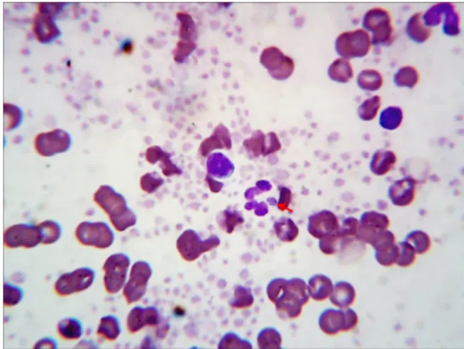
Fig. 1.Perpheral blood smear showing intracytoplasmic morula in the neutrophil. Wright-Giemsa stain, ×100 objective.
(Illumina Inc., San Diego, CA, USA). Multiplex PCR kit (QuantiTect Multiplex PCR kit, Qiagen, Valencia, CA, USA) was used by following the manufacturer’s recommended protocols in a reaction volume of 20 L.
The primers and probes for the Ehrlichia species were adopted from published information (Doyle et al, 2005).
A 65-bp oligonucleotide internal control (IC) was de- signed to monitor for false negative result due to failure of the PCR process causing the presence of inhibitory substances in reactions. The IC contained a non-specific 16S ribosomal RNA (rRNA) gene sequence flanked by the uvrC gene sequence of Mycobacterium bovis to min- imize cross-reactivitiy with M. bovis. For every reaction, 0.001 M of IC was added, which resulted in the pos- itive signal with Ct values of 36 to 38 if PCR in- hibitory substances were not present in the reaction.
Recombinant vectors for each pathogen were used as positive controls.
For the PCR of Babesia and Thelieria species, the genomic DNA was extracted by using a DynabeadsⓇ DNA DIRECTTM Universal Kit (Invitrogen Life Techno- logies, Inc., Carlsbad, CA, USA). The genomic DNA encoding the small subunit rRNA gene (18S rDNA) re- gion was amplified using primers RIB-19 and RIB-20, as described previously (Zahler et al, 2000). Field iso- lates of Babesia gipsoni or Theileria spp. were used as positive controls.
CASE PRESENTATION
A 2-year-old intact female Beagle dog was presented with left hindlimb lameness. The symptom was firstly detected 1 week previously, and was slowly progressive.
Before the onset of clinical signs, the dog was found af- ter spending 6 days lost in a nearby hill. At the rescue, the dog had ixodid tick infestation, thus had received antiparasitic treatment (FrontlineⓇ, Merial, Duluth, GA, USA) in a local hospital. On presentation, the dog ap- peared to be normal otherwise, except for mild fever (39.8°C) and lameness. The dog tested negative to anti- bodies for E. canis, B. burgdorferi, and A. phag- ocytophilum (4DxⓇ kit, IDEXX Laboratories). CBC showed mild neutrophilia (1.74×10⁴ cells/L, reference
interval [RI] 0.6−1.69×10⁴ cells//L) and thrombocy- tosis (5.52×10¹¹ cells/L, RI 1.75−5.00×10¹¹ cells/L).
Serum biochemistry profiles, electrolytes, urinalysis and radiographic examination were unremarkable.
The CBC findings were consistent with inflammation.
The blood smear contained basophilic intracellular or- ganisms (Fig. 1). The organisms, ranging in diameter from 2 to 3 m, had a mulberry-shaped clustered body with serrated margin and were frequently observed in the cytoplasm of neutrophils (1∼2 parasites/50 neu- trophils). The organisms were compatible with morula stage of Ehrlichia or Anaplasma spp., and a diagnosis of canine rickettsial disease was made. To identify the organism, real-time quantitative PCR (qPCR) and gel-based PCR analyses for medically important Ehrlichia species, Anaplasma phagocytophilum, Babesia and Theileria spe- cies were performed as described previously (Zahler et al, 2000; Doyle et al, 2005; Santos et al, 2011). While PCRs for E. canis, E. chaffeensis, A. phagocytophilum, Babesia and Theileria species were negative, qPCR for E. ewingii was positive, indicating E. ewingii infection in the dog.
The dog was treated with oral doxycycline twice dai- ly for 2 months. A month after treatment, the dog was fully recovered from the fever, lameness and CBC abnormalities. After 8 months of the treatment, qPCR for E. ewingii was negative.
Ehrlichia ewingii infection in a dog from South Korea 279
Korean J Vet Serv, 2018, Vol. 41, No. 4
DISCUSSION
This report describes a case of E. ewingii infection in a dog with acute lameness. Due to the history of tick infestation, the dog was initially confirmed negative to antibodies for E. canis, B. burgdorferi, and A. phag- ocytophilum, common tick-borne pathogens in South Korea. Based on the mild inflammatory changes and no other abnormality in laboratory and radiographic exami- nations, the dog was suspected of suffering a muscu- loskeletal disorder because of highly dynamic person- ality of the dog. However, blood smear examination re- vealed unexpected intracytoplasmic morula in neu- trophils and molecular analyses confirmed E. ewingii infection. The dog was treated with oral doxycycline for 2 months and the lameness was completely resolved.
After 8 months of the treatment, qPCR for E. ewingii was negative.
In 1992, E. ewingii was firstly identified as a patho- gen of ehrlichiosis in dogs on the basis of 16S rRNA gene sequence differences between the 2 most closely related species, E. canis and E. chaffeensis (Anderson et al, 1992). Since then, canine infection of E. ewingii has been detected by PCR of 16S rRNA gene in south cen- tral and southeastern USA, as well as a recent detection in Cameroon (Stockham et al, 1992; Dawson et al, 1996; Goldman et al, 1998; Murphy et al, 1998; Liddell et al, 2003; Ndip et al, 2005). In contrast to E. canis that parasitizes mononuclear cells, E. ewingii invades granulocytes causing fever, acute lameness or poly- arthritis called as canine granulocytic ehrlichiosis (CGE).
The long star tick (Amblyomma americanum) has been known as the primary vector. In South Korea, the tick has not been found yet; however, a recent study has demonstrated the presence of another ixodid ticks or wild rodents containing E. ewingii in South Korea, in- dicating that E. ewingii may have high prevalence in wild environment in South Korea (Kim et al, 2006). To the authors’ knowledge, this is the first report of natural E. ewingii infection in dogs in South Korea.
ACKNOWLEDGEMENTS
This subject is supported by Korea Ministry of Environment (MOE) as “Public Technology Program based on Environmental Policy (No. 2016000210002)”.
REFERENCES
Anderson BE, Greene CE, Jones DC, Dawson JE. 1992. Ehrlichia ewingii sp. nov., the etiologic agent of canine gran- ulocytic ehrlichiosis. Int J Syst Bacteriol 42: 299-302.
Dawson JE, Biggie KL, Warner CK, Cookson K, Jenkins S, Levine JF, Olson JG. 1996. Polymerase chain reaction evidence of Ehrlichia chaffeensis, an etiologic agent of human ehrlichiosis, in dogs from southeast Virginia. Am J Vet Res 57: 1175-1179.
Doyle CK, Labruna MB, Breitschwerdt EB, Tang YW, Corstvet RE, Hegarty BC, Bloch KC, Li P, Walker DH, McBride JW. 2005. Detection of medically important Ehrlichia by quantitative multicolor taqman real-time polymerase chain reaction of the dsb gene. J Mol Diag 7: 504-510.
Goldman EE, Breitschwerdt EB, Grindem CB, Hegarty BC, Walls JJ, Dumler JS. 1998. Granulocytic ehrlichiosis in dogs from North Carolina and Virginia. J Vet Intern Med 12:
61-70.
Kim CM, Yi YH, Yu DH, Lee MJ, Cho MR, Desai AR, Shringi S, Klein TA, Kim HC, Song JW, Baek LJ, Chong ST, O’Guinn ML, Lee JS, Lee IY, Park JH, Foley J, Chae JS. 2006. Tick-borne rickettsial pathogens in ticks and small mammals in Korea. Appl Environ Microbiol 72:
5766-5776.
Liddell AM, Stockham SL, Scott MA, Sumner JW, Paddock CD, Gaudreault-Keener M, Arens MQ, Storch GA. 2003.
Predominance of Ehrlichia ewingii in Missouri dog. J Clin Microbiol 41: 4617-4622.
Murphy GL, Ewing SA, Whitworth LC, Fox JC, Kocan AA.
1998. A molecular and serologic survey of Ehrlichia canis, E. chaffeensis, and E. ewingii in dogs and ticks from Oklahom. Vet Parasitol 79: 325-339.
Ndip LM, Ndip RN, Esemu SN, Dickmu VL, Fokam EB, Walker DH, McBride JW. 2005. Ehrlichial infection in Cameroonian canines by Ehrlichia canis and Ehrlichia ewingii. Vet Microbiol 111: 59-66.
Rikihisa Y. 2006. New findings on members of the family Anaplasmataceae of veterinary importance. Ann N Y Acad Sci 1078: 438-445.
Santos HA, Pires MS, Vilela JAR, Santos TM, Faccini JLH, Baldani CD, Thomé SMG, Sanavria A, Massard CL.
2011. Detection of Anaplasma phagocytophilum in Brazilian dogs by real-time polymerase chain reaction. J Vet Diagn Invest 23: 770-774.
Schaefer JJ, Needham GR, Bremer WG, Rikihisa Y, Ewing SA,
280 Jae-Ik Han, Ki-Jeong Na
Korean J Vet Serv, 2018, Vol. 41, No. 4
Stich RW. 2007. Tick acquisition of Ehrlichia canis from dogs treated with doxycycline hyclate. Antimicrob Agents Chemother 9: 3394-3396.
Stockham SL, Schmidt DA, Curtis KS, Schauf BG, Tyler JW, Simpson ST. 1992. Evaluation of granulocytic ehrlichio- sis in dogs of Missouri, including serologic status of
Ehrlichia canis, Ehrlichia equi, and Borrelia burgodorferi. Am J Vet Res 53: 63-68.
Zahler M, Rinder H, Gothe R. 2000. Detection of a new patho- genic Babesia microti-like species in dogs. Vet Parasitol 89: 241-248.