Inhibitory Effect of Scopoletin Isolated from Sorbus commixta on TNF-α-Induced Inflammation in Human Vascular Endothelial EA.hy926 Cells through NF-κB Signaling Pathway Suppression
Hye Ryung Kang
1, Hyo Jung Kim
1, Bomi Kim
1, Sun-Gun Kim
1, Jai-Hyun So
1, Soo Jeong Cho
2and Hyun Sook Kwon
1*
1
National Institute for Korean Medicine Development, Gyeongsan 38540, Korea
2
Department of Pharmaceutical Engineering, Gyeongnam National University of Science and Technology, Jinju 52725, Korea Received January 6, 2020 /Revised February 24, 2020 /Accepted February 24, 2020
Sorbus commixta Hedl. has traditionally been used as a remedy for cough, asthma, and other bronchial disorders. In this study, three major triterpenoids—lupeol, β-sitosterol, and ursolic acid and a coumar- in, scopoletin, were isolated from a CHCl
3-soluble fragment of the bark of S. commixta. Their structures were identified by spectroscopic analyses, including mass spectrometry (MS), 1D-, and 2D- nuclear magnetic resonance spectroscopy (NMR), as well as by comparing the data with data reported in the literature. Scopoletin was isolated from this plant for the first time. It is a nutraceutical compound con- tained in many plants that has been reported to exert diverse biological activities, including anti-in- flammatory effects. This study examined the inhibitory effect of scopoletin on TNF-α-induced vascular endothelial inflammation. Unlike the marginal impact of other compounds against low-density lip- oprotein (LDL) oxidation and vascular endothelial inflammation, scopoletin showed remarkable activ- ity on LDL oxidation (IC
50= 10.2 μM) and exerted vascular anti-inflammatory effects in EA.hy926 hu- man endothelial cells activated by TNF-α. It suppressed the expression of adhesion molecules, such as ICAM-1, VCAM-1, and E-selectin, and blocked the adhesion between THP-1 monocytes and EA.
hy926 endothelial cells. It also inhibited TNF-α-induced NF-κB translocation from the cytosol to the nucleus. Moreover, IκBα phosphorylation, which was increased by TNF-α treatment, was reduced after treatment with scopoletin. Thus, scopoletin inhibited TNF-α-induced vascular inflammation in endo- thelial cells by suppressing the NF-κB signaling pathway. These results demonstrate that owing to its anti-inflammatory activity in the vascular endothelium, scopoletin has the potential to inhibit athero- sclerosis development.
Key words : Adhesion molecules, inflammation, scopoletin, Sorbus commixta
*Corresponding author
*Tel : +82-53-810-0389, Fax : +82-53-810-0301
*E-mail : [email protected]
This is an Open-Access article distributed under the terms of the Creative Commons Attribution Non-Commercial License (http://creativecommons.org/licenses/by-nc/3.0) which permits unrestricted non-commercial use, distribution, and reproduction in any medium, provided the original work is properly cited.
Journal of Life Science 2020 Vol. 30. No. 4. 343~351 DOI : https://doi.org/10.5352/JLS.2020.30.4.343
Introduction
Atherosclerosis is a chronic vascular disease described as endothelial dysfunction, increase of cell adhesion molecules, and accumulation of foam calls, smooth muscle cells, and fibrous tissue in the intima area. The process of athero- genesis is not fully understood yet, but it is well known that inflammation plays a crucial role in all stages of athero- sclerosis [10, 19, 20]. Previous studies reported that adhesion of circulating monocytes to the injured endothelial layer, in- vasion of monocytes into the vessel wall, and differentiation
into macrophages are early events in the development of atherosclerosis [10, 19, 20]. The adhesion of monocytes onto endothelial cells can be controled by the expression of cell adhesion molecules, including intercellular cell adhesion molecule-1 (ICAM-1), vascular cell adhesion molecule-1 (VCAM-1), and E-selectin [10]. Furthermore, the expressions are upregulated by a proinflammatory cytokine such as tu- mor necrosis factor-α (TNF-α) [10]. Besides, nuclear factor-κB (NF-κB) is a central mediator in adhesion molecules ex- pression and monocyte adhesion. In normal condition, NF-κ B is localized in the cytoplasm and bind to its inhibitor pro- tein, IκB. When it is activated by a variety of external stimuli, such as TNF-α, the IκB is phosphorylated and degraded in proteasome [2, 4, 7]. This action results in release of NF-κ B, which then translocates to the nucleus and binds to its promoter κB binding site and transcribes a number of in- flammatory genes [2, 4, 7].
Sorbus commixta Hedl. (Rosaceae) is well known as medic-
inal plant in Korea, China, and Japan. The diverse pharma- cological effects of S. commixta on antioxidative [1], anti-in- flammatory [24], anti-lipid peroxidative [14], anti-athero- genic [21, 22], and vasorelaxant activities [23] have been reported. Scopoletin (6-methoxy-7-hydroxycomarin), an ac- tive component of S. commixta, showed anti-inflammatory [13], anti-allergy [5] and anti-angiogenic properties [17]. The effect of scopoletin on the expressions of proinflammatory mediators has been estimated by several studies. Scopoletin reduced expression levels of inducible nitric oxide synthase (iNOS), cyclooxygenase-2 (COX-2), interleukin-1β (IL-1β), interleukin-6 (IL-6), and TNF-α in Raw264.7 cells stimulated with lipopolysaccharide [13]. Although the prior reports fo- cused on anti-inflammatory effects of scopoletin in several cell types, its anti-inflammatory effects in EA.hy926 human vascular endothelial cells have not been clarified. In the pres- ent study, we did isolate pure compound, scopoletin, from bark MeOH extracts of S. commixta, and evaluated whether scopoletin inhibits the expression of cellular adhesion mole- cules and monocyte adhesion onto EA.hy926 human vas- cular endothelial cells and nuclear NF-κB are targets for the inhibitory actions of scopoletin on adhesion molecule ex- pression.
Materials and Methods
Plant material
The S. commixta Hedl. was collected on December 2017, in Gyeongsangnam-do Agricultural Research & Extension Service, Medicinal Source Research Institute, Hamyang dis- trict of Korea.
Instruments
Melting points were measured on a Thomas Scientific Capillary Melting Point Apparatus and are uncorrected. IR spectra were recorded on a Bruker IFS66 infrared Fourier transform spectrophotometer (KBr). NMR experiments were conducted on Bruker AM 300 and 500 (
1H-NMR at 300 and 500 MHz,
13C-NMR at 75 and 125 MHz) spectrometer with tetramethylsilane (TMS) as the internal standard. EIMS were recorded on a Jeol JMS-700 instrument operated at 70 eV.
TLC analysis was performed on Kieselgel 60 F
254(Merk, Darmstadt, Germany) plates. Silica gel (Merck, 70-230 and 230-240 mesh) and Sephadex LH-20 (Amersham Biosciences, Uppsala, Sweden) were used for column chromatography.
Extraction and isolation
The dried bark of S. commixta (500 g) was extracted with MeOH (5000 mL) at room temperature for three days. The MeOH extracts (20 g) was evaporated to dryness and sus- pended in H
2O, then it was partitioned with CHCl
3(10 g) and BuOH (5.5 g). The CHCl
3-soluble fraction was chromato- graphed on a silica gel column eluted with a gradient of 100% CHCl
3to 100% MeOH to afford ten fractions (F1 to F10). F1 (2 g) was chromatographed over silica gel using n-hexane:EtOAc (19:1→1:1) to give ten major subfractions (F1-1 to F1-10). F1-3 (900 mg) was subjected to column chro- matography on Sephadex LH-20 to yield five fractions (F1-3-1 to F1-3-5). F1-3-3 was recrystallized from MeOH to give compound 1 (108.2 mg). F1-7 (0.95 g) was purified by repeated silica gel column chromatography using n-hex- ane:EtOAc gradient (19:1→1:1) to afford eight fractions (F1-9-1 to F1-9-8). Compound 2 (94.1 mg) was crystallized in CHCl
3from F1-9-2. TLC analysis of F1-9-8 indicated the presence of only one major component. Compound 3 (105.6 mg) was purified by sephadex LH-20 column chromatog- raphy in 100% MeOH solvent condition. F5 (0.94 g) was chromatographed over silica gel using n-hexane:EtOAc gra- dient (4:1→1:1) to afford ten fractions (F5-1 to F5-10). Of these, F5-9 (0.8 g) was chromatographed over silica gel with CHCl
3:Me
2CO gradient (99:1→1:1) to produce six fractions (F5-9-1 to F5-9-6). Further chromatographic separation of F5-9-5 was carried out by preparative TLC to give com- pound 4 (50 mg).
Lupeol (1)
White amorphous powder; mp 210℃; EI/MS m/z 426 [M]
+; IR λ
max3300, 1650 and 1500 cm
-1;
1H-NMR (300 MHz, CDCl
3) δ: 0.76 (3H, s, H-24) 0.79 (3H, s, H-28), 0.83 (3H, s, H-25), 0.95 (3H, s, H-27), 0.96 (3H, s, H-23), 1.03 (3H, s, H-26), 1.68 (3H, s, H-30), 1.92 (1H, m, H-21), 2.40 (1H, m, H-19), 3.20 (1H, dd, J=5.4, 9.9 Hz, H-3), 4.57 (1H, s, H-29b), 4.69 (1H, s, H-29a);
13C-NMR (75 MHz, CDCl
3) see Table 1.
β-sitosterol (2)
White amorphous powder; mp 140°C; EI/MS m/z 414 [M]
+; IR λ
max3421, 2937, 1643, 1462 and 1380 cm
-1;
1H-NMR (500 MHz, CDCl
3) δ: 0.70 (3H, s, H-18), 0.82 (3H, s, H-27), 0.84 (3H, s, H-26), 0.86 (3H, s, H-29), 0.95 (3H, s, H-21), 1.0 (3H, s, H-19), 3.54 (1H, m, H-3), 5.35 (1H, d, J=5.2 Hz, H-6);
13
C-NMR (125 MHz, CDCl
3) see Table 1.
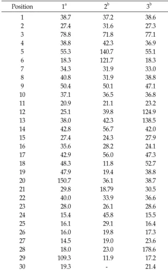
Table 1.
13C-NMR data for compound 1-3
Position 1
a2
b3
b1 2 3 4 5 6 7 8 9 10 11 12 13 14 15 16 17 18 19 20 21 22 23 24 25 26 27 28 29 30
38.7 27.4 78.8 38.8 55.3 18.3 34.3 40.8 50.4 37.1 20.9 25.1 38.0 42.8 27.4 35.6 42.9 48.3 47.9 150.7 29.8 40.0 28.0 15.4 16.1 16.0 14.5 18.0 109.3 19.3
37.2 31.6 71.8 42.3 140.7 121.7 31.9 31.9 50.1 36.5 21.1 39.8 42.3 56.7 24.3 28.2 56.0 11.8 19.4 36.1 18.79 33.9 26.1 45.8 29.1 19.8 19.0 23.0 11.9 -
38.6 27.3 77.1 36.9 55.1 18.3 33.0 38.8 47.1 36.8 23.2 124.9 138.5 42.0 27.9 24.1 47.3 52.7 38.8 38.7 30.5 36.6 28.6 15.5 16.4 17.3 23.6 178.6 17.2 21.4
a
75MHz in CDCl
3at 25℃.
b125MHz in CDCl
3at 25℃.
Ursolic acid (3)
White amorphous powder; mp 279℃; EI/MS m/z 456 [M]
+; IR λ
max3420, 2920, 1690, 1450 and 1376 cm
-1;
1H-NMR (500 MHz, DMSO-d
6) δ: 0.68 (3H, s, H-25), 0.76 (3H, s, H-29), 0.82 (3H, d, J=6.3 Hz, H-30), 0.87 (3H, s, H-24), 0.92 (6H, s, H-26, 27), 1.05 (3H, s, H-23), 3.02 (1H, dd, J=4.9, 10.0 Hz, H-3), 5.13 (1H, s, H-12);
13C-NMR (125 MHz, DMSO-d
6) see Table 1.
Scopoletin (4)
Yellow amorphous powder; mp 204℃; EI/MS m/z 192 [M]
+; IR λ
max3339, 1704 and 1566 cm
-1;
1H-NMR (500 MHz, CD
3OD) δ: 3.88 (3H, s, OCH
3), 6.14 (1H, d, J=9.3 Hz, H-3), 6.70 (1H, s, H-8), 7.03 (1H, s, H-5), 7.82 (1H, d, J=9.3 Hz, H-4);
13C-NMR (125 MHz, CD
3OD) δ: 57.0 (C-6, OCH
3), 104.5 (C-8), 109.9 (C-5), 111.6 (C-3), 112.0 (C-4a), 146.6 (C-4), 148.3 (C-6), 152.4 (C-8a), 155.9 (C-7), 164.8 (C-2).
All compounds (1-4) were dissolved in DMSO at the con- centration 100 mM as the stock solution, which were stored at -20℃ until their anti-vascular inflammatory analysis.
LDL oxidation assay
Assay of LDL oxidation was measured at the microplate reader from the absorbance of thiobarbituric acid reactive substances (TBARS) products. Briefly, 0.1 ml human LDL (Merck, Darmstadt, Germany) of 0.1 mg/ml (diluted in 10 mM PBS) was mixed with samples (0-0.1 mM), followed by addition of copper sulfate (CuSO
4, final concentration 0.05 mM) as a LDL oxidation generator. After incubation at 37℃
for 6 hr, mixture was added with 50 μl of 20% tricarboxylic acid (TCA) and 0.67% thiobarbituric acid (TBA, diluted in 0.05 M NaOH), and then heated at 37℃ for 40 min, cooled.
The absorbance of mixture was measured at 532 nm by mi- croplate reader (Sunrise; Tecan, Grödig, Austria).
Cell culture and treatments
Human vascular endothelial cells (EA.hy926) and human THP-1 monocytes were obtained from American Type of Culture Collection (Manassas, USA). The cells maintained in Dulbecco’s modified eagle medium (Hyclone, Logan, UT, USA) supplemented with 10% fetal bovine serum (Hyclone, Logan, UT, USA) at 37℃ in 5% CO
2/95% air and passaged every other day. For cell-based experiments, EA.hy926 cells (1.5×10
4cells/well) were treated with control (con., non- treated), TNF-α (30 ng/ml) and TNF-α plus samples (0-40 μM). Cells were preincubated with samples for 2 hr before addition of TNF-α (Enzo, NY, USA) for the indicated times.
Western blotting
For whole cell lysates preparation, EA.hy926 cells were
washed with cold PBS and lysed in RIPA lysis buffer (Wako,
Osaka, Japan) and for nuclear and cytosolic extractions, cells
were lysed using NE-PER
®Nuclear and Cytoplasmic Extrac-
tion Reagents (Thermo Scientific, Waltham, MA, USA) ac-
cording to manufacturer’s protocol. Protein concentration
was determined using Pierce
™BCA Protein Assay Kit
(Thermo Scientific, Waltham, MA, USA). Lysates were sepa-
rated by electrophoresis on 10% SDS-polyacrylamide gel and
transferred onto PVDF membrane. Membranes were in-
cubated with anti-ICAM-1, anti-NF-κB, anti-phospho-IκBα,
anti-β-actin or anti-Lamin B antibodies (Cell Signaling
Technology, Danvers, MA, USA) overnight at 4℃, and then
incubated horse radish peroxidase-linked secondary anti-
HO HO
COOH HO
O H
3CO
HO O
1 2
3 4
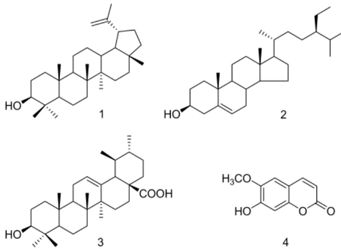
Fig. 1. Structures of compounds 1-4.
bodies (Santa Cruz, Dallas, TX, USA) for 2 hr. Target protein bands were detected using a SuperSignal
TMWest Pico PLUS Chemiluminescent Substrate (Thermo Scientific, Waltham, MA, USA) and chemiluminescence image analyzer (Fuji Film LAS-400, Tokyo, Japan). Densitometry of the bands was analyzed by Image Studio Software (LI-COR, Inc., Lincoln, NE, USA).
Real-time polymerase chain reaction (PCR) Total RNA was extracted from cultured cells with the Qiazol lysis reagent (Qiagen, Hilden, Germany) and RNA extracts (0.2 μg) were reverse-transcribed into cDNA using Revert Aid
TMFirst cDNA Kit (Thermo Scientific, Waltham, MA, USA) as described in the manufacturer’s directions. The reaction was amplified with StepOnePlus
TMreal-time PCR system (Applied Biosystems, CA, USA) for 40 cycles with denaturing at 95℃ for 15 sec, annealing at 60℃ for 60 sec and elongation at 95℃ for 60 sec. Primer sequences were as follows: ICAM-1 (forward, 5’-TATGGCAACGACTCCTT CT-3’; reverse, 5’- CATTCAGCGTCACCTTGG-3’), VCAM-1 (forward, 5’-AGTTGAAGGATGCGG GAGTA-3’; reverse, 5’- AGAGCACGAGAAGCTCAGGA-3’), E-selectin (forward, 5’- GAGGCCAGTGCTTATTGTCA-3’; reverse, 5’-CATTCAGCG TCACCTTGG-3’) and GAPDH (forward, 5’-CAACGGATTT GGTCGTATTG-3’; reverse, 5’-GATGACAAGC TTCCCGTT CT-3’). The levels of gene expression were normalized to GAPDH as an internal control and quantified using the com- parative threshold cycle (Ct) method.
Adhesion assay
THP-1 cells were stimulated with TNF-α for 18 hr and then were labeled with 10 μM BCECF-AM (Invitrogen, Pais-
ley, UK) for 1 hr. The labeled THP-1 cells (7×10
4cells/well) were then added to the EA.hy926 cells which were treated with TNF-α and/or samples, followed by incubated for 45 min at 37℃. After incubating, non-adherent cells were re- moved by washing with PBS and quantification of adherent cells was detected by fluorescence intensity at 485 and 535 nm of excitation and emission in a fluorescence microplate reader (Victor X5; PerkinElmer, Waltham, MA, USA).
Statistical analysis
All data values are expressed as mean ± standard devia- tion (SD). Significant differences between groups were ana- lyzed using analysis of variance (ANOVA) by PASW sta- tistics (SPSS Inc., Chicago, IL, USA) followed by Duncan’s multiple range test to determine statistical difference among groups. Statistical significance was defined as p<0.05.
Results and Discussion
Isolation and structural elucidation of compounds Three triterpenoids, lupeol (1), β-sitosterol (2), ursolic acid (3), and a coumarin, scopoletin (4) were isolated from a CHCl
3-soluble fraction of S. commixta bark by repeated silica gel column chromatography.
Compound 1 was obtained as white amorphous powder and a molecular ion peak at m/z 426 [M]
+. The molecular formula C
30H
50O was deduced from its EIMS and NMR. The IR spectrum exhibited a hydroxyl (3,300 cm
-1) absorption band. The
1H-NMR spectrum of compound 1 showed two olefinic methines [δ 4.69 (1H, s, H-29a) and 4.57 (1H, s, H- 29b)], an oxygenated methine δ 3.20 (1H, dd, J=5.4, 9.9 Hz, H-3) and seven methyl groups [δ 1.68 (3H, s, H-30), 1.03 (3H, s, H-26), 0.96 (3H, s, H-23), 0.95 (3H, s, H-27), 0.83 (3H, s, H-25), 0.79 (3H, s, H-28) and 0.76 (3H, s, H-24)]. The
13C- NMR and DEPT spectra showed thirty signals and exhibited two olefinic methines [δ 150.7 (C-20) and 109.3 (C-29)], an oxygenated methine [δ 78.8 (C-3)], and seven methyl groups [δ 28.0 (C-23), 19.3 (C-30), 18.0 (C-28), 16.1 (C-25), 16.0 (C-26), 15.4 (C-24) and 14.5 (C-27)]. As a result, compound 1 was identified as 3β-hydroxylup-20 (29)-ene (lupeol) by compar- ing its spectroscopic data with the previously reported data [12, 15].
Compound 2 was identified as stigmast-5-en-3-β-ol (β-si- tosterol) through the comparison of spectroscopic data with the previously reported data [8, 11].
Compound 3 was obtained as white amorphous powder
Table 2. Inhibitory activity of CuSO
4-induced LDL oxidation of compounds 1-4
Compounds IC
50(μM)
1 2 3 4 Ascorbic acid
>80
>80
>80 10.2±0.1 84.1±0.3
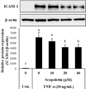
Fig. 2. Effects of scopoletin on TNF-α-induced ICAM-1 expression.
EA.hy926 cells were pretreated with scopoletin for 2 hr and stimulated with TNF-α for an additional 18 hr.
Whole cell extracts (20 μg) were subjected to Western blotting for ICAM-1. Bar graph (lower part) shows densi- tometric evaluation of ICAM-1 relative to β-actin. Results are expressed as mean ± SD (n=3). Different letters in- dicate a significant difference according to the ANOVA (p<0.05).
and a molecular ion peak at m/z 456 [M]
+. The molecular formula C
30H
48O
3was deduced from its EIMS and NMR.
The IR spectrum exhibited a hydroxyl (3,420 cm
-1) and an olefine (1,690 cm
-1) absorption band. The
1H-NMR spectrum of compound 3 showed an olefinic methine [δ 5.13 (1H, s, H-12)], an oxygenated methine [δ 3.02 (1H, dd, J=4.9, 10.0 Hz, H-3)] and seven methyl groups [δ 1.05 (3H, s, H-23), 0.92 (6H, s, H-26, 27), 0.87 (3H, s, H-24), 0.82 (3H, d, J=6.3 Hz, H-30), 0.76 (3H, s, H-29) and 0.68 (3H, s, H-25)]. The
13