1
Department of Periodontology, Research Institute for Periodontal Regeneration, Yonsei University College of Dentistry, Seoul, Republic of Korea
Young Woo Song, Ui-Won Jung, Jae-Kook Cha
Soft tissue volume changes following gingival grafting for labial gingival recession in the
mandibular anterior area: a case report
Soft tissue volume changes following gingival grafting for labial gingival recession in the mandibular anterior area: a case report
Department of Periodontology, Research Institute for Periodontal Regeneration, Yonsei University College of Dentistry, Seoul, Republic of Korea
Young Woo Song, Ui-Won Jung, Jae-Kook Cha
This case report presents results for gingival recession coverage following gingival grafting and for gingival biotype enhance- ments by visualizing soft tissue volume changes using intraoral three-dimensional scanning. A 28 year old female patient with mul- tiple gingival recessions and a 19 year old female patient with a single gingival recession on mandibular anterior area were treated.
Root coverage was performed in both cases using autogenous subepithelial connective tissue harvested from palate. Intraoral 3D scan data were obatained presurgery and at 3 months, 1 year, and 2 years postsurgery.
The recession areas were recovered successfully by subepithelial connective tissue graft combined with pedicle flap repositioning, and the patients showed neither further recurrence nor post-operative complication. Soft tissue biotype changes were identified by superimposing and analyzing scan data, revealing that gingival biotype was enhanced in both cases.
These cases suggest that SCTG could be advantageous in terms of the gingival biotype enhancement, as well as gingival recession coverage, and intraoral 3D scanning might be suitable for assessing post-surgical gingival biotype change.
Keywords: gingival recession, gingival biotype, subepithelial connective tissue graft, intraoral 3D scanning
Corresponding Author Jae-Kook Cha, DDS, PhD.
Department of Periodontology, Yonsei University College of Dentistry 50-1 Yonsei-ro, Seodaemun-gu, Seoul 03722, Republic of Korea
E-mail: [email protected], Fax: +82-2-392-0398 Tel.: +82-2-2228-3191
ACKNOWLEDGMENT This work was supported by the National Research Foundation of Korea (NRF) grant funded by the Korea government
ABSTRACT
ollowing gingival grafting for labial gingival recession in the mandibular anterior area: a case report
Ⅰ. Introduction
Gingival recession frequently appears in patients with fair or poor oral hygiene1-3). In patients with poor oral hygiene, recession occurs as a result of periodontal attachment destruction, which makes it difficult for the clinician to recover the recession owing to the reduced clinical attachment level in proximal areas 4,5). On the other hand, there are several reasons why recession presents in patients with fair oral hygiene and a healthy periodon- tal status, which results in alveolar deficiency on the labial/buccal side such as in cases of labially/
buccally tilted root apex following orthodontic treatment or the congenital condition of a par- tial absence of the labial/buccal bone plate 6). The sufficient blood supply from the adjacent alveolar bone and gingival flap in these cases means that the exposed root surfaces can be predictably cov- ered by gingival grafts combined with pedicle flap repositioning, such as when using a coronally ad- vanced flap (CAF) or a laterally repositioned flap (LRF) 6-9).
Gingival grafting not only covers the recession but also improves the gingival biotype 10). Hwang and Wang reported that the enhanced biotype serves as a critical prognostic factor for recession coverage from the following aspects: (i) greater amounts of extracellular matrix and collagen fi- bers improve biomechanical properties, which may prevent relapse of gingival recession, and (ii) a more-abundant blood supply enhances the drain- age of toxic substances that accumulate in the gin-
giva and the migration of growth factors to where they are needed.
Following up the change in the gingival thick- ness after gingival grafting is as critical as evaluat- ing the change in the marginal gingival level. Previ- ous studies have employed various ways to assess the gingival biotype, such as piercing the gingiva tissue with endodontic files, placing a periodon- tal probe in the gingival sulcus to determine how much of it is visible through the sulcus, and using wax gauge calipers 11-13). Even though these meth- ods may be faster than others, there is a possibil- ity of damaging the gingiva and a need for local anesthesia. Computed tomography (CT) has been used to observe the thickness of soft tissue 14), but the increased exposure to radiation during period- ic CT represents a disadvantage. To minimize the discomfort of patients associated with these pre- vious methods, three-dimensional (3D) intraoral scanning has been recently introduced, which has been found to provide the data that are as accurate as data obtainable using other methods 15-18).
This report presents two cases of a root cover- age procedure accompanied by a subepithelial connective tissue graft (SCTG), and describes the changes in the gingival biotype between the pre- surgical and postsurgical states as assessed by ana- lyzing 3D intraoral scanned data.
Ⅱ. Materials and Methods
This investigation received ethical approval from
the Institutional Review Board of Yonsei University Dental Hospital (Approval No. 2-2018-0005).
1. Surgical intervention
1) Case 1. Labial gingival recession coverage of the left mandibular central and lateral incisors by an LRF with an SCTG
A female patient aged 28 years visited the clinic with a chief complaint of hypersensitivity on the exposed labial root surface of the left lower central and lateral incisors. The patient had neither a his-
tory of orthodontic treatment nor any past medical history. Labial soft tissue recession was observed:
6 mm on the central incisor and 3 mm on the later- al incisor. The adjacent proximal bone levels were intact, and the recession did not exceed mucogin- gival junction (MGJ). The recession could therefore be classified as Miller Class I according to the clas- sification proposed by Miller 4), or Recession Type 1 based on the more recent classification suggested by Cairo et al. 5). The probing depth was 2 mm at all sites, and the oral hygiene state was good (Fig. 1a to 1c).
Figure 1. Clinical photographs and a standard periapical radiograph of case 1 obtained at the first visit (a–c) and on the day of surgery (d–i).
ollowing gingival grafting for labial gingival recession in the mandibular anterior area: a case report To minimize coronal displacement of the MGJ
on the labial side, the use of an LRF with an SCTG was planned. Scaling and plaque control were conducted 1 month before the surgery. Local in- filtration anesthesia was applied to the surgical site with lidocaine (2% lidocaine HCL with epinephrine 1:80,000; Kwangmyung Pharm., Seoul, Korea). The recession on the left lateral incisor was recovered by making two incisions—a vertical incision at 5 mm distal to the distal marginal gingiva of the lateral incisor and an oblique incision parallel to the mesial marginal gingiva of the adjacent ca- nine—to allow elevation of a partial-thickness flap (Fig. 1d). To ensure coverage of the central inci- sor, two horizontal incisions were made at 3 mm from the top of the mesial and distal interdental papilla of the right central incisor, and a crevicu- lar incision was made from the mesial side of the left central incisor to the distal side of the right lat- eral incisor for elevating a partial-thickness flap (Fig. 1e). The marginal gingiva on the mesial side of the left lateral incisor and on the distal side of the left central incisor were de-epithelialized using a surgical blade, where two partial-thickness pedicle flaps were going to be repositioned (Fig. 1f). The denuded root surfaces were meticulously debrided using a periodontal curette and root planing bur, and conditioned with a tetracycline-soaked cot- ton pellet. Subepithelial connective tissue obtained from the palate on the left side was trimmed (Fig.
1g) and fixed at the recipient site using a periosteal suture with resorbable 6-0 glyconate monofila- ment sulture material (Monosyn®, B. Braun, Tut-
tlingen, Germany) (Fig. 1h). Two pedicle flaps were moved laterally to cover the grafted tissue and then sutured (Fig. 1i). The sutures were removed 10 days after the surgery.
2) Case 2. Labial gingival recession coverage of the right mandibular central incisor by a CAF with an SCTG
A female patient aged 19 years presented with 3 mm of labial gingival recession and hypersen- sitivity of the right lower central incisor. Since the apicocoronal range of the recession did not exceed the MGJ and adjacent interproximal bone levels were intact in a radiograph, this case could be clas- sified as Miller Class I 4) or Recession Type I 5). The patient had received orthodontic treatment at a lo- cal dental clinic 10 years previously, and there was no atypical past medical history. Her overall oral hygiene state was fair, but mild gingival swelling and redness in the mandibular anterior area with supragingival calculus were observed. The probing depths were 3 to 4 mm on the labial side and 2 to 3 mm on the lingual side (Fig. 2a to 2c).
The use of a CAF with an SCTG was planned for root coverage. Scaling and plaque control were performed 1 month before the surgery, and the in- terdental papilla in the lower anterior area present- ed with 1 mm of recession on the day of surgery (Fig. 2d). After applying local infiltration anesthesia with lidocaine (2% lidocaine HCL with epinephrine 1:80,000; Kwangmyung Pharm.), two vertical inci- sions were made on each side of the labial gingiva of the central incisor to form a partial-thickness
flap, and both interdental papilla were de-epithe- lialized using a surgical blade (Fig. 2d). After thor- ough planing of the exposed root surface using a periodontal curette and a root planing bur, and root conditioning with a tetracycline-soaked cot- ton pellet, subepithelial connective tissue was ob- tained from the left palate (Fig. 2e) and fixed at the recipient site using periosteal sutures with resorb- able 6-0 glyconate monofilament suture material (Monosyn®, B. Braun) (Fig. 2f). The grafted site was covered by a CAF, and interrupted sutures were in- serted to adapt the flap to the interdental papilla
on each side (Fig. 2g). The sutures were removed 10 days after the surgery.
2. Volumetric analysis by superimposition of intraoral 3D scans
The surgical sites of both cases were scanned with a 3D intraoral scanner (Trios®, 3Shape, Co- penhagen, Denmark) presurgery and at 3 months, 1 year, and 2 years postsurgery. The scanned files of each patient were superimposed using comput- er software (SMOP®, Swissmeda, Zurich, Switzer-
Figure 2. Clinical photographs and a standard periapical radio- graph of case 2 obtained at the first visit (a–c) and on the day of surgery (d–g).
ollowing gingival grafting for labial gingival recession in the mandibular anterior area: a case report
land), and the postsurgical volume data were com- pared to the presurgery data (Fig. 3a). The region of interest (ROI) was set as follows at each grafted site in accordance with previous studies (Fig. 3b) 16-18).
Apicocoronal dimension: 0.5 mm apically from the gingival margin and extending 3.0 mm in an apical direction.
Mesiodistal dimension: 0.5 mm from the mesial and distal adjacent teeth.
The mean distances (MD, mm) within the ROI were measured using the computer software for comparing the volume changes over time.
Ⅱ. Results
1. Clinical and radiographic findings
Postsurgical healing was uneventful in both cases, and it took 2 to 3 months for the texture and color
of the grafted sites to become more harmonized with the surrounding tissue (Fig. 4a, 4e, 5a, and 5e).
During the follow-up period of 2 years, neither case demonstrated any atypical clinical (Fig. 4b, 4c, 4f, 4g, 5b, 5c, 5f, and 5g) or radiographic findings (Fig. 4d and 5d), and the patients no longer com- plained of hypersensitivity. Both cases presented full recession coverage, and they were well main- tained for 2 years without relapses. However, case 2 exhibited 1 mm of interdental papilla recession at the sites, where mild gingival swelling had been present at the first visit (Fig. 5a to 5c). The labial keratinized gingival width (apicocoronal) at the grafted sites was 3 mm in case 1 and 5 mm in case 2, with a probing depth of 2 mm at all sites.
2. Volumetric analysis
Comparing the scanned files between the post- surgical and presurgical states revealed labial gin- Figure 3. Three-dimensional volume analysis. Scanned images were superimposed, and the region of interest
(ROI) was defined as the area in orange color at the labial gingiva (a). The gap between the scanned images and the coronoapical dimension of the ROI are shown in the sliced section of the grafted site (b).
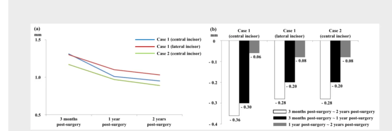
gival thickness gains after 3 months, 1 year, and 2 years of 1.31, 1.01, and 0.95 mm, respectively, for the central incisor of case 1, 1.30, 1.10, and 1.03 mm for the lateral incisor of case 1, and 1.17, 0.97, and 0.89 mm for case 2 (Table 1 and Fig. 6a). The gingival thickness decreased from 3 months post- surgery to 2 years postsurgery by 0.36 mm for the central incisor of case 1 and by 0.28 mm for the lat-
eral incisor of case 1 and the central incisor of case 2 (white bars in Fig. 6b). During the first 9 months (from 3 months postsurgery to 1 year postsurgery) the gingival thickness of the central incisor of case 1 decreased by 0.30 mm, while that of the lateral incisor of case 1 and the central incisor of case 2 each decreased by 0.20 mm (black bars in Fig. 6b).
During the following year (from 1 year postsurgery Figure 4. Clinical photographs of case 1 obtained at 3 months (a, e), 1 year (b, f), and 2 years (c, g) postsurgery.
A standard periapical radiograph of case 1 obtained at 2 years postsurgery (d).
Figure 5. Clinical photographs of case 2 obtained at 3 months (a, e), 1 year (b, f), and 2 years (c, g) postsurgery.
A standard periapical radiograph of case 2 obtained at 2 years postsurgery (d).
ollowing gingival grafting for labial gingival recession in the mandibular anterior area: a case report to 2 years postsurgery) the gingival thickness of the
central incisor of case 1 decreased by 0.06 mm, while that of the lateral incisor of case 1 and the central incisor of case 2 each decreased by 0.08 mm (gray bars in Fig. 6b).
Ⅳ. Discussion
The findings of this study demonstrate the ef- fectiveness of an SCTG combined with pedicle flap repositioning for recession coverage and gingival
biotype thickening. The labial recession was com- pletely recovered in both cases, but recession of the interdental papilla was observed in case 2. During the 2-year follow-up period, labial gingival width (apicocoronal) was well maintained in both cas- es, and labial gingival thickness (labiolingual) in- creased by 0.95 mm (central incisor) and 1.03 mm (lateral incisor) in case 1 and by 0.89 mm in case 2.
These increases in labial gingival thickness in both cases were more favorable compared to a previous report of a mean increase of approximately 0.72 mm at 1 year after an SCTG 19). These may have
Figure 6. Volume gains at each postsurgical time point compared to presurgery (a). Volume shrinkage over time (b).
Case 1
(central incisor) Case 1
(lateral incisor) Case 2
3 months postsurgery vs. presurgery 1.31 1.30 1.17
1 year postsurgery vs. presurgery 1.01 1.10 0.97
2 years postsurgery vs. presurgery 0.95 1.03 0.89
Table 1. Volume gains at 3 months, 1 year and 2 years postsurgery compared to presurgery (mm).
been due to the presurgical states of both cases be- ing classified as Miller Class I or Recession Type 1
4,5,20).
Complete gingival recession coverage has been known to depend on the height of the interden- tal alveolar bone and the coronoapical position of the MGJ 4). Both of the present cases had intact interdental bone heights, and the apical margins of recession did not exceed the MGJ. Since the in- terproximal periodontal tissue was intact, it is likely that the SCTG received a sufficient blood supply.
While both cases exhibited full recession coverage, 1 mm of interdental papilla recession appeared in case 2 compared to the state observed at the first visit. This papilla recession was found throughout the dentition in the anterior mandible when gin- gival inflammation was controlled by scaling and plaque control before the surgery, which means that the papilla-filled state observed at the first visit was the consequence of gingival swelling. Tarnow et al. found that approximately 98% of papilla re- cession could be resolved when the distance be- tween the interproximal bone crest and interdental contact point did not exceed 5 mm 21); however, the papilla heights were not increased in the pres- ent case 2.
In terms of volume, the gingival thickness had increased by more than 1 mm in both cases at 3 months postsurgery, similar to a previous report
22). Compared to the volume measured at 3 months, shrinkage was observed at 1 and 2 years after the surgery in both cases. The volume decreases in the central incisor and lateral incisor of case 1 and
the central incisor of case 2 were 22.9, 15.4, and 17.1%, respectively, at 1 year postsurgery, and 27.5, 21.5, and 23.9% at 2 years postsurgery compared to the volume gained at 3 months postsurgery. These rates are similar to a previous report of a volume decrease of 20.5% at 1 year after an SCTG associ- ated with a CAF 23). Since most of the soft tissue remodeling takes place within the first 3 months after gingival grafting, followed by a volume reduc- tion of approximately 50%, it might be reasonable to consider that the results in both cases were sta- ble 24).
An enhanced gingival biotype may prevent the relapse of gingival recession after a root coverage, via increasing the extracellular matrix volume and blood supply 10). Whether an SCTG should be com- bined with pedicle flap repositioning for root cov- erage has been controversial. Recent studies have found that the root coverage with an SCTG pro- duced significantly more favorable results than the procedure without an SCTG, in terms of both gin- gival thickening and recession coverage 25,26). While free gingival grafting may guarantee a thicker and wider keratinized tissue, blood supply might be less sufficient since it only comes from the supraperi- osteal tissue of the recipient bed, whereas vascular supply to an SCTG originates from both the recipi- ent bed and the inner surface of the pedicle flap
6,27).
Previous studies have performed long-term eval- uations of root coverage by an SCTG. The recently published long-term study by Rasperini et al. found that using an SCTG with a CAF did not produce
ollowing gingival grafting for labial gingival recession in the mandibular anterior area: a case report superior results for complete recession coverage
compared to using only a CAF; however, it provid- ed a greater increase in the thickness of keratinized tissue 28). Pini Prato et al. showed that the short- term soft tissue defect reduction and keratinized tissue enhancement were both well preserved over a follow-up period of 20 years 29). Based on these reports, it could be expected that the results ob- tained in present two cases would remain stable over a long time period.
Ⅴ. Conclusion
Root coverage combined with an SCTG can be a predictable treatment modality for gingival reces-
sion coverage and gingival biotype improvement.
Complete coverage of the denuded root surface and enhanced gingival thickness were successfully achieved in both of the present cases. Long-term follow-up is needed, and intraoral 3D scanning can be a suitable way to observe changes in the gingival thickness over time.
1. Loe H, Anerud A, Boysen H. The natural history of periodontal dis- ease in man: prevalence, severity, and extent of gingival recession. J Periodontol 1992;63(6):489-495
2. Sangnes G, Gjermo P. Prevalence of oral soft and hard tissue lesions related to mechanical toothcleansing procedures. Community Dent Oral Epidemiol 1976;4(2):77-83
3. Susin C, Haas AN, Oppermann RV, et al. Gingival recession: epide- miology and risk indicators in a representative urban Brazilian popula- tion. J Periodontol 2004;75(10):1377-1386
4. Miller PD, Jr. A classification of marginal tissue recession. Int J Peri- odontics Restorative Dent 1985;5(2):8-13
5. Cairo F, Nieri M, Cincinelli S, et al. The interproximal clinical attach- ment level to classify gingival recessions and predict root coverage outcomes: an explorative and reliability study. J Clin Periodontol 2011;38(7):661-666
6. Zucchelli G, Mounssif I. Periodontal plastic surgery. Periodontol 2000 2015;68(1):333-368
7. Borghetti A, Gardella JP. Thick gingival autograft for the coverage of gingival recession: a clinical evaluation. Int J Periodontics Restorative Dent 1990;10(3):216-229
8. Jung UW, Kim CS, Choi SH, Kim S. Gingival coverage of iatrogenically denuded labial bone resulting from thermal trauma. Int J Periodontics Restorative Dent 2013;33(5):635-639
9. Jung UW, Um YJ, Choi SH. Histologic observation of soft tissue ac- quired from maxillary tuberosity area for root coverage. J Periodontol 2008;79(5):934-940
10. Hwang D, Wang HL. Flap thickness as a predictor of root coverage:
a systematic review. J Periodontol 2006;77(10):1625-1634 11. Memon S, Patel JR, Sethuraman R, et al. A comparative evaluation
of the reliability of three methods of assessing gingival biotype in dentate subjects in different age groups: An in vivo study. J Indian Prosthodont Soc 2015;15(4):313-317
12. Zawawi KH, Al-Zahrani MS. Gingival biotype in relation to incisors' inclination and position. Saudi Med J 2014;35(11):1378-1383
참 고 문 헌
13. Pietruska M, Skurska A, Podlewski L, et al. Clinical evaluation of Miller class I and II recessions treatment with use of modified coro- nally advanced tunnel technique with either collagen matrix (CM) or subepithelial connective tissue graft (SCTG): a randomized clinical study. J Clin Periodontol 2018
14. Gurlek O, Sonmez S, Guneri P, Nizam N. A novel soft tissue thick- ness measuring method using cone beam computed tomography.
J Esthet Restor Dent 2018
15. Thoma DS, Jung RE, Schneider D, et al. Soft tissue volume aug- mentation by the use of collagen-based matrices: a volumetric analysis. J Clin Periodontol 2010;37(7):659-666
16. Sanz-Martin I, Sailer I, Hammerle CH, Thoma DS. Soft tissue sta- bility and volumetric changes after 5 years in pontic sites with or without soft tissue grafting: a retrospective cohort study. Clin Oral Implants Res 2016;27(8):969-974
17. Sanz Martin I, Benic GI, Hammerle CH, Thoma DS. Prospective randomized controlled clinical study comparing two dental implant types: volumetric soft tissue changes at 1 year of loading. Clin Oral Implants Res 2016;27(4):406-411
18. Song YW, Kim S, Waller T, et al. Soft tissue substitutes to increase gingival thickness: Histologic and volumetric analyses in dogs. J Clin Periodontol 2019;46(1):96-104
19. Zucchelli G, Mounssif I, Mazzotti C, et al. Does the dimension of the graft influence patient morbidity and root coverage out- comes? A randomized controlled clinical trial. J Clin Periodontol 2014;41(7):708-716
20. Jepsen S, Caton JG, Albandar JM, et al. Periodontal manifestations of systemic diseases and developmental and acquired conditions:
Consensus report of workgroup 3 of the 2017 World Workshop on the Classification of Periodontal and Peri-Implant Diseases and Conditions. J Clin Periodontol 2018;45 Suppl 20:S219-s229 21. Tarnow DP, Magner AW, Fletcher P. The effect of the distance from
the contact point to the crest of bone on the presence or absence of
the interproximal dental papilla. J Periodontol 1992;63(12):995-996 22. Zeltner M, Jung RE, Hammerle CH, et al. Randomized controlled
clinical study comparing a volume-stable collagen matrix to au- togenous connective tissue grafts for soft tissue augmentation at implant sites: linear volumetric soft tissue changes up to 3 months.
J Clin Periodontol 2017;44(4):446-453
23. Azaripour A, Kissinger M, Farina VS, et al. Root coverage with con- nective tissue graft associated with coronally advanced flap or tun- nel technique: a randomized, double-blind, mono-centre clinical trial. J Clin Periodontol 2016;43(12):1142-1150
24. Akcali A, Schneider D, Unlu F, et al. Soft tissue augmentation of ridge defects in the maxillary anterior area using two different methods: a randomized controlled clinical trial. Clin Oral Implants Res 2015;26(6):688-695
25. Stefanini M, Jepsen K, de Sanctis M, et al. Patient-reported out- comes and aesthetic evaluation of root coverage procedures: a 12-month follow-up of a randomized controlled clinical trial. J Clin Periodontol 2016;43(12):1132-1141
26. Cairo F, Nieri M, Pagliaro U. Efficacy of periodontal plastic surgery procedures in the treatment of localized facial gingival recessions.
A systematic review. J Clin Periodontol 2014;41 Suppl 15:S44-62 27. Langer B, Langer L. Subepithelial connective tissue graft technique
for root coverage. J Periodontol 1985;56(12):715-720
28. Rasperini G, Acunzo R, Pellegrini G, et al. Predictor factors for long- term outcomes stability of coronally advanced flap with or without connective tissue graft in the treatment of single maxillary gingival recessions: 9 years results of a randomized controlled clinical trial. J Clin Periodontol 2018;45(9):1107-1117
29. Pini Prato GP, Franceschi D, Cortellini P, Chambrone L. Long-term evaluation (20 years) of the outcomes of subepithelial connective tissue graft plus coronally advanced flap in the treatment of maxillary single recession-type defects. J Periodontol 2018;89(11):1290- 1299
참 고 문 헌