An effective cough is an important host defense mechanism to clear the airway.1 The phases of cough are classified as 1) inspiratory, 2) compressive, and 3) expi- ratory.1-4During the inspiration phase, normal subjects have pre-cough volumes that are about 85% to 90% of their inspiratory capacity and have a total cough volume of about 2.3 L to obtain optimal cough flows.5After enough air volume is inhaled, the glottis automatically closes and prevents any outflow of inhaled air.6,7 The contraction of the expiratory muscles while the glottis is open initiates the
How Respiratory Muscle Strength Correlates with Cough Capacity in Patients
with Respiratory Muscle Weakness
Jung Hyun Park,
1Seong-Woong Kang,
2Sang Chul Lee,
3Won Ah Choi,
2and Dong Hyun Kim
21Department of Rehabilitation Medicine, Eulji University Hospital, Daejeon;
2Department of Rehabilitation Medicine and Rehabilitation Institute of Muscular Disease, Yonsei University College of Medicine, Seoul;
3Department of Physical Medicine and Rehabilitation, Myongji Hospital, Kwandong University College of Medicine, Seoul, Korea.
Purpose:The purpose of this study is to investigate how respiratory muscle strength correlates to cough capacity in patients with respiratory muscle weakness. Materials and Methods:Forty-five patients with amyotrophic lateral sclerosis (ALS), 43 with cervical spinal cord injury (SCI), and 42 with Duchenne muscular dystrophy (DMD) were recruited. Pulmonary function tests including forced vital capacity (FVC) and respiratory muscle strength (maximal expiratory pressure, MEP;
maximal inspiratory pressure, MIP) were performed. The correlation between respiratory muscle strength and cough capacity was analyzed. Results:In the SCI group, FVC in a supine position (2,597 ± 648 mL) was significantly higher than FVC in a sitting position (2,304 ± 564 mL, p < 0.01). Conversely, in the ALS group, FVC sitting (1,370 ± 604 mL) was significantly higher than in supine (1,168
± 599 mL, p < 0.01). In the DMD group, there was no statistically significant difference between FVC while sitting (1,342 ± 506 mL) and FVC while supine (1,304 ± 500 mL). In addition, the MEP and MIP of all three groups showed a significant correlation with peak cough flow (PCF) (p < 0.01, Pearson’s correlation analysis). In the SCI group, MIP was more closely correlated with PCF, while in the ALS and DMD groups, MEP was more closely correlated with PCF (p < 0.01, multiple regression analysis). Conclusion:To generate cough flow, inspiratory muscle strength is significantly more important for SCI patients, while expiratory muscle function is significantly more important for ALS and DMD patients.
Key Words: Amyotrophic lateral sclerosis, muscular dystrophy, duchenne, respiratory function tests, spinal cord injuries
Received: July 7, 2009 Revised: August 11, 2009 Accepted: August 13, 2009
Corresponding author: Dr. Seong-Woong Kang, Department of Rehabilitation Medicine and Rehabilitation Institue of Muscular Disease, Yonsei University College of Medicine, 612 Eonju-ro, Gangnam-gu,
Seoul 135-720, Korea.
Tel: 82-2-2019-3492, Fax: 82-2-2019-3499 E-mail: [email protected]
∙The authors have no financial conflicts of interest.
© Copyright:
Yonsei University College of Medicine 2010 This is an Open Access article distributed under the terms of the Creative Commons Attribution Non- Commercial License (http://creativecommons.org/
licenses/by-nc/3.0) which permits unrestricted non- commercial use, distribution, and reproduction in any medium, provided the original work is properly cited.
INTRODUCTION
expulsion phase during which air from the lungs is forcibly expelled.4An effective cough cannot be obtained if one of these phases fails.
Respiratory muscle weakness and decreased cough capacity are the main causes of pulmonary complications that result in morbidity and mortality in patients with neuromuscular disorders (NMDs) and in tetraplegics. To prevent pulmonary complications in these patients, it is necessary to understand the mechanics of the respiratory system and to accurately evaluate pulmonary function.
In patients with Duchenne muscular dystrophy (DMD), diaphragmatic function is relatively well preserved despite generalized muscle dysfunction.8However, patients with amyotrophic lateral sclerosis (ALS) experience profound diaphragm weakness, early in the disease process.9-11Finally, in many patients with spinal cord injury (SCI) if injured at high cervical levels, the functions of intercostal and abdo- minal muscles are impaired, while diaphragm function is preserved.8-10,12,13
Previous studies have examined respiratory function in patients with NMDs by analyzing the differences between vital capacity measured while patients are in various posi- tions.8-10,13Several researchers have also studied the correla- tion between expiratory muscle strength and cough.4,14 How- ever, the relationship between cough capacity and differen- tial respiratory muscle weakness patterns has rarely been discussed. Although expiratory muscle function is crucial for an effective cough, the lung volume attained before expiratory muscle contraction also plays an important role.
If insufficient pre-cough volume is obtained due to inspira- tory muscle weakness, cough capacity decreases in spite of functional expiratory muscles.15As a result, a lack of deep breathing leads to atelectasis, where the lung and chest walls lose their elasticity and compliance in the chronic stage.16-18 In addition, a loss of lung compliance may exacerbate cough weakness by restricting dynamic airway compression.15
The correlation between cough capacity and the inspira- tory muscle strength necessary to obtain adequate lung volume has rarely been studied.19,20For this reason, we studied patients with clinically common diseases that accompany restrictive lung disease (RLD) to establish how weakened respiratory muscles affect cough capacity. We evaluated patients with ALS, cervical SCI, and DMD that had different but well-defined patterns of respiratory mus- cles weakness.
Subjects
Forty-two patients with DMD were involved in this study;
DMD diagnosis was established according to an internatio-
nally accepted criteria21: progressive muscular weakness, increased plasma muscle enzymes, muscle biopsy showing fiber degeneration and the absence of the dystrophin pro- tein, and alterations in the dystrophin gene (e.g., deletions).
Forty-five patients with probable or definitive ALS accord- ing to El Escorial criteria22were also included in the study.
Finally, 43 patients with traumatic cervical SCI, who sustain- ed complete motor injuries as defined by the American Spinal Injury Association (ASIA) criteria, were also includ- ed. The recruitment period was from February 2000 to May 2007. Patients who had concomitant intrinsic lung diseases, who could not hold breaths due to vocal cord paralysis, who were intubated or had a tracheostomy, and who took medi- cations that affect pulmonary function were excluded.
The institutional review board of our institute approved our study. Informed consent was obtained from all subjects.
Measurements
Forced vital capacity (FVC)
Forced vital capacity (FVC) was measured in sitting and supine positions using a spirometer (Wright spirometer;
Ferraris Development & Engineering Co. Ltd., London, England). This process was repeated at least three times;
the highest value was selected as the FVC. We calculated predicted FVC values (FVCpre) based on age, height, and weight.23Relative FVC values were reported as FVC/
FVCpre (%).
Respiratory muscle strength
Maximal respiratory pressure and reflecting respiratory muscle strength was measured by mouth pressure-meter (MicroRPM; Micro Medical Ltd., Rochester, England) in a sitting position. To measure maximal expiratory pressure (MEP), subjects performed maximal expiratory effort after maximal inspiration. Maximal inspiratory pressure (MIP) was measured by exerting maximal inspiratory effort after maximal expiration. To measure these pressures, effort was maintained for at least 1 second. The highest positive MEP value and the lowest negative MIP value in three or more attempts were chosen. We calculated predicted MEP (MEPpre) and MIP (MIPpre) values based on age, height, and weight.23Relative MEP and MIP values were presented as MEP/MEPpre (%) and MIP/MIPpre (%).
Peak cough flow (PCF)
Peak cough flow (PCF) was measured by a peak flow-meter (ASSESS®; Health Scan Products Inc., Cedar Grove, NJ, USA). Unassisted PCF was measured by having the patient cough as much as possible through a peak flow-meter. The highest value in each test was used by running at least three trials. Cough capacity, defined as the ability to clear
MATERIALS AND METHODS
airway secretion, can be represented as PCF.2,14,19 Statistical analysis
A paired t-test was used to compare the FVC in sitting and supine positions of patients with ALS, cervical SCI, and DMD. The relationships between MIP and PCF and between MEP and PCF were analyzed in each patient group using Pearson’s correlation and multiple regression analysis. All data were analyzed using Statistical Package for the Social Sciences (SPSS) 12.0 (SPSS, Inc; Chicago, IL, USA).
Table 1 lists patient characteristics. All 43 SCI patients showed complete motor paralysis at the cervical level; 32 were classified as having ASIA-A. Twenty-one of these patients were injured at C6, six patients were injured at C7,
and five patients were injured at C8. Eleven patients had ASIA-B tetraplegia; seven of these patients had levels of injury at C6, three patients were injured at C7, and one patient was injured at C8.
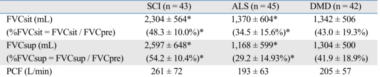
Table 2 shows that in the cervical SCI group, FVC and
%FVC while supine was significantly greater than FVC and %FVC while sitting (p < 0.01). In the ALS group, however, FVC and %FVC while sitting was significantly greater than while supine (p < 0.01). In the DMD group, there was no statistically significant difference between the mean FVCs while sitting or supine. The mean PCF and
%FVC is also noted in Table 2.
The SCI group had a relative MIP value much greater than the relative MEP value (p < 0.01) (Table 3). In the ALS group, the relative MEP value was lower than the relative MIP value, but there was no significant difference between the two values (Table 3). In DMD patients, the relative MIP value was significantly higher than the relative MEP value (p < 0.01) (Table 3).
RESULTS
Table 1. Subject Characteristics
SCI (n = 43) ALS (n = 45) DMD (n = 42)
Sex Men 35 35 42
Women 8 10 0
Age (yr) 35.0 ± 12.6 54.4 ± 11.1 16.1 ± 4.0
Height (cm) 107.4 ± 7.7 165.9 ± 10.7 152.5 ± 11.4
Body weight (kg) 62.3 ± 9.2 54.9 ± 9.1 42.7 ± 12.7
SCI, spinal cord injury; ALS, amyotrophic lateral sclerosis; DMD, Duchenne muscular dystrophy.
Data presented as mean±SD.
Table 3. Comparison between MIP and MEP
SCI (n = 43) ALS (n = 45) DMD (n = 42)
MIP (cmH2O) 63.8 ± 25.0* 24.3 ± 10.6 32.4 ± 11.3*
(%MIP = MIP / MIPpre) (63.8 ± 21.3%)* (30.1 ± 13.6%) (40.2 ± 16.7%)*
MEP (cmH2O) 36.2 ± 17.2* 31.6 ± 15.2 28.3 ± 10.4*
(%MEP = MEP / MEPpre) (26.4 ± 11.4%)* (26.9 ± 13.1%) (23.8 ± 10.1%)*
%MIP / %MEP 2.42 1.12 1.69
MIP, maximum inspiratory pressure; MEP, maximum expiratory pressure; MIPpre, predicted maximum inspiratory pressure; MEPpre, predicted maximum expiratory pressure; SCI, spinal cord injury; ALS, amyotrophic lateral sclerosis; DMD, Duchenne muscular dystrophy.
Data presented as mean±SD.
*Paired t-test, p < 0.01.
Table 2. Pulmonary Function Test Results
SCI (n = 43) ALS (n = 45) DMD (n = 42)
FVCsit (mL) 2,304 ± 564* 1,370 ± 604* 1,342 ± 506
(%FVCsit = FVCsit / FVCpre) (48.3 ± 10.0%)* (34.5 ± 15.6%)* (43.0 ± 19.3%)
FVCsup (mL) 2,597 ± 648* 1,168 ± 599* 1,304 ± 500
(%FVCsup = FVCsup / FVCpre) (54.2 ± 10.4%)* (29.2 ± 14.93%)* (41.9 ± 18.9%)
PCF (L/min) 261 ± 72 193 ± 63 205 ± 57
FVCsit, forced vital capacity in sitting position; FVCpre, predicted value of forced vital capacity; FVCsup, forced vital capacity in supine position; PCF, peak cough flow; SCI, spinal cord injury; ALS, amyotrophic lateral sclerosis; DMD, Duchenne muscular dystrophy.
Data presented as mean±SD.
*Paired t-test, p < 0.01.
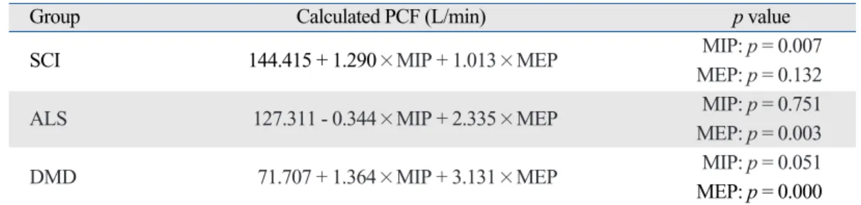
Respiratory muscle strength and PCF were significantly correlated in all three groups. In the SCI group, both MEP (Pearson’s coefficient of correlation r = 0.534, p < 0.01) and MIP (r = 0.609, p < 0.01) (Table 4) were significantly cor- related with PCF, but MIP (p = 0.007) was more strongly correlated than MEP (p = 0.132) (Table 5) with PCF. The results of the ALS group were somewhat different: again, both MEP (r = 0.528, p < 0.01) and MIP (r = 0.339, p < 0.05) (Table 4) were significantly correlated with PCF, but in this group, MEP (p = 0.003) was more strongly correlated than MIP (p = 0.751) (Table 5). The DMD group showed similar results to the ALS group: MEP (r = 0.742, p < 0.01) and MIP (r = 0.637, p < 0.01) (Table 4) were significantly correlated with PCF, but MEP (p = 0.000) was more stro- ngly correlated than MIP (p = 0.051) (Table 5).
In this study, we compared the FVC of subjects in both sitting and supine positions. In the SCI group, the FVC was larger in the supine position than in the sitting posi- tion, which reflects preserved diaphragmatic function but impaired function of the intercostal and abdominal muscles.
However, in ALS patients the FVC in the supine position was much smaller than that in the sitting position, suggest- ing both rapidly progressing muscle weakness with pro- found diaphragm weakness. In contrast, because in DMD patients the diaphragm retains its function as the primary inspiratory muscle, there is scant difference in vital capa- city when the patient’s position changes. These results are similar to those of previous studies.8-13,24We confirmed that measuring FVC in different positions is important to fully
understand the weakness patterns of inspiratory and ex- piratory muscles in patients with RLD.
In this study, the cervical SCI group showed markedly decreased MEP, moderately decreased MIP, and high
%MIP/%MEP (2.42) (Table 3), indicating expiratory mus- cle weakness as the predominant respiratory dysfunction in the SCI group. In the ALS group, both MIP and MEP were markedly decreased, in addition to a lower %MIP/%MEP value (1.12) (Table 3), which suggests that the inspiratory and expiratory muscles had similar and profound levels of weakness. In the DMD group, both MIP and MEP were low, but the ratio of %MIP/%MEP (1.67) (Table 3) was midway between that of the SCI and ALS groups. This indicates that the DMD group exhibited both inspiratory and expiratory muscle weakness, but retained more inspi- ratory muscle function than the ALS group.
In our correlation study, both MIP and MEP showed significant correlations with PCF in all three groups. How- ever, in the SCI group, multiple regression analysis showed that MIP (p = 0.007) was more highly correlated with PCF than MEP (p = 0.132) (Table 5). This may indicate that inspiratory muscles are more important for cough produc- tion in motor complete cervical SCI patients who have lost a large percentage of expiratory muscle power. Conversely, in the ALS group, MEP (p = 0.003) was more highly cor- related with PCF than MIP (p = 0.751) (Table 5). Due to the inspiratory and bulbar muscle weakness in patients with ALS, it is difficult to attain sufficient pre-cough volume and to hold the breath just before coughing. Thus, in ALS patients, cough may depend predominantly on expiratory muscle strength. Finally, in the DMD group, MEP (p = 0.000) was more highly correlated with PCF than MIP (p = 0.051) (Table 5). This indicates that although patients with
DISCUSSION
Table 4. Correlation between PCF and MIP, MEP
SCI (n = 43) ALS (n = 45) DMD (n = 42)
MIP MEP MIP MEP MIP MEP
PCF r 0.609 0.534 0.339 0.528 0.637 0.742
p < 0.01 < 0.01 0.023 < 0.01 < 0.01 < 0.01 PCF, peak cough flow; MIP, maximum inspiratory pressure; MEP, maximum expiratory pressure; SCI, spinal cord injury; ALS, amyotrophic lateral sclerosis; DMD, Duchenne muscular dystrophy; r, Pearson correlation coefficient.
Table 5. Results of Multiple Regression Analysis
Group Calculated PCF (L/min) p value
SCI 144.415 + 1.290×MIP + 1.013×MEP MIP: p = 0.007
MEP: p = 0.132
ALS 127.311 - 0.344×MIP + 2.335×MEP MIP: p = 0.751
MEP: p = 0.003
DMD 71.707 + 1.364×MIP + 3.131×MEP MIP: p = 0.051
MEP: p = 0.000 PCF, peak cough flow; SCI, spinal cord injury; ALS, amyotrophic lateral sclerosis; DMD, Duchenne muscular dystrophy; MIP, maximum inspiratory pressure; MEP, maximum expiratory pressure.
DMD retain more of their inspiratory muscle strength, both inspiratory and expiratory muscle weakness are pro- found. Because inspiratory muscle strength is insufficient to generate enough pre-cough volume, cough in patients with DMD depends more upon expiratory muscle strength than in patients with SCI.
It is easily understood that coughing is a kind of exhala- tion process, which leads to the conclusion that expiratory muscle strength is more strongly correlated than inspira- tory muscle strength with cough capacity. In light of our results, however, this assumption can be misleading, de- pending on the disease process and its effects on respiratory muscle weakness. Like the SCI cases in this study, ins- piratory muscle strength can correlate more strongly with cough capacity. Thus, we conclude that both expiratory and inspiratory muscle function are important to build an effective cough. Given this assertion, it is difficult to achieve a sufficiently strong cough by assisting only expiratory muscles without assisting inspiratory muscles in RLD patients whose pre-cough volumes decrease.15 Therefore, an adequate cough can be generated by not only expiratory phase assistance, but also by pre-cough volume support.25,26
This study has several limitations. We think it is worthy to note that inspiratory muscle strength may be important for producing an effective cough in patients with predo- minantly expiratory muscle dysfunction, in light of the fact that inspiratory muscle strength is one of the determinants of pre-cough lung volume. However, we were unable to determine the pre-cough lung volume directly. We believe that the functional residual capacity (FRC) is an indicator of pre-cough lung volume, but our study would be streng- thened by obtaining additional lung volume data (total lung capacity, functional residual capacity, residual vol- ume, etc.). With this information, the correlation between FRC and PCF could be analyzed in detail. However, in our hospital, those parameters are not included in routine pulmonary evaluation of patients with severe respiratory compromise.
In conclusion, 1) inspiratory muscle strength is impor- tant for producing an effective cough in certain patients with respiratory muscle dysfunction, even though cough- ing is a type of exhalation process; and 2) pre-cough lung volume may play an important role in establishing an effective cough in patients with respiratory muscle dys- function whose expiratory muscles are highly impaired.
Authors would like to thank Dr. ImHee Shin for her con- tribution to data analysis in this study.
This study was performed at Department of Rehabili-
tation Medicine, Yonsei University College of Medicine.
1. McCool FD. Global physiology and pathophysiology of cough:
ACCP evidence-based clinical practice guidelines. Chest 2006;
129(1 Suppl):48S-53.
2. Kang SW, Kang YS, Sohn HS, Park JH, Moon JH. Respiratory muscle strength and cough capacity in patients with Duchenne muscular dystrophy. Yonsei Med J 2006;47:184-90.
3. McCool FD, Leith DE. Pathophysiology of cough. Clin Chest Med 1987;8:189-95.
4. Schramm CM. Current concepts of respiratory complications of neuromuscular disease in children. Curr Opin Pediatr 2000;12:
203-7.
5. Brain JD, Proctor DF, Reid L. Respiratory defense mechanisms.
New York: M. Dekker; 1977.
6. Braun SR, Giovannoni R, O’Connor M. Improving the cough in patients with spinal cord injury. Am J Phys Med 1984;63:1-10.
7. Sivasothy P, Brown L, Smith IE, Shneerson JM. Effect of manu- ally assisted cough and mechanical insufflation on cough flow of normal subjects, patients with chronic obstructive pulmonary disease (COPD), and patients with respiratory muscle weakness.
Thorax 2001;56:438-44.
8. Inkley SR, Oldenburg FC, Vignos PJ Jr. Pulmonary function in Duchenne muscular dystrophy related to stage of disease. Am J Med 1974;56:297-306.
9. Lechtzin N, Wiener CM, Shade DM, Clawson L, Diette GB.
Spirometry in the supine position improves the detection of dia- phragmatic weakness in patients with amyotrophic lateral sclerosis.
Chest 2002;121:436-42.
10. Varrato J, Siderowf A, Damiano P, Gregory S, Feinberg D, McCluskey L. Postural change of forced vital capacity predicts some respiratory symptoms in ALS. Neurology 2001;57:357-9.
11. Wade OL, Gilson JC. The effect of posture on diaphragmatic movement and vital capacity in normal subjects with a note on spirometry as an aid in determining radiological chest volumes.
Thorax 1951;6:103-26.
12. Baydur A, Adkins RH, Milic-Emili J. Lung mechanics in indi- viduals with spinal cord injury: effects of injury level and posture.
J Appl Physiol 2001;90:405-11.
13. Winslow C, Rozovsky J. Effect of spinal cord injury on the res- piratory system. Am J Phys Med Rehabil 2003;82:803-14.
14. Szeinberg A, Tabachnik E, Rashed N, McLaughlin FJ, England S, Bryan CA, et al. Cough capacity in patients with muscular dystrophy. Chest 1988;94:1232-5.
15. Smith PE, Calverley PM, Edwards RH, Evans GA, Campbell EJ.
Practical problems in the respiratory care of patients with mus- cular dystrophy. N Engl J Med 1987;316:1197-205.
16. Estenne M, De Troyer A. The effects of tetraplegia on chest wall statics. Am Rev Respir Dis 1986;134:121-4.
17. Estenne M, Heilporn A, Delhez L, Yernault JC, De Troyer A.
Chest wall stiffness in patients with chronic respiratory muscle weakness. Am Rev Respir Dis 1983;128:1002-7.
18. McCool FD, Tzelepis GE. Inspiratory muscle training in the patient with neuromuscular disease. Phys Ther 1995;75:1006-14.
19. Kang SW, Shin JC, Park CI, Moon JH, Rha DW, Cho DH. Rela- tionship between inspiratory muscle strength and cough capacity
ACKNOWLEDGEMENTS
REFERENCES
in cervical spinal cord injured patients. Spinal Cord 2006;44:242-8.
20. Koessler W, Wanke T, Winkler G, Nader A, Toifl K, Kurz H, et al.
2 Years’ experience with inspiratory muscle training in patients with neuromuscular disorders. Chest 2001;120:765-9.
21. Griggs RC, Bushby K. Continued need for caution in the diag- nosis of Duchenne muscular dystrophy. Neurology 2005;64:
1498-9.
22. Brooks BR, Miller RG, Swash M, Munsat TL; El Escorial revisited: revised criteria for the diagnosis of amyotrophic lateral sclerosis. Amyotroph Lateral Scler Other Motor Neuron Disord 2000;1:293-9.
23. Wilson SH, Cooke NT, Edwards RH, Spiro SG. Predicted normal values for maximal respiratory pressures in caucasian adults and children. Thorax 1984;39:535-8.
24. Gozal D. Pulmonary manifestations of neuromuscular disease with special reference to Duchenne muscular dystrophy and spinal muscular atrophy. Pediatr Pulmonol 2000;29:141-50.
25. Kang SW. Pulmonary rehabilitation in patients with neuromu- scular disease. Yonsei Med J 2006;47:307-14.
26. Simonds AK, Muntoni F, Heather S, Fielding S. Impact of nasal ventilation on survival in hypercapnic Duchenne muscular dys- trophy. Thorax 1998;53:949-52.