나, 염색체마이크로어레이(chromosomal microarray)를 이용한 연 구에서 공격성 NK세포 백혈병과 림프절외 NK/T 세포 림프종(ex- tranodal NK/T cell lymphoma) 사이의 상당한 차이를 발견하였 고, NK세포 백혈병에서 호발하는 이상 소견은 1q 획득과 7p와 17p의 결손이며, 6q의 결손은 림프절 외 NK/T 세포 림프종에서 더 흔하다고 보고되었다[2, 4]. 이러한 세포 유전학적 차이는 두 질 환의 임상적 차이를 반영하는 것으로 생각된다[5]. 연구자들은 염 색체마이크로어레이 검사를 통해 1q 획득과 결손이 함께 동반되 고 3번 염색체의 3배체가 확인된 공격성 NK세포 백혈병 1예를 경 험하여 보고하고자 한다.
증 례
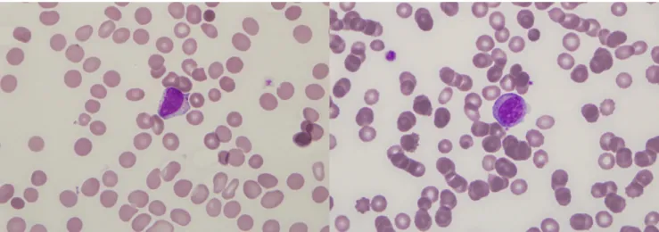
환자는 57세 여자로 심신허약과 안구통증과 안구부종으로 내 원하였다. 처음에는 열이 없었고 비장종대나 말초 림프절병증도 없었다. 그녀는 심하지 않은 범혈구 감소증을 보였는데 헤모글로빈 9.5 g/dL, 백혈구 수 2,070/μL, 혈소판 수 117,000/μL이었다. 항생제 치료 5일 후 범혈구감소증은 심화되었다. 말초혈액도말 소견에서 비전형적 림프구가 60% 관찰되었다(Fig. 1). 게다가 페리틴(ferritin)
서 론
공격성 NK세포 백혈병은 매우 드문 전신성 질환으로 자주 Ep- stein-Barr virus (EBV)와 연관되어 있으며 공격적인 임상적 경과를 보인다[1]. 다른 인종보다는 아시아인에서 호발하며 주로 젊은 층 에서 발병하는 것으로 알려져 있다[2]. NK세포 관련 종양에 대한 염색체 검사에서 다양한 세포유전학적 클론성 이상이 발견되었으 며 6번 염색체 장완의 결손 등이 가장 흔하게 보고되었다[3]. 그러
혈액종양질환에서 염색체 마이크로어레이의 유용성:
염색체 1q 이상을 동반한 공격성 NK세포 백혈병 1예
Usefulness of Chromosomal Microarray in Hematologic Malignancies: A Case of Aggressive NK-cell Leukemia with 1q Abnormality
정유나1·김하늬1·이세련2·성화정2·남명현1
Yoo Na Chung, M.D.
1, Ha-Nui Kim, M.D.
1, Se-Ryeon Lee, M.D.
2, Hwa Jung Sung, M.D.
2, Myung-Hyun Nam, M.D.
1고려대학교 의과대학 진단검사의학과1, 내과2
Departments of Laboratory Medicine
1and Internal Medicine
2, Korea University Medical School, Seoul, Korea Vol. 9, No. 3: 189-193, July 2019
https://doi.org/10.3343/lmo.2019.9.3.189 진단유전학
Corresponding author: Myung-Hyun Nam, M.D.
https://orcid.org/0000-0002-8737-6902
Department of Laboratory Medicine, Korea University Ansan Hospital, 123 Jeokgeum-ro, Danwon-gu, Ansan 15355, Korea
Tel: +82-31-412-5300, Fax: +82-31-412-4228, E-mail: yuret@korea.ac.kr Received: August 30, 2018
Revision received: October 26, 2018 Accepted: October 26, 2018
This article is available from http://www.labmedonline.org 2019, Laboratory Medicine Online
This is an Open Access article distributed under the terms of the Creative Commons Attribution Non-Commercial License (http://creativecommons.org/licenses/by-nc/4.0/) which permits unrestricted non-commercial use, distribution, and reproduction in any medium, provided the original work is properly cited.
A variety of clonal cytogenetic abnormalities have been reported in aggressive natural killer (NK)-cell lymphoma and leukemia. Recent chromo- somal microarray studies have shown both gain and loss of 1q and loss of 7p as recurrent abnormalities in aggressive NK-cell leukemia. Here, we report a case of aggressive NK-cell leukemia with complex chromosomal gains and losses, as confirmed by chromosomal microarray analysis. The patient showed an aggressive clinical course, which was complicated by hemophagocytic lymphohistiocytosis. Conventional cytogenetic analysis revealed trisomy 3 and 1q gain only. However, chromosomal microarray analysis detected an additional gain of 1q21.1-q24.2 and a loss of 1q24.2- q31.3. These abnormal lesions might play a role in the pathogenesis of aggressive NK-cell leukemia by inactivating tumor suppressor genes or by activating oncogenes. These results suggest that chromosomal microarray analysis may be used to provide further genetic information for pa- tients with hematological malignancies, including aggressive NK-cell leukemia.
Key Words: Aggressive NK-cell leukemia, Chromosomal microarray, 1q abnormality
2017-03-16 https://crossmark-cdn.crossref.org/widget/v2.0/logos/CROSSMARK_Color_square.svg
수치가 급격히 상승하였으며(>40,000 ng/mL) 38.6˚C의 열이 발생 하였다. 복부컴퓨터단층촬영(Abdomen computed tomography, CT) 결과에서는 처음에는 관찰되지 않았던 간비장종대가 발견되
었다. 골수저하(bone marrow suppression)와 혈구탐식성림프구조 직구증식증(hemophagocytic lymphohistiocytosis)을 판단하기 위 해 골수검사를 시행하였다. 환자는 접착도말(touch imprint) 슬라
Fig. 1. In the peripheral blood smear, abnormal lymphoid cells resembling large granular lymphocytes are observed (Wright stain, ×1,000).
Fig. 2. Immunophenotype analysis of leukemic cells using flow cytometry. Gated cells are 76.2% on side scatter vs. CD45 plot. Leukemic cells are
positive for CD2, CD7 and CD56 and negative for surface CD3 (Cytomics FC500, Beckmann Coulter).1,023
0
SSLin
CD45-PC5
10
010
110
210
3A 10
310
210
110
0CD13-PE
CD2-FITC
10
010
110
210
3C
CD1 17-PE
CD7-FITC
10
010
110
210
3B 10
310
210
110
010
310
210
110
0CD56-PE
CD3-FITC
10
010
110
210
3D
이드에서 혈구 탐식이 관찰되어 혈구탐식성림프구조직구증식증 으로 진단받았다. 골수흡인검체는 희석되어서 판독이 불가능하였 다. 말초혈액 검체로 면역표현형검사를 실시하였고 CD2, CD7, CD56 양성이고 CD3 음성을 보여 NK세포 관련 종양으로 확인되었다 (Fig. 2). 골수생검에서 백혈병성 세포들의 전체적인 침범이 관찰되 었다(Fig. 3). 이 백혈병성 세포들은 Epstein-Barr encoding region (EBER) 동소보합검사(Fig. 4)와 CD56에 양성이었으며 부분적으로 CD3과 CD30에 양성이었다. 이 모든 소견들이 공격성 NK세포백혈 병(aggressive NK cell leukemia)과 일치하였다. 염색체 결과는 47, XX,+3[2]/47,idem,der(22)t(1;22)(q21;p11.2)[14]/46,XX[4]로 3번 염색 체 3배체와 1q 획득의 소견을 보였다(Fig. 5). Cytoscan 750K array (Affymetrix, Santa Clara, CA, USA)로 시행한 염색체마이크로어레 이 결과는 1q21.1–q24.2에서 24 MB 획득과 1q24.2–q31.3에서 26 MB
결손이 3번 염색체의 획득과 함께 발견되었다(arr[hg19] 1q21.1q24.2 (143932349-167430174)x3,1q24.2q31.3(170086902-196698230)x1,(3) x3). 진단 즉시 바로 화학요법을 시작했으나 상태는 악화되었고 입 원 후 12일만에 사망하였다.
고 찰
종양 유전자의 활성화와 종양 억제 유전자의 억제는 암으로의
Fig. 3. Bone marrow biopsy slide showing infiltration of leukemic cells
(H&E stain, ×100).
Fig. 4. Epstein-Barr encoding region (EBER)-positive cells in bone
marrow biopsy (×100).Fig. 5. Karyotyping and chromosomal microarray analysis. (A) The
karyotypes of peripheral blood cells at diagnosis by Giemsa banding.Arrows indicate abnormal chromosomes showing trisomy 3 and der(22)t(1;22)(q21;p11.2). (B) Idiogram of chromosomal microarray showing 1q gain and loss and trisomy 3. (C) Chromosomal microarray analysis of chromosome 1 showing 1q21.2-q24.2 gain of 24 Mb and 1q24.2-q31.3 loss of 26 Mb.
A
B
C
전환에 중요한 역할을 한다[6]. 하지만 NK cell 관련 암에서는 연구 가 많이 부족한 실정이다[6]. NK세포 백혈병에서는 1q 획득과 7p 와 17p의 결손이 호발하며, 6q의 결손은 림프절외 NK/T 세포 림 프종에서 더 흔하다고 알려져 있다. 이 중에서 가장 많이 알려진 염색체 이상은 6q21의 결손이다[5]. 이 부위에는 4개의 종양 억제 유전자로 PRDM1, ATG5, AIM1와 HACE1이 확인되었다[7].
Nakashima 등[1]의 연구에 따르면 1q의 획득이 NK세포 백혈병 에서 높은 빈도로 발견되었고 관련 유전자도 밝혀졌으나 구체적인 작용기전은 알려지지 않았다. 그 부위에 존재하는 유전자로는 SELENBP1, SLAMF1, PTPRC, KCNH1, RAB4A와 SMYD3가 있다 [1]. SELENBP1은 1q21.3에 위치하며 selenium-binding protein을 발현한다. Selenium은 암의 예방에 관여하므로 selenium binding protein gene의 획득은 암 예방효과가 있을 것으로 생각된다[8].
SLAMF1은 1q23.3에 위치하며 signaling lymphocyte activation molecule (SLAM)을 발현하여 T 세포 활성화에 관여한다[9]. SAP (signaling lymphocytic activation molecule [SLAM] associated protein) 유전자는 T & NK cell specific protein을 만들며 X-linked lymphoproliferative syndrome에서 이상 또는 소실된다. 그러므로 이 유전자가 1q 획득에 관여할 가능성이 가장 크다고 할 수 있겠 다[10]. PTPRC는 1q31.3–q32.1에 위치하며 protein tyrosine phos- phatase receptor type C를 발현하며 severe combined immuno- deficiency의 발병 원인 유전자로 알려져 있다[11]. KCNH1은 1q32.2 에 위치하며 신경계에 존재하는 voltage-gated potassium channel 을 발현한다[12]. RAB4A는 1q42.13에 위치하며 RAS-associated protein을 발현한다[13]. SMYD3는 1q44에 위치하며 histone meth- yltransferase를 발현하며 전사 조절에 관여한다[14]
일부 염색체 획득과 소실이 공격성 NK세포 백혈병과 연관되어 있다는 것이 밝혀졌지만 호발하는 유전자 이상은 확립되지 않았 다[15]. Huang 등[15]의 연구에 의하면 공격성 NK세포 백혈병 환자 8명에서 전장 유전체 시퀀싱(whole genome sequencing)을 한 결과 JAK-STAT cytokine 신호 전달에 관여하는 유전자인 JAK2, JAK3, STAT3, STAT5B에서 유전적 이상을 주로 보고하였다. 또한 전사체 시퀀싱(transcriptome sequencing)으로 전사 요소인 MYC 유전자 의 활성화를 증명하였다[15]. JAK-STAT cytokine 신호 전달이 활성 화되면 MYC 발현이 증가되며 또한 이는 JAK-STAT 신호 관련 유전 자의 이상 없이 TET2 또는 TP53 돌연변이에 의해서도 MYC 발현 은 증가할 수 있다[15]. Dufva 등[16]의 연구에 의하면 공격성 NK세 포 백혈병 환자 14명을 전장 엑솜 시퀀싱(whole exome sequensc- ing)한 결과 DDX3X와 STAT3가 가장 호발하는 유전자 변이로 확 인되었다. 또한 염색체 이상으로는 JAK2가 위치하는 9p24와 STAT3, STAT5A, STAT5B가 위치하는 17q21.1의 획득이 공격성 NK세포 백혈병과 림프절외 NK/T 세포 림프종(extranodal NK/T cell lym-
phoma)에서 모두 보고되었음을 여러 문헌을 통해 확인하였다[16].
앞의 서론에서 언급하였듯이 본 증례에서는 NK세포 백혈병에서 주로 발견되는 1q gain 이외에도 3번 염색체 획득이 발견되었다.
공격성 NK세포 백혈병과 관련된 3번 염색체 이상은 지금까지 문헌 보고에서 찾을 수 없었다. Liu 등[17]의 연구자들이 시행한 후 향적인 연구에 따르면 3번 염색체 획득은 300명의 혈액종양질환 환자 중 형질세포골수종(plasma cell myeloma)에서 그 빈도가 가 장 높았고 그 외에 만성림프구백혈병과 림프종 환자에서 주로 발 견되었다. 예후 측면에서는 3번 염색체 획득이 3번 염색체 소실보 다 통계학적으로 유의하게 생존율이 높았다[17]. 공격성 NK세포 백혈병에서의 3번 염색체 획득의 의미는 병인과 예후 측면에서 앞 으로의 연구가 필요할 것이다.
마지막으로 여러 문헌 고찰을 하였지만 혈액종양질환과 t(1;22) (q21;p11.2)의 염색체 전위와 연관된 보고를 찾을 수 없었다. 이는 본 질환의 유전적 이상이 위 염색체 전위에 의한 융합유전자의 발 현의 가능성보다는 전위 경계 부위의 염색체 획득과 결실에 의한 것임을 시사한다.
염색체 획득과 소실은 유전자 변이와 함께 종양의 발생과 증식 의 유전학적 배경으로 생각되고 있다. 염색체마이크로어레이는 고 식적 염색체검사보다 더 민감하게 염색체 이상을 검출할 수 있고 차세대염기서열분석으로 검사가 어려운 커다란 삽입/결실을 검출 할 수 있다. 따라서 염색체마이크로어레이를 통해 염색체 획득과 소실을 확인하는 것은 차세대염기서열분석으로 종양유전체를 검 사하는 것과 더불어 혈액종양질환 발생의 유전적 기초를 연구하 는데 도움을 줄 수 있고, 이를 통해 향후 감별진단, 예후판정, 치료 방향결정 등에 역할을 함으로써 질환에 대한 유용한 임상적 효용 성을 보일 것으로 기대된다.
요 약
공격성 NK세포 백혈병에서 다양한 세포유전학적 클론성 이상 이 보고되었다. 최근 염색체마이크로어레이(chromosomal micro- array)를 이용한 연구를 통해서 공격성 NK세포 백혈병에서 호발 하는 이상으로 1q 획득과 결손 그리고 7p의 결손이 보고되었다. 연 구자들은 염색체마이크로어레이법으로 확인된 복잡한 염색체 획 득과 소실을 보인 공격성 NK세포 백혈병 1 증례를 경험하여 보고 하고자 한다. 이환자는 혈구탐식성림프조직구증식증을 동반하는 공격적인 경과를 보였다. 염색체 검사에서는 3번 염색체 3배체와 1q 획득만이 발견되었으나 염색체 마이크로어레이법을 통해 1q21.1- q24.2의 획득과 1q24.2-q31.3의 결손이 추가로 발견되었다. 이러한 이상 부위는 NK세포 백혈병 발병에 역할을 하는 종양 억제 유전 자의 비활성화 또는 종양 유도 유전자의 활성화에 관여할 것으로
생각된다. 염색체마이크로어레이 검사는 공격성 NK세포 백혈병을 포함한 혈액종양질환에서 추가적인 유전적 정보를 제공할 수 있 을 것으로 기대된다.
이해관계
저자들은 본 연구와 관련하여 어떠한 이해관계도 없음을 밝힙 니다.
감사의 글
본 연구는 고려대학교 특별연구비에 의하여 수행되었습니다.
REFERENCES
1. Nakashima Y, Tagawa H, Suzuki R, Karnan S, Karube K, Ohshima K, et al. Genome-wide array-based comparative genomic hybridization of natural killer cell lymphoma/leukemia: different genomic alteration patterns of aggressive NK-cell leukemia and extranodal NK/T-cell lymphoma, nasal type. Genes Chromosomes Cancer 2005;44:247-55.
2. Chan JKC, Jaffe ES, Ko YH. Aggressive NK cell leukaemia. In: Swerd- low SH, Campo E, et al. eds. WHO classification of tumours of haema- topoietic and lymphoid tissues. Revised 4th ed. Lyon: International Agency for Research on Cancer, 2017:353.
3. Siebert R. Mature B- and T-Cell neoplasms and Hodgkins lymphoma.
In: Heim S, Mitelman F, eds. Cancer Cytogenetics. 3rd ed. Hoboken (N.J.): Wiley-Blackwell, 2010:336.
4. Siu LL, Wong KF, Chan JK, Kwong YL. Comparative genomic hybrid- ization analysis of natural killer cell lymphoma/leukemia. Recognition of consistent patterns of genetic alterations. Am J Pathol 1999;155:1419- 25.
5. Karube K, Nakagawa M, Tsuzuki S, Takeuchi I, Honma K, Nakashima Y, et al. Identification of FOXO3 and PRDM1 as tumor-suppressor gene candidates in NK-cell neoplasms by genomic and functional analyses.
Blood 2011;118:3195-204.
6. Iqbal J, Kucuk C, Deleeuw RJ, Srivastava G, Tam W, Geng H, et al. Ge- nomic analyses reveal global functional alterations that promote tu- mor growth and novel tumor suppressor genes in natural killer-cell
malignancies. Leukemia 2009;23:1139-51.
7. Sako N, Dessirier V, Bagot M, Bensussan A, Schmitt C. HACE1, a po- tential tumor suppressor gene on 6q21, is not involved in extranodal natural killer/T-cell lymphoma pathophysiology. Am J Pathol 2014;184:
2899-907.
8. Chang PW, Tsui SK, Liew C, Lee CC, Waye MM, Fung KP. Isolation, characterization, and chromosomal mapping of a novel cDNA clone encoding human selenium binding protein. J Cell Biochem 1997;64:
217-24.
9. Chan B, Lanyi A, Song HK, Griesbach J, Simarro-Grande M, Poy F, et al. SAP couples Fyn to SLAM immune receptors. Nat Cell Biol 2003;5:
155-60.
10. Engel P, Eck MJ, Terhorst C. The SAP and SLAM families in immune responses and X-linked lymphoproliferative disease. Nat Rev Immu- nol 2003;3:813-21.
11. Kung C, Pingel JT, Heikinheimo M, Klemola T, Varkila K, Yoo LI, et al.
Mutations in the tyrosine phosphatase CD45 gene in a child with se- vere combined immunodeficiency disease. Nat Med 2000;6:343-5.
12. Simons C, Rash LD, Crawford J, Ma L, Cristofori-Armstrong B, Miller D, et al. Mutations in the voltage-gated potassium channel gene KCNH1 cause Temple-Baraitser syndrome and epilepsy. Nat Genet 2015;47:73-7.
13. Goueli BS, Powell MB, Finger EC, Pfeffer SR. TBC1D16 is a Rab4A GT- Pase activating protein that regulates receptor recycling and EGF re- ceptor signaling. Proc Natl Acad Sci U S A 2012;109:15787-92.
14. Hamamoto R, Furukawa Y, Morita M, Iimura Y, Silva FP, Li M, et al.
SMYD3 encodes a histone methyltransferase involved in the prolifera-
tion of cancer cells. Nat Cell Biol 2004;6:731-40.15. Huang L, Liu D, Wang N, Ling S, Tang Y, Wu J, et al. Integrated genomic analysis identifies deregulated JAK/STAT-MYC-biosynthesis axis in aggressive NK-cell leukemia. Cell Res 2018;28:172-86.
16. Dufva O, Kankainen M, Kelkka T, Sekiguchi N, Awad SA, Eldfors S, et al. Aggressive natural killer-cell leukemia mutational landscape and drug profiling highlight JAK-STAT signaling as therapeutic target. Nat Commun 2018;9:1567.
17. Liu D, Zhang Y, Chen S, Pan J, He X, Liang J, et al. Retrospective evalu- ation of the clinical and laboratory data from 300 patients of various hematological malignancies with chromosome 3 abnormalities. Can- cer Genet 2015;208:333-40.