ORIGINAL ARTICLE
위식도 역류질환이 있는 기관지확장증 환자에서 양성자 펌프 억제제의 효과
안병규1, 이동호1,2, 이창민1, 황재진1, 윤 혁1, 신철민1, 박영수1, 김나영1,2
분당서울대학교병원 내과1, 서울대학교 의과대학 간연구소2
Effect of Proton Pump Inhibitors in Bronchiectatic Patients with Gastroesophageal Reflux Disease
Byungkyu Ahn1, Dong Ho Lee1,2, Chang Min Lee1, Jae Jin Hwang1, Hyuk Yoon1, Cheol Min Shin1, Young Soo Park1, and Nayoung Kim1,2
Department of Internal Medicine, Seoul National University Bundang Hospital, Seongnam1, Liver Research Institute, Seoul National University College of Medicine, Seoul2, Korea
Background/Aims: Bronchiectasis is aggravated by gastroesophageal reflux disease (GERD) owing to micro aspiration. Some researchers note the effect of antireflux surgery in bronchiectasis with GERD. However, few have investigated the effects of medical antireflux therapy. We investigated the effect of proton pump inhibitors (PPIs) in bronchiectasis with GERD.
Methods: From March 2003 to May 2015, the clinical records of patients who had bronchiectasis with GERD were reviewed.
Patients underwent an initial pulmonary function test (PFT) and chest computed tomography when diagnosed with bronchiectasis.
One group with typical GERD symptoms was treated with PPIs, while the other group was not. Both groups underwent PFTs within six months after completing PPI therapy. Population characteristics and associations were compared between the groups.
Results: Two hundred and fifty-seven patients (124 male, 133 female; mean age 67.6±10.0 years) were included. There were no significant differences between the groups in terms of forced vital capacity (FVC; p=0.239), forced expiratory volume in one second (FEV1; p=0.555), or FEV1/FVC (p=0.374) after PPI therapy. However, there were significant improvements in FVC (p=0.002) and FEV1 (p=0.006) in patients with high BMI in the PPI treatment group.
Conclusions: PPIs have no effect on the pulmonary function in patients with bronchiectasis and GERD. However, PPIs were noted to produce improvements in lung function in patients with bronchiectasis and high BMI. (Korean J Gastroenterol 2016;68:10-15) Key Words: Bronchiectasis; Gastroesophageal reflux; Proton pump inhibitors
Received February 1, 2016. Revised April 11, 2016. Accepted May 9, 2016.
CC This is an open access article distributed under the terms of the Creative Commons Attribution Non-Commercial License (http://creativecommons.org/licenses/
by-nc/4.0) which permits unrestricted non-commercial use, distribution, and reproduction in any medium, provided the original work is properly cited.
Copyright © 2016. Korean Society of Gastroenterology.
교신저자: 이동호, 13620, 성남시 분당구 구미로 173번길 82, 분당서울대학교병원 소화기내과
Correspondence to: Dong Ho Lee, Department of Gastroenterology, Seoul National University Bundang Hospital, 82 Gumi-ro 173beon-gil, Bundang-gu, Seongnam 13620, Korea. Tel: +82-31-787-7006, Fax: +82-31-787-4051, E-mail: [email protected]
Financial support: None. Conflict of interest: None.
INTRODUCTION
Bronchiectasis is a chronic dilatation of the airways with thickening of the bronchial walls, which typically presents with chronic cough and sputum hypersecretion.1 Studies from the USA estimate a prevalence of 4.2 per 100,000 people among
those aged 18 to 34 years, which increases to 271.8 per 100,000 among people aged more than 75 years.2 Several eti- ologies, ranging from idiopathic to congenital diseases, sys- temic diseases (e.g., autoimmune disease such as rheumatoid arthritis), and post-infective causes, have been implicated.3
The clinical presentation of bronchiectasis may be compli-
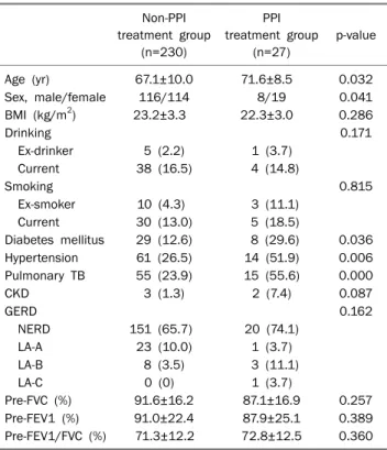
Table 1. Baseline Characteristics of the Non-PPI Treatment Group and the PPI Treatment Group
Non-PPI treatment group
(n=230)
PPI treatment group
(n=27)
p-value
Age (yr) 67.1±10.0 71.6±8.5 0.032
Sex, male/female 116/114 8/19 0.041
BMI (kg/m2) 23.2±3.3 22.3±3.0 0.286
Drinking 0.171
Ex-drinker 5 (2.2) 1 (3.7)
Current 38 (16.5) 4 (14.8)
Smoking 0.815
Ex-smoker 10 (4.3) 3 (11.1)
Current 30 (13.0) 5 (18.5)
Diabetes mellitus 29 (12.6) 8 (29.6) 0.036
Hypertension 61 (26.5) 14 (51.9) 0.006
Pulmonary TB 55 (23.9) 15 (55.6) 0.000
CKD 3 (1.3) 2 (7.4) 0.087
GERD 0.162
NERD 151 (65.7) 20 (74.1)
LA-A 23 (10.0) 1 (3.7)
LA-B 8 (3.5) 3 (11.1)
LA-C 0 (0) 1 (3.7)
Pre-FVC (%) 91.6±16.2 87.1±16.9 0.257
Pre-FEV1 (%) 91.0±22.4 87.9±25.1 0.389
Pre-FEV1/FVC (%) 71.3±12.2 72.8±12.5 0.360 Values are presented as mean±SD or n (%).
PPI, proton pump inhibitor; TB, tuberculosis; CKD, chronic kidney disease; GERD, gastroesophageal reflux disease; NERD, non- erosive reflux disease; FVC, forced vital capacity; FEV1, forced expiratory volume in 1 second.
cated by the coexistence of other conditions, including gas- troesophageal reflux disease (GERD).4,5 The prevalence of GERD in patients with bronchiectasis was approximately 40% according to Lee et al.6 in 2014, compared to 18% in the control group. GERD has an inverse relationship with lung function tests in bronchiectatic patients.4
Bronchiectasis is aggravated by GERD due to vagally medi- ated reflex bronchoconstriction and pulmonary micro aspiration.7 Research on the effect of antireflux surgery in bron- chiectasis patients with GERD8,9 finds that after surgery, respi- ratory symptoms and pulmonary function improves.8,9 However, few studies have investigated the effects of antireflux medication. The purpose of this study was to determine the ef- fect of proton pump inhibitors (PPIs) among the antireflux medi- cal therapies in patients with bronchiectasis and GERD.
SUBJECTS AND METHODS
This retrospective study received institutional review board approval from Seoul National University Bundang Hospital (IRB No. B-1512-326-105), and informed patient consent was waived. We collected data from electronic medi- cal records for patients who had bronchiectasis with GERD between March 2003 and May 2015. Heartburn, regur- gitation, and chest soreness were considered suggestive of GERD. We also evaluated computed tomography scans of the chest in order to identify bronchiectasis.
Patients underwent initial pulmonary function tests in our institution when diagnosed with bronchiectasis. One group with typical GERD symptoms was treated with PPIs, while the other group was not. Both groups underwent pulmonary func- tion testing within six months after finishing antireflux medi- cal therapy. Several lung function parameters, specifically forced vital capacity (FVC), forced expiratory volume in one second (FEV1), and FEV1/FVC, were analyzed. We excluded patients treated with PPIs for less than 21 days and those di- agnosed with erosive gastritis or peptic ulcer on esoph- ago-gastro-duodenoscopy. Twenty-seven patients who un- derwent antireflux medical therapy and 230 patients who did not undergo antireflux medical therapy were included.
Statistical analysis was performed using IBM SPSS Statistics ver. 22.0 (IBM Co., Armonk, NY, USA). We compared the two groups by sex, drinking habits, smoking habits, and presence of diabetes mellitus, hypertension, pulmonary tu-
berculosis, chronic kidney disease, and GERD using the χ2 test for to reduce confounding. Continuous variables are pre- sented as means±SD. Continuous variables including age, BMI, and lung function were analyzed using the Mann-Whitney U test, as the data were not normally distributed. Correlations among age, BMI, severity of obesity, drinking, smoking, diabetes mellitus, hypertension, pulmo- nary tuberculosis history, chronic kidney disease, and lung function were analyzed using Spearman’s rho. Null hypoth- eses of no difference were rejected if p-values were less than 0.05.
RESULTS
Two hundred and fifty-seven patients (124 male, 133 fe- male; mean age: 67.6±10.0 years [24-92 years]) were en- rolled in this study. The PPI treatment group comprised 27 in- dividuals, including eight males with a mean±SD age of 71.6±8.5 years. The non-PPI treatment group comprised
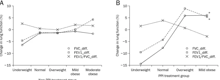
Fig. 1. Severity of obesity and lung function in patients with bronchiectasis and gastroesophageal reflux disease. (A) Differences in lung function between before and after antireflux treatment in the proton pump inhibitor (PPI) treatment group. (B) Differences in lung function between before and after antireflux treatment in the non-PPI treatment group. *p<0.05.
FVC, forced vital capacity; FEV1, forced expiratory volume in 1 second; diff., difference in lung function parameters before and after anti-reflux treatment.
Underweight: BMI <18.5; normal: BMI ≥18.5, <23; overweight: ≥23, <25; mildly obese: ≥25, <30; moderately obese: ≥30, <35;
severely obese: BMI ≥35.
Table 2. Comparison of Lung Function between Groups after PPI Treatment
Non-PPI treatment group
(n=230)
PPI treatment group
(n=27)
p-value
Post-FVC (%) 90.3±17.8 84.5±20.7 0.239
Post-FEV1 (%) 90.3±22.9 87.9±28.0 0.555 Post-FEV1/FVC (%) 72.1±14.8 74.5±15.8 0.374 FVC diff. (%) −1.3±11.0 −2.6±13.0 0.975
FEV1 diff. (%) −0.7±11.2 0±11.9 0.611
FEV1/FVC diff. (%) 0.9±9.7 1.7±12.3 0.757 Values are presented as mean±SD.
PPI, proton pump inhibitor; diff., difference in lung function parameters before and after PPI treatment; FVC, forced vital capacity; FEV1, forced expiratory volume in 1 second.
230 individuals, which included 116 males with a mean±SD age of 67.1±10.0 years. The baseline characteristics of the patients are summarized in Table 1.
The mean±SD pre-treatment FVCs (%) for the PPI treat- ment group was 87.1±16.9%, and for the non-PPI treatment group 91.6±16.2%. The mean±SD post-treatment FVCs (%) for the PPI treatment group was 84.5±20.7%, and non-PPI treatment group was 90.3±17.8%. The mean±SD difference between the pre-treatment FVC and the post- treatment FVC for the PPI treatment group was −2.6±13.0%, while for the non-PPI treatment group it was −1.3±11.0%. There were no
statistically significant differences between the groups in terms of post-treatment FVC (p=0.239) or changes between pre-treatment and post- treatment FVC (p=0.975) (Table 2, Fig. 1).
The mean±SD pre-treatment FEV1s (%) for the PPI treat- ment group was 87.9±25.1%, and the non-PPI treatment group was 91.0±22.4%. The mean±SD post-treatment FEV1s (%) for the PPI treatment group was 87.9±28.0%, and the non-PPI treatment group 90.3±22.9%. The mean±SD dif- ferences of the pre-treatment FEV1 and post-treatment FEV1 for the PPI treatment group was 0±11.9% and the non-PPI treatment group were −0.7±11.2%. There were no statisti- cally significant differences between the groups concerning post-treatment FEV1 (p=0.555) or changes between pre- treatment and post-treatment FEV1 (p=0.611) (Table 2, Fig.
1).
The mean±SD pre-treatment FEV1/FVCs (%) for the PPI treatment group was 72.8±12.5%, and for the non-PPI treat- ment group was 71.3±12.2%. The mean±SD post-treatment FEV1/FVCs (%) for the PPI treatment group was 74.5±15.8%, and non-PPI treatment group 72.1±14.8%. The mean±SD difference between pre-treatment FEV1/FVC and post-treat- ment FEV1/FVC for the PPI treatment group was 1.7±12.3%, and for the non-PPI treatment group was 0.9±9.7%. There were no statistically significant differences between the
Table 4. Linear Regression Analysis of Lung Function with BMI and with Severity of Obesity in the PPI Treatment Group
FVC diff. FEV1 diff.
B R2 p-value B R2 p-value
BMI 2.522 0.348 0.001 2.004 0.262 0.006
Severity of obesity 7.729 0.282 0.004 6.075 0.208 0.017
PPI, proton pump inhibitor; FVC, forced vital capacity; FEV1, forced expiratory volume in 1 second; diff., difference in lung function parameters before and after PPI treatment.
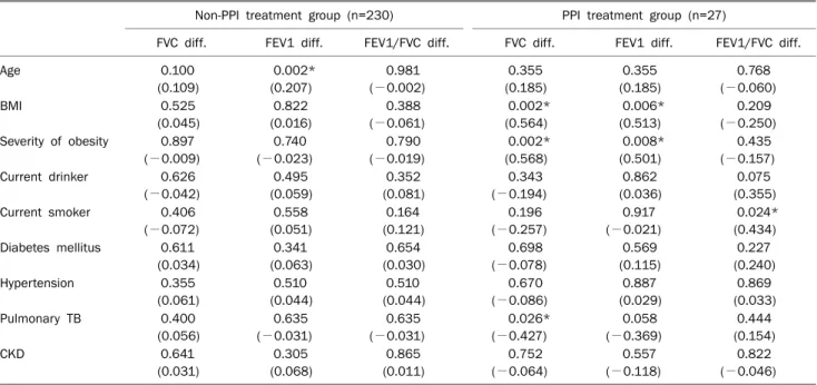
Table 3. Spearman Correlation between Lung Function Tests for the Two Groups
Non-PPI treatment group (n=230) PPI treatment group (n=27)
FVC diff. FEV1 diff. FEV1/FVC diff. FVC diff. FEV1 diff. FEV1/FVC diff.
Age 0.100
(0.109)
0.002*
(0.207)
0.981 (−0.002)
0.355 (0.185)
0.355 (0.185)
0.768 (−0.060)
BMI 0.525
(0.045)
0.822 (0.016)
0.388 (−0.061)
0.002*
(0.564)
0.006*
(0.513)
0.209 (−0.250) Severity of obesity 0.897
(−0.009)
0.740 (−0.023)
0.790 (−0.019)
0.002*
(0.568)
0.008*
(0.501)
0.435 (−0.157) Current drinker 0.626
(−0.042)
0.495 (0.059)
0.352 (0.081)
0.343 (−0.194)
0.862 (0.036)
0.075 (0.355)
Current smoker 0.406
(−0.072)
0.558 (0.051)
0.164 (0.121)
0.196 (−0.257)
0.917 (−0.021)
0.024*
(0.434) Diabetes mellitus 0.611
(0.034)
0.341 (0.063)
0.654 (0.030)
0.698 (−0.078)
0.569 (0.115)
0.227 (0.240)
Hypertension 0.355
(0.061)
0.510 (0.044)
0.510 (0.044)
0.670 (−0.086)
0.887 (0.029)
0.869 (0.033)
Pulmonary TB 0.400
(0.056)
0.635 (−0.031)
0.635 (−0.031)
0.026*
(−0.427)
0.058 (−0.369)
0.444 (0.154)
CKD 0.641
(0.031)
0.305 (0.068)
0.865 (0.011)
0.752 (−0.064)
0.557 (−0.118)
0.822 (−0.046) Values are presented as p-value (rho).
PPI, proton pump inhibitor; FVC, forced vital capacity; FEV1, forced expiratory volume in 1 second; diff., difference in lung function parameters before and after PPI treatment; TB, tuberculosis; CKD, chronic kidney disease.
*p<0.05.
groups in terms of post-treatment FEV1/FVC (p=0.374) or changes between pre-treatment and post-treatment FEV1/FVC (p=0.757; Table 2).
We assessed the associations of factors such as age, BMI, and comorbid conditions, with the patients’ pulmonary func- tion parameters. In the PPI treatment group, FVC (rs=0.564, p=0.002) and FEV1 (rs=0.513, p=0.006) improved sig- nificantly from the pre-treatment values in patients with in- creased BMI. Improvements of changes in FVC (rs=0.568, p=0.002) and FEV1 (rs=0.501, p=0.008) in the PPI treat- ment group were significantly related to severity of obesity.
This was not observed in the non-PPI treatment group.
However, it was also noted that changes in FVC (rs=−0.427, p=0.026) were significantly worse in patients in the PPI treat- ment group with a history of pulmonary tuberculosis (Table
3, Fig. 1).
Changes in FVC (R2=0.348, p=0.001) and FEV1 (R2=0.262, p=0.006) were significantly associated with in- creased BMI. Changes in FVC (R2=0.282, p=0.004) and FEV1 (R2=0.208, p=0.017) also significantly associated with the severity of obesity (Table 4).
DISCUSSION
The role of GERD in the pathogenesis of respiratory symp- toms and diseases has been discussed. There is a high preva- lence of reflux in asthma and chronic cough, which may be induced through different mechanisms, including micro as- piration and reflex bronchoconstriction.7,10,11 Refluxed gas- tric material reaches the proximal esophagus and moves into
the hypopharynx with the potential to enter the trachea.12 GERD may also cause bronchiectasis in the adult population.4 In a study of four patients with severe bron- chiectasis who completed dual-channel esophageal pH monitoring, the prevalence of distal reflux in 75% and prox- imal reflux in 50% suggests that patients with more severe bronchiectasis may be more likely to have GERD.13
Helicobacter pylori has been identified in tracheobron- chial secretions, and a high seroprevalence is noted in pa- tients with bronchiectasis.14 H. pylori produces a wide range of toxins including urease, phospholipidases, alcohol de- hydrogenase, hemolysin, platelet-activating factor, and mu- colytic factor.15 These toxins are harmful to the stomach and duodenum, and act by generation of an intense immune re- sponse, resulting in submucosal lymphocyte and neutrophil infiltration. They can likewise interact with other tissues.16 Aspiration or inhalation of H. pylori exotoxins may contribute to chronic airway inflammation in patients with idiopathic bronchiectasis.14 Longstanding respiratory disease related to GERD has negative effects on lung functions.9,12
Active antireflux interventions, such as laparoscopic fun- doplication, Stretta radiofrequency, and cruroraphy, can pre- vent aggravation of chronic lung disease with bronchiec- tasis.8,9 Several studies have noted improvements in lung function after these surgical procedures.8,9
Pasteur et al.17 found GERD to be a cause in 4% of patients, based on gastrointestinal symptoms and symptomatic im- provement after antireflux therapy. In the present study, we assessed the effect of PPIs among the antireflux medications on the lung function of patients with bronchiectasis. We com- pared the differences in lung function parameters before and after PPIs between the treatment and the control groups.
There were no statistically significant differences in lung function tests between groups. However, we observed stat- istically significant improvements between the pre-PPI and the post-PPI FVC and between the pre-PPI and the post-PPI FEV1 based on BMI. Thus, although PPIs did not significantly change pulmonary function parameters in patients with bronchiectasis and GERD, there were statistically significant improvements in certain subgroups, specifically obese individuals. This is believed to result from obesity causing in- creased esophageal acid exposure time compared to the non-obese population.18
As antireflux surgery is an anatomical repair, it prevents
the reflux of both acid and non-acid materials. PPIs, however, only prevent the reflux of acids. This may be why the lung pa- rameters did not differ by PPI treatment.
This study has some limitations. First, this study is retrospective. The retrospective study design was non-rando- mized study. Furthermore, the PPI treatment duration varied from 22 days to 1,784 days in the PPI treatment group.
In conclusion, we were unable to reject the null hypothesis that PPIs have no effect on the pulmonary function in patients with bronchiectasis and GERD. We attribute this to the fact that unlike antireflux surgery, PPIs are unable to prevent non- acid materials from refluxing. However, PPIs do produce im- provements in lung function in patients with bronchiectasis and higher BMI in accordance with the severity of obesity.
This could be because obesity causes increased esophageal acid exposure time. Future studies are required with larger sample sizes and a randomized prospective design to de- termine the effects of antireflux medical therapy in obese pa- tients with bronchiectasis and GERD.
REFERENCES
1. Ilowite J, Spiegler P, Chawla S. Bronchiectasis: new findings in the pathogenesis and treatment of this disease. Curr Opin Infect Dis 2008;21:163-167.
2. Weycker D, Edelsberg J, Oster G, Tino G. Prevalence and econom- ic burden of bronchiectasis. Clin Pulm Med 2005;12:205-209.
3. Chang AB, Bilton D. Exacerbations in cystic fibrosis: 4--Non-cyst- ic fibrosis bronchiectasis. Thorax 2008;63:269-276.
4. Tsang KW, Lam WK, Kwok E, et al. Helicobacter pylori and upper gastrointestinal symptoms in bronchiectasis. Eur Respir J 1999;14:1345-1350.
5. Kim JH. Are proton pump inhibitors effective in asthmatics with gastroesophageal reflux disease? Korean J Gastroenterol 2011;58:169-170.
6. Lee AL, Button BM, Denehy L, et al. Proximal and distal gas- tro-oesophageal reflux in chronic obstructive pulmonary dis- ease and bronchiectasis. Respirology 2014;19:211-217.
7. Lee AL, Button BM, Denehy L, Wilson JW. Gastro-oesophageal re- flux in noncystic fibrosis bronchiectasis. Pulm Med 2011;
2011:395020.
8. Ozaydin I, Annakkaya AN, Ozaydin C, Aydın M. Effects of cruro- raphy and laparoscopic Nissen fundoplication procedures on pulmonary function tests in gastroesophageal reflux patients.
Int J Clin Exp Med 2014;7:431-434.
9. Hu ZW, Wang ZG, Zhang Y, et al. Gastroesophageal reflux in bron- chiectasis and the effect of anti-reflux treatment. BMC Pulm Med 2013;13:34.
10. Vakil N, van Zanten SV, Kahrilas P, Dent J, Jones R; Global
Consensus Group. The montreal definition and classification of gastroesophageal reflux disease: a global evidence-based consensus. Am J Gastroenterol 2006;101:1900-1920; quiz 1943.
11. Sweet MP, Patti MG, Hoopes C, Hays SR, Golden JA. Gastro-oeso- phageal reflux and aspiration in patients with advanced lung disease. Thorax 2009;64:167-173.
12. Schan CA, Harding SM, Haile JM, Bradley LA, Richter JE.
Gastroesophageal reflux-induced bronchoconstriction. An intra- esophageal acid infusion study using state-of-the-art techno- logy. Chest 1994;106:731-737.
13. Sweet MP, Herbella FA, Leard L, et al. The prevalence of distal and proximal gastroesophageal reflux in patients awaiting lung transplantation. Ann Surg 2006;244:491-497.
14. Tsang KW, Lam SK, Lam WK, et al. High seroprevalence of
Helicobacter pylori in active bronchiectasis. Am J Respir Crit Care Med 1998;158:1047-1051.
15. Mégraud F. Toxic factors of Helicobacter pylori. Eur J Gastroenterol Hepatol 1994;6 Suppl 1:S5-S10.
16. Goodwin CS, Armstrong JA, Marshall BJ. Campylobacter pylo- ridis, gastritis, and peptic ulceration. J Clin Pathol 1986;39:
353-365.
17. Pasteur MC, Helliwell SM, Houghton SJ, et al. An investigation in- to causative factors in patients with bronchiectasis. Am J Respir Crit Care Med 2000;162:1277-1284.
18. Shah SL, Lacy BE, DiBaise JK, Vela MF, Crowell MD. The impact of obesity on oesophageal acid exposure time on and off proton pump inhibitor therapy. Aliment Pharmacol Ther 2015;42:
1093-1100.