Received:June 2, 2017, Revised:June 30, 2017, Accepted:June 30, 2017
Corresponding to:Young Ho Lee, Division of Rheumatology, Department of Internal Medicine, Korea University Anam Hospital, Korea University College of Medicine, 73 Inchon-ro, Seongbuk-gu, Seoul 02841, Korea. E-mail:[email protected]
Copyright ⓒ 2017 by The Korean College of Rheumatology. All rights reserved.
This is a Open Access article, which permits unrestricted non-commerical use, distribution, and reproduction in any medium, provided the original work is properly cited.
Association of Neutrophil to Lymphocyte Ratio, Platelet to Lymphocyte Ratio, and Mean Platelet Volume with Systemic Lupus Erythematosus Disease Activity: A Meta-analysis
Young Ho Lee, Gwan Gyu Song
Division of Rheumatology, Department of Internal Medicine, Korea University Medical Center, Korea University College of Medicine, Seoul, Korea
Objective. A series of common blood tests neutrophil to lymphocyte ratio (NLR), platelet to lymphocyte ratio (PLR), and mean platelet volume (MPV) could provide a measure of systemic lupus erythematosus (SLE) activity. Methods. We searched the Medline, Embase, and Cochrane databases and performed a meta-analysis comparing NLR, PLR, and MPV in patients with SLE to controls, and examined correlation coefficients between NLR, PLR, and MPV and SLE activity based on SLE Disease Activity Index (SLEDAI) using random-effects models. Results. Nine studies were included in this meta-analysis. Meta-analysis revealed that NLR was significantly higher in the SLE group than in the control group (standard mean difference [SMD]=2.747, 95% con- fidence interval [CI]=1.241∼4.254, p<0.001). PLR was also significantly higher in the SLE group (SMD=1.564, 95%
CI=0.122∼3.006, p=0.034). Meta-analysis of correlation coefficients showed that both NLR and PLR were positively asso- ciated with SLEDAI (correlation coefficient=0.404, 95% CI=0.299∼0.500, p<0.001; correlation coefficient=0.378, 95%
CI=0.234∼0.505, p<0.001). The pooled sensitivity and specificity of NLR for diagnosis of lupus nephritis were 75.1% (95%
CI, 68.5∼81.0) and 72.9% (95% CI, 64.9∼80.0), respectively. The area under the curve of NLR were 0.794. However, meta-analysis indicated no elevated MPV in the SLE group and no correlation between MPV and SLE activity. Conclusion. This meta-analysis demonstrated that both NLR and PLR are higher in patients with SLE, a significantly positive correlation exists be- tween NLR/PLR and SLE activity. (J Rheum Dis 2017;24:279-286)
Key Words. Blood cell count, Systemic lupus erythematosus
INTRODUCTION
Systemic lupus erythematosus (SLE) is a prototypic au- toimmune disease characterized by aberrant immune reg- ulation, activation of T cells and polyclonal B cells, and ex- cessive production of autoantibodies and cytokines lead- ing to intense inflammation and multiple organ damage [1]. Disrupted immune regulation caused by the dereg- ulation of B- and T-cell activation and aberrant pro- duction of cytokines is considered to play a key role in the pathogenesis of SLE [2].
Recent studies have reported the numbers and ratios of complete blood cell (CBC) subgroups in rheumatic dis- eases [3,4]. Neutrophil-to-lymphocyte ratio (NLR), pla-
telet-to-lymphocyte ratio (PLR), and mean platelet vol- ume (MPV) have recently been investigated as new in- flammatory markers for the assessment of inflammation in many inflammatory, cardiovascular, and malignant dis- eases [5,6]. NLR is calculated as the absolute count of neutrophils divided by the absolute count of lympho- cytes, and PLR is calculated as the absolute platelet count divided by the absolute lymphocyte count. As a novel marker for inflammation, NLR may be useful to estimate the activity of autoimmune and inflammatory diseases [7]. PLR is also used as an index for inflammatory status in diverse diseases [8]. The MPV is the volume of the average circulating platelets in femtoliters, and it is a marker of platelet activation known to be associated with
inflammation [9]. NLR, PLR, and MPV are inexpensive and easily obtainable laboratory markers for systemic inflammation. However, their roles in SLE remain unclear.
Studies on NLR, PLR, and MPV in SLE patients com- pared to healthy controls, on the relationship between NLR, PLR, and MPV levels and SLE activity, and on the as- sociation of NLR with renal involvement in SLE have re- ported controversial results [3,10-17]. This may be be- cause of the small sample sizes, low statistical power, and/or the presence of clinical heterogeneity. We per- formed the present meta-analysis to overcome the limi- tations of individual studies and resolve inconsistencies [18-20]. The aim of this meta-analysis was to systemati- cally review the evidence concerning the relationship be- tween hematologic indices and SLE, to establish a corre- lation between NLR, PLR, and MPV and SLE activity, and to evaluate the diagnostic value of NLR for differentiating renal involvement from SLE.
MATERIALS AND METHODS
Identification of eligible studies and data extraction
We performed a literature search for studies that exam- ined NLR, PLR, or MPV in patients with SLE and healthy controls. The Medline, Embase, and Cochrane databases were searched to identify all available previous articles (until April 2017). The keywords and subject terms used in the search were “neutrophil to lymphocyte ratio,”
“mean platelet volume,” “neutrophil to lymphocyte ra- tio,” “platelet to lymphocyte ratio,” and “systemic lupus erythematosus.” All references cited in the identified ar- ticles were also reviewed to identify additional studies not covered by the abovementioned electronic databases.
Studies were considered eligible if: (1) they were case-con- trol, cross-sectional, or cohort studies; (2) they provided data on NLR, PLR, or MPV in SLE and controls; (4) they provided data on the correlation coefficient between NLR, MPV, or PLR and SLE activity based on SLE Disease Activity Index (SLEDAI); or (5) they included sufficient data to calculate the sensitivity and specificity of NLR for the diagnosis of lupus nephritis (LN). Studies were ex- cluded if: (1) they contained overlapping or insufficient data; or (2) they were reviews or case reports. Data con- cerning methods and results were extracted from original studies by two independent reviewers. Discrepancies in findings between the reviewers were resolved by consensus.
The meta-analysis was conducted in accordance with the
Preferred Reporting Items for Systematic reviews and Meta-Analyses guidelines [21]. The following in- formation was extracted from each study: primary author, year of publication, country, number of participants, mean and standard deviation (SD) of NLR, PLR, or MPV, and correlation coefficients between NLR, PLR, or MPV and disease activity. Raw data on NLR were extracted from primary studies to fill four cell values (true positive, false positive, true negative, and false negative) in a diag- nostic 2×2 table. When the data given were medians, in- terquartile ranges, or ranges, the mean and SD values were obtained using previously described formulae [22,23]. We scored the quality of each included study based on the Newcastle-Ottawa Scale [24]. The highest score was nine. Scores ranging from 6 to 9 were consid- ered to indicate high methodological quality.
Evaluation of statistical associations
We performed a meta-analysis examining NLR, PLR, or MPV in patients with SLE and healthy controls; correla- tion coefficients between NLR, PLR, or MPV and SLEDAI;
and the diagnostic accuracy of NLR for LN. For continuity of data, results were presented as standardized mean dif- ferences (SMDs) and 95% confidence intervals (CIs). We assessed within-study and between-study variations and heterogeneity using Cochran’s Q test [25]. The hetero- geneity test was used to assess the null hypothesis that all studies were evaluating the same effect. When the sig- nificant Q statistic (p<0.10) indicated heterogeneity across studies, the random effects model was used for the meta-analysis [26]. When the significant Q statistic (p
<0.10) did not indicate heterogeneity across studies, the fixed-effects model was used. The model assumed that all studies estimated the same underlying effect, and it con- sidered within-study variations only [25]. We quantified the effect of heterogeneity using I2=100%×(Q-df)/Q [27], where I2 measured the degree of inconsistency be- tween studies and determined whether the percentage of total variation across studies was due to heterogeneity rather than chance. I2 ranged from 0% to 100%; I2 values of 25%, 50%, and 75% were referred to as low, moderate, and high estimates, respectively [27]. We combined sen- sitivity, specificity, positive and negative likelihood ratios (PLR and NLR), and diagnostic odds ratio estimates and analyzed summary receiver operating characteristic (SROC) curves for diagnosing LN. Area under the curve (AUC) (in this case, area under the SROC curve) provides an overall summary of test performance and shows the
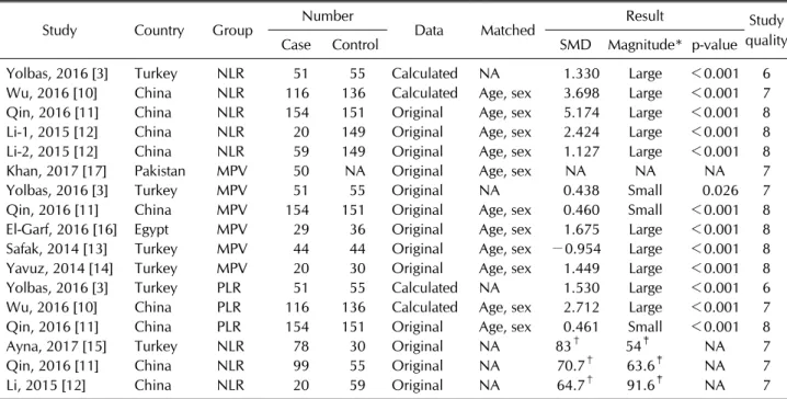
Table 1. Characteristics of the individual studies included in the meta-analysis
Study Country Group Number
Data Matched Result Study
quality
Case Control SMD Magnitude* p-value
Yolbas, 2016 [3] Turkey NLR 51 55 Calculated NA 1.330 Large <0.001 6
Wu, 2016 [10] China NLR 116 136 Calculated Age, sex 3.698 Large <0.001 7
Qin, 2016 [11] China NLR 154 151 Original Age, sex 5.174 Large <0.001 8
Li-1, 2015 [12] China NLR 20 149 Original Age, sex 2.424 Large <0.001 8
Li-2, 2015 [12] China NLR 59 149 Original Age, sex 1.127 Large <0.001 8
Khan, 2017 [17] Pakistan MPV 50 NA Original Age, sex NA NA NA 7
Yolbas, 2016 [3] Turkey MPV 51 55 Original NA 0.438 Small 0.026 7
Qin, 2016 [11] China MPV 154 151 Original Age, sex 0.460 Small <0.001 8
El-Garf, 2016 [16] Egypt MPV 29 36 Original Age, sex 1.675 Large <0.001 8
Safak, 2014 [13] Turkey MPV 44 44 Original Age, sex −0.954 Large <0.001 8
Yavuz, 2014 [14] Turkey MPV 20 30 Original Age, sex 1.449 Large <0.001 8
Yolbas, 2016 [3] Turkey PLR 51 55 Calculated NA 1.530 Large <0.001 6
Wu, 2016 [10] China PLR 116 136 Calculated Age, sex 2.712 Large <0.001 7
Qin, 2016 [11] China PLR 154 151 Original Age, sex 0.461 Small <0.001 8
Ayna, 2017 [15] Turkey NLR 78 30 Original NA 83† 54‡ NA 7
Qin, 2016 [11] China NLR 99 55 Original NA 70.7† 63.6‡ NA 7
Li, 2015 [12] China NLR 20 59 Original NA 64.7† 91.6‡ NA 7
SMD: standardized mean difference, NLR: neutrophil to lymphocyte ratio, MPV: mean platelet volume, PLR: platelet to lymphocyte ratio, NA: not available. *Magnitude of Cohen’s d effect size: 0.2∼0.5, small effect; 0.5∼0.8, medium effect; ≥0.8, large effect. †Sensitivity for diagnosis of lupus nephritis, ‡specificity for diagnosis of lupus nephritis.
trade-off between sensitivity and specificity [28]. Q* in- dex is another useful global estimate of test accuracy for comparing SROC curves [28]. In the present meta-analy- sis, statistical manipulations were undertaken using the Comprehensive Meta-Analysis computer program (Biostat, Englewood, NJ, USA) and Meta-DiSc version 1.4 (Hospital Universitario Ramón y Cajal, Madrid, Spain) [29].
Evaluation of publication bias
Although funnel plots are often used to detect pub- lication bias, they require diverse study types of varying sample sizes, and their interpretation involves subjective judgment. Therefore, we assessed publication bias using Egger’s linear regression test [30], which measured fun- nel plot asymmetry using a natural logarithm scale of SMDs.
RESULTS
Studies included in the meta-analysis
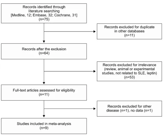
We identified 75 studies using electronic and manual search methods (Supplementary data). Ten of the studies were selected for full-text review on the basis of their ti- tles and abstracts. One of these was excluded because
they included other diseases [31]. Thus, nine articles met the inclusion criteria [3,10-17] (Table 1, Figure 1). One report contained data on two different groups [12], so we analyzed these studies independently. There were five comparison studies on NLR in SLE and controls, three on PLR, five on MPV, two on correlation coefficients be- tween NLR, PLR, or MPV and SLE, and three on the diag- nosis of LN (Table 1). The quality assessment score of each study ranged between 6 and 8. The characteristic features of the studies included in the meta-analysis are summarized in Table 1.
Meta-analysis comparing NLR, PLR, and MPV in SLE patients and controls
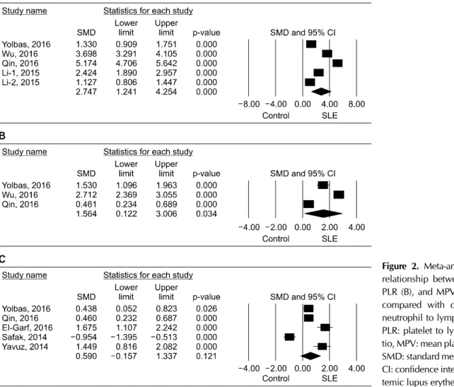
The present meta-analysis revealed that NLR was sig- nificantly higher in the SLE group than that in the control group (SMD=2.747, 95% CI=1.241∼4.254, p<0.001) (Table 2, Figure 2). PLR was significantly higher in the SLE group than that in the control group (SMD=1.564, 95% CI=0.122∼3.006, p=0.034) (Table 2, Figure 2).
However, the meta-analysis showed no evidence of elevated MPV in the SLE group (SMD=0.590, 95% CI=−0.157∼
1.337, p=0.121) (Table 2, Figure 2).
Table 2. Meta-analysis of the association between NLR, PLR, and MPV and SLE
Group Population No. of studies Test of association Test of heterogeneity
SMD 95% CI p-value Model p-value I2
NLR Overall 5 2.747 1.241∼4.254 <0.001 R <0.001 98.4
PLR Overall 3 1.564 0.122∼3.006 <0.001 R <0.001 98.2
MPV Overall 5 0.590 −0.157∼1.337 0.121 R <0.001 93.9
NLR: neutrophil to lymphocyte ratio, PLR: platelet to lymphocyte ratio, MPV: mean platelet volume, SLE: systemic lupus erythematosus, SMD: standard mean difference, CI: confidence interval, R: random effects model.
Figure 1. Flow diagram of the study selection process. SLE:
systemic lupus erythematosus.
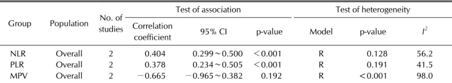
Meta-analysis of the relationship between NLR, PLR, and MPV and SLE activity
Meta-analysis of correlation coefficients identified that NLR was positively associated with SLE activity based on SLEDAI (correlation coefficient=0.404, 95% CI=0.299∼
0.500, p<0.001) (Table 3). PLR was positively associated with SLE activity (correlation coefficient=0.378, 95%
CI=0.234∼0.505, p<0.001) (Table 3). However, the meta-analysis showed no correlation between MPV and SLE activity (correlation coefficient=−0.665, 95% CI=
−0.965∼0.382, p=0.192) (Table 3).
Diagnostic accuracy of NLR for LN
The pooled sensitivity and specificity of NLR were 75.1% (95% CI, 68.5∼81.0) and 72.9% (95% CI, 64.9∼
80.0), respectively (Table 3). The PLR and NLR were
2.575 (95% CI, 1.403∼4.726) and 0.407, respectively (95% CI, 0.309∼0.537) (Table 3). Figure 3 shows the performance of the NLR test in the form of SROC curves.
The AUC and Q* index of NLR were 0.794 and 0.731, re- spectively (Table 3, Figure 3).
Heterogeneity and publication bias
Between-study heterogeneity was identified during the meta-analyses of NLR, PLR, and MPV in patients with SLE (Tables 2 and 3). However, all of the studies showed the same direction of the effect size, except for MPV.
Publication bias results in a disproportionate number of positive studies, and poses a problem for meta-analyses.
However, we found no evidence of publication bias in the meta-analysis performed in this study (Egger’s regression test p-values>0.1), indicating low probability of pub-
Figure 2. Meta-analysis of the relationship between NLR (A), PLR (B), and MPV (C) and SLE compared with control. NLR:
neutrophil to lymphocyte ratio, PLR: platelet to lymphocyte ra- tio, MPV: mean platelet volume, SMD: standard mean difference, CI: confidence interval, SLE: sys- temic lupus erythematosus.
lication bias (Tables 2 and 3).
DISCUSSION
In this meta-analysis, we combined the evidence for NLR, PLR, and MPV in SLE, the correlation between NLR, PLR, and MPV and SLE activity, and diagnostic val- ues of NLR for LN. The meta-analysis revealed that NLR and PLR were significantly higher in the SLE group than in the control group, and that NLR and PLR, were pos- itively correlated with SLE activity measured by SLEDAI.
It is needed to be explained why NLR and PLR are high in SLE. The cause of increased NLR and PLR may be in- creased cytokines and the inflammatory processes asso- ciated with SLE. The inflammatory process in SLE in- volves inflammatory cells and molecules that cause changes in the number, shapes, and sizes of bone marrow cells and peripheral blood cells [2]. SLE is characterized by B-cell activation and resultant autoimmunity with the
production of numerous cytokines [32]. Cytokines play a very important role in the pathogenesis of SLE [33].
Neutrophils and platelets are involved in the production of these cytokines, which contribute to the activation of neutrophils and platelets [34]. Leukocytes play a major role in inflammatory processes, and neutrophils are the most abundant type of leukocytes. Platelet activation is observed in patients with SLE [35]. Lymphocyte count is usually decreased in SLE, and platelet count is decreased in SLE patients very often [36]. High correlation may sug- gest that NLR and PLR would be conditional relations of cytokines or inflammatory products from high SLE activity.
ESR and CRP level are the most widely used markers for measuring acute-phase response to indicate inflammation in RA. ESR and CRP are influenced by several factors un- related to inflammation such as age, sex, anemia, and re- nal failure [37]. However, NLR and PLR are not affected by age, gender, and hemoglobin level [38]. In addition,
Table 3. Meta-analysis of the correlation coefficients between NLR, PLR, and MPV and SLE activity (SLEDAI) (A) and of the diagnostic accuracy of NLR for lupus nephrtitis (B)
A.
Group Population No. of studies
Test of association Test of heterogeneity Correlation
coefficient 95% CI p-value Model p-value I2
NLR Overall 2 0.404 0.299∼0.500 <0.001 R 0.128 56.2
PLR Overall 2 0.378 0.234∼0.505 <0.001 R 0.191 41.5
MPV Overall 2 −0.665 −0.965∼0.382 0.192 R <0.001 98.0
NLR: neutrophil to lymphocyte ratio, PLR: platelet to lymphocyte ratio, MPV: mean platelet volume, SLEDAI: systemic lupus erythematosus disease activity index, CI: confidence interval, R: random effects model.
B.
Population No. of studies Sensitivity (95% CI)
Specificity (95% CI)
PLR (95% CI)
NLR (95% CI)
AUC (SE)
Q*
(SE)
Overall 3 0.751
(0.685∼0.810)
0.729 (0.649∼0.800)
2.575 (1.403∼4.726)
0.407 (0.309∼0.537)
0.794 (0.046)
0.731 (0.039) CI: confidence interval, PLR: positive likelihood ratio, NLR: negative likelihood ratio, AUC: area under the curve, SE: standard error.
Figure 3. Summary receiver-operating characteristic curves for neutrophil to lymphocyte ratio for the diagnosis of lupus nephritis. Solid circles represent individual studies included in this meta-analysis. The curve shown is a regression line that summarizes the overall diagnostic accuracy. SE (AUC): stand- ard error of the area under the curve, Q*: an index defined by the point on the S receiver operating characteristics curve where the sensitivity and specificity are equal, and SE (Q*): Q*
index standard error. SROC: summary receiver operating characteristic, AUC: area under the curve, SE: standard error.
NLR and PLR are relatively stable compared to individual white blood cell parameters [11]. NLR and PLR are cost effective and easily obtained indicators from CBC tests.
As easily measurable and available laboratory parame- ters, NLR and PLR could be considered as new bio-
markers for inflammatory response or disease activity in SLE patients. MPV is another marker used in the assess- ment of inflammation. However, we failed to observe high or low levels of MPV in SLE or a correlation of MPV with disease activity. The association between MPV and SLE remains unclear.
The present study has certain shortcomings that should be considered. First, a small number of studies were in- cluded in this meta-analysis, most of the included studies had small sample sizes, and only a small number of stud- ies evaluated the correlation coefficients between the hematological indices and SLE severity and their diag- nostic value for LN. Thus, the meta-analysis may be underpowered. Second, the studies included patients with heterogeneous demographic characteristics and clinical features. NLR, PLR, and MPV values may be af- fected by multiple factors. Heterogeneity and confound- ing factors such as drugs used (e.g., immunosuppressive agents, hydroxychloroquine, and corticosteroids) may have affected the present results. For example, gluco- corticoids may affect the count, size, and function of neutrophils, lymphocytes, and platelets. However, this meta-analysis also has strengths. First, to the best of our knowledge, this meta-analysis is the first study to com- bine evidence of NLR, MPV, and PLR in SLE according to disease activity. Second, compared with individual stud- ies, this study should provide more reliable data on the re- lationship between NLR, PLR, and MPV and SLE by in-
creasing the level of statistical power and resolution through the pooling of the results of independent analyses.
CONCLUSION
The present meta-analysis demonstrated that NLR and PLR are higher in patients with SLE, and that a sig- nificantly positive correlation exists between NLR/PLR and SLE activity. These findings suggest that NLR and PLR may be useful indices for determining the extent of inflammation of SLE. Although there is high correlation between NLR, PLR and SLE activity, these ratios from CBC profile cannot totally replace SLE activity such as SLEDAI. Further studies are needed to elucidate whether NLR and PLR can serve as biomarkers for monitoring SLE activity.
CONFLICT OF INTEREST
No potential conflict of interest relevant to this article was reported.
SUPPLEMENTARY DATA
Supplementary data can be found with this article online at http://www.jrd.or.kr and at https://doi.org/10.4078/
jrd.2017.24.5.279.
REFERENCES
1. Lisnevskaia L, Murphy G, Isenberg D. Systemic lupus erythematosus. Lancet 2014;384:1878-88.
2. Ohl K, Tenbrock K. Inflammatory cytokines in systemic lu- pus erythematosus. J Biomed Biotechnol 2011;2011:
432595.
3. Yolbas S, Yildirim A, Gozel N, Uz B, Koca SS. Hematological indices may be useful in the diagnosis of systemic lupus er- ythematosus and in determining disease activity in Behçet's disease. Med Princ Pract 2016;25:510-6.
4. Mercan R, Bitik B, Tufan A, Bozbulut UB, Atas N, Ozturk MA, et al. The association between neutrophil/lymphocyte ratio and disease activity in rheumatoid arthritis and anky- losing spondylitis. J Clin Lab Anal 2016;30:597-601.
5. Liu JF, Ba L, Lv H, Lv D, Du JT, Jing XM, et al. Association between neutrophil-to-lymphocyte ratio and differentiated thyroid cancer: a meta-analysis. Sci Rep 2016;6:38551.
6. Leader A, Pereg D, Lishner M. Are platelet volume indices of clinical use? A multidisciplinary review. Ann Med 2012;
44:805-16.
7. Okyay GU, Inal S, Oneç K, Er RE, Paşaoğlu O, Paşaoğlu H, et al. Neutrophil to lymphocyte ratio in evaluation of in-
flammation in patients with chronic kidney disease. Ren Fail 2013;35:29-36.
8. Turkmen K, Erdur FM, Ozcicek F, Ozcicek A, Akbas EM, Ozbicer A, et al. Platelet-to-lymphocyte ratio better predicts inflammation than neutrophil-to-lymphocyte ratio in end-stage renal disease patients. Hemodial Int 2013;17:
391-6.
9. Bath PM, Butterworth RJ. Platelet size: measurement, phys- iology and vascular disease. Blood Coagul Fibrinolysis 1996;7:157-61.
10. Wu Y, Chen Y, Yang X, Chen L, Yang Y. Neutrophil-to-lym- phocyte ratio (NLR) and platelet-to-lymphocyte ratio (PLR) were associated with disease activity in patients with sys- temic lupus erythematosus. Int Immunopharmacol 2016;
36:94-9.
11. Qin B, Ma N, Tang Q, Wei T, Yang M, Fu H, et al. Neutrophil to lymphocyte ratio (NLR) and platelet to lymphocyte ratio (PLR) were useful markers in assessment of inflammatory response and disease activity in SLE patients. Mod Rheumatol 2016;26:372-6.
12. Li L, Xia Y, Chen C, Cheng P, Peng C. Neutrophil-lympho- cyte ratio in systemic lupus erythematosus disease: a retro- spective study. Int J Clin Exp Med 2015;8:11026-31.
13. Safak S, Uslu AU, Serdal K, Turker T, Soner S, Lutfi A.
Association between mean platelet volume levels and in- flammation in SLE patients presented with arthritis. Afr Health Sci 2014;14:919-24.
14. Yavuz S, Ece A. Mean platelet volume as an indicator of dis- ease activity in juvenile SLE. Clin Rheumatol 2014;33:
637-41.
15. Ayna AB, Ermurat S, Coşkun BN, Harman H, Pehlivan Y.
Neutrophil to lymphocyte ratio and mean platelet volume as inflammatory indicators in systemic lupus erythematosus nephritis. Arch Rheumatol 2017;32:21-5.
16. El-Garf K, Marzouk H, Farag Y, Rasheed L, El-Garf A. Mean platelet volume is a marker of inflammation but not a mark- er of disease activity in children with juvenile SLE. Egypt Rheumatol 2016;38:35-9.
17. Khan A, Haider I, Ayub M, Khan S. Mean Platelet Volume (MPV) as an indicator of disease activity and severity in lupus. F1000Res 2017;6:126.
18. Lee YH, Woo JH, Choi SJ, Ji JD, Song GG. Induction and maintenance therapy for lupus nephritis: a systematic re- view and meta-analysis. Lupus 2010;19:703-10.
19. Lee YH, Woo JH, Choi SJ, Ji JD, Song GG. Association of pro- grammed cell death 1 polymorphisms and systemic lupus erythematosus: a meta-analysis. Lupus 2009;18:9-15.
20. Lee YH, Woo JH, Choi SJ, Ji JD, Song GG. Associations be- tween osteoprotegerin polymorphisms and bone mineral density: a meta-analysis. Mol Biol Rep 2010;37:227-34.
21. Moher D, Liberati A, Tetzlaff J, Altman DG; PRISMA Group.
Preferred reporting items for systematic reviews and meta-analyses: the PRISMA statement. PLoS Med 2009;
6:e1000097.
22. Hozo SP, Djulbegovic B, Hozo I. Estimating the mean and variance from the median, range, and the size of a sample.
BMC Med Res Methodol 2005;5:13.
23. Ridout KK, Ridout SJ, Price LH, Sen S, Tyrka AR.
Depression and telomere length: a meta-analysis. J Affect Disord 2016;191:237-47.
24. Wells G, Shea B, O’Connell D, Peterson J, Welch V, Losos
M, et al. The Newcastle-Ottawa Scale (NOS) for assessing the quality if nonrandomized studies in meta-analyses [Internet]. Ottawa (ON): Ottawa Hospital Research Institute, 2009. [cited 2009 Oct 19]. Available from: http://
www.ohri.ca/programs/clinical_epidemiology/oxford.
htm.
25. Egger M, Smith GD, Phillips AN. Meta-analysis: principles and procedures. BMJ 1997;315:1533-7.
26. DerSimonian R, Laird N. Meta-analysis in clinical trials.
Control Clin Trials 1986;7:177-88.
27. Higgins JP, Thompson SG. Quantifying heterogeneity in a meta-analysis. Stat Med 2002;21:1539-58.
28. Walter SD. Properties of the summary receiver operating characteristic (SROC) curve for diagnostic test data. Stat Med 2002;21:1237-56.
29. Zamora J, Abraira V, Muriel A, Khan K, Coomarasamy A.
Meta-DiSc: a software for meta-analysis of test accuracy data. BMC Med Res Methodol 2006;6:31.
30. Egger M, Davey Smith G, Schneider M, Minder C. Bias in meta-analysis detected by a simple, graphical test. BMJ 1997;315:629-34.
31. Şahin A, Yetişgin A, Şahin M, Durmaz Y, Cengiz AK. Can mean platelet volume be a surrogate marker of in-
flammation in rheumatic diseases? West Indian Med J 2015;65:165-9.
32. Lipsky PE. Systemic lupus erythematosus: an autoimmune disease of B cell hyperactivity. Nat Immunol 2001;2:764-6.
33. Wong CK, Ho CY, Li EK, Lam CW. Elevation of proin- flammatory cytokine (IL-18, IL-17, IL-12) and Th2 cytokine (IL-4) concentrations in patients with systemic lupus erythematosus. Lupus 2000;9:589-93.
34. Cassatella MA. The production of cytokines by poly- morphonuclear neutrophils. Immunol Today 1995;16:21-6.
35. Boilard E, Blanco P, Nigrovic PA. Platelets: active players in the pathogenesis of arthritis and SLE. Nat Rev Rheumatol 2012;8:534-42.
36. Bashal F. Hematological disorders in patients with systemic lupus erythematosus. Open Rheumatol J 2013;7:87-95.
37. Talstad I, Scheie P, Dalen H, Röli J. Influence of plasma pro- teins on erythrocyte morphology and sedimentation. Scand J Haematol 1983;31:478-84.
38. Qin B, Ma N, Tang Q, Wei T, Yang M, Fu H, et al. Neutrophil to lymphocyte ratio (NLR) and platelet to lymphocyte ratio (PLR) were useful markers in assessment of inflammatory response and disease activity in SLE patients. Mod Rheuma- tol 2016;26:372-6.