www.krspine.org
Impact of the Severity of Endplate Fracture on the Development of Intervertebral Disc Degeneration in Patients Treated with Instrumented Fusion for Unstable
Traumatic Thoracic and Lumbar Fractures
Chang-Hoon Jeon, M.D., Ph.D., Nam-Su Chung, M.D., Hee-Woong Chung, M.D., Han-Dong Lee, M.D.
J Korean Soc Spine Surg 2021 Mar;28(1):22-31.
Originally published online March 31, 2021;
https://doi.org/10.4184/jkss.2021.28.1.22
Korean Society of Spine Surgery
SMG-SNU Boramae Medical Center, 20, Boramae-ro 5-gil, Dongjak-gu, Seoul 07061, Korea Tel: +82-2-831-3413 Fax: +82-2-831-3414
©Copyright 2017 Korean Society of Spine Surgery pISSN 2093-4378 eISSN 2093-4386
The online version of this article, along with updated information and services, is located on the World Wide Web at:
http://www.krspine.org/DOIx.php?id=10.4184/jkss.2021.28.1.22
This is an Open Access article distributed under the terms of the Creative Commons Attribution Non-Commercial License (http://
creativecommons.org/licenses/by-nc/4.0) which permits unrestricted non-commercial use, distribution, and reproduction in any medium, provided the original work is properly cited.
Spine Surgery
22
J Korean Soc Spine Surg. 2021 Mar;28(1):22-31. https://doi.org/10.4184/jkss.2021.28.1.22
Original Article
© Copyright 2021 Korean Society of Spine Surgery
Journal of Korean Society of Spine Surgery. www.krspine.org. pISSN 2093-4378 eISSN 2093-4386
This is an Open Access article distributed under the terms of the Creative Commons Attribution Non-Commercial License (http://creativecommons.org/licenses/by-nc/4.0/) which permits unrestricted non-commercial use, distribution, and reproduction in any medium, provided the original work is properly cited.
Impact of the Severity of Endplate Fracture on the Development of Intervertebral Disc Degeneration in Patients Treated with Instrumented Fusion for Unstable Traumatic Thoracic and Lumbar Fractures
Chang-Hoon Jeon, M.D., Ph.D., Nam-Su Chung, M.D., Hee-Woong Chung, M.D., Han-Dong Lee, M.D.
Department of Orthopaedic Surgery, Ajou University School of Medicine, Suwon, Korea Study Design: Retrospective analysis.
Objectives: To determine the impact of the severity of endplate fracture (EF) on intervertebral disc degeneration (DD) in patients treated with instrumented fusion for unstable traumatic thoracic and lumbar fractures.
Summary of Literature Review: The relationship between the severity of EF and DD has not been established.
Materials and Methods: This study analyzed 90 levels of intervertebral discs adjacent to EF and 180 adjacent vertebral endplates.
We enrolled 34 consecutive patients who had suffered a traumatic thoracic or lumbar fracture and were treated surgically. Magnetic resonance imaging (MRI) was used to assess the Pfirrmann grade of the intervertebral discs adjacent to the fractured vertebra at injury (baseline) and follow-up (mean, 16.1±3.9 months from baseline). MRI at baseline was used to evaluate the severity of EF using the total endplate defect sore (TEPS). Multivariate logistic regression analysis was used to identify independent risk factors among baseline parameters for predicting the development of DD (Pfirrmann grade ≥ III) at follow-up.
Results: All discs were grade II at baseline and changed to grade III in 20 (21.2%) discs and grade IV in 4 (4.3%) discs at follow-up. TEPS at baseline had the strongest association with the development of DD at follow-up in the analysis. Receiver operating characteristic curve analysis indicated that the optimal cut-off value of TEPS for the development of DD was 6.
Conclusions: The severity of EF at the time of the injury was associated with the development of DD. Severe EF (TEPS ≥6) at the time of the injury resulted in DD.
Key words: Intervertebral disc degeneration, Dndplate defect; Endplate fracture, Total endplate defect score, Spinal fracture
Received: November 19, 2020 Revised: December 2, 2020 Accepted: February 16, 2021 Published Online: March 31, 2021 Corresponding author: Han-Dong Lee, M.D.
ORCID ID: Chang-Hoon Jeon: https//orcid.org/0000-0003-0092-5022 Nam-Su Chung: https//orcid.org/0000-0003-2790-765X Hee-Woong Chung: https//orcid.org/0000-0001-9865-0623 Han-Dong Lee: https//orcid.org/0000-0001-6604-7715 Department of Orthopaedic Surgery, Ajou University School of Medicine Address: 164, World Cup road, Yeongtong-gu, Suwon, 16499 Korea TEL: +82-31-219-5220, FAX: +82-31-219-5229
E-mail: [email protected]
Introduction
Intervertebral disc degeneration (DD) is a core pathophysiology in spinal disorder and could be a possible risk factor for back pain in adults.1,2) DD has commonly been observed after fractures of the thoracolumbar spine.3-6) It has been associated with progressive kyphosis and pain in thoracolumbar fracture patients treated conservatively, or with recurrent kyphosis and pain even after spinal instrumentation and fusion.7-10)
The correlation between thoracolumbar fracture and DD is still controversial.3-6,11,12) Some studies have reported that the endplate fracture (EF) was related with the degenerative change of adjacent intervertebral discs.3-6,13,14) The vertebral endplate
is a thin structure composed of cartilaginous and bony layers
that lie between the vertebral body and intervertebral disc. It helps to stabilize disc structures and plays a role in metabolic transport to the intervertebral disc. Therefore, some argued that DD is affected by the instability and metabolic dysfunction caused by EF, not by a simple external force.3-6,13,14)
However, DD does not always occur when there is EF.
Nevertheless, most studies focused only on the relationship between the presence of EF and DD, and the impact of the severity of EF on DD was not known. Rajasekaran et al.15) graded the severity of endplate defects using the total endplate defect score (TEPS), and reported a relationship with the DD.
Rade et al.16) proved the usefulness of TEPS again by proving the correlation between severity of ED and DD in large-scale twin studies.
We hypothesized that since EF is a type of endplate defect,17) TEPS can be used to assess the severity of EF and predict the development of DD. In this study, the incidence of DD and related factors, especially the severity of EF according to TEPS, were investigated in patients with unstable thoracic and lumbar fractures treated with spinal instrumentation and fusion.
Materials and Methods
1. Subjects
This retrospective study has been approved by our Institute’s Ethics Committee. (AJIRB-MED-MDB-18-180).
This was a retrospective analysis of prospectively recruited subjects. The study population consisted of 34 consecutive patients, treated with long-level (two levels above and below) stabilization and posterior fusion using auto iliac bone (all segments included in instrumentation) for unstable thoracolumbar fractures (AO Spine Thoracolumbar Spine Injury Classification System grade A4-C) and underwent removal surgery after successful fusion, confirmed on dynamic radiograph and computed tomography, in a single institute between June 2013 and April 2018. All patients wanted to undergo implant removal surgery and were provided with informed consent forms that included information pertaining to the limited evidence regarding any benefits, and complications, of pedicle screw removal. We excluded patients with previous spinal disease or DD (Pfirrmann grade >II), previous spine surgery, history of genetic or metabolic disease, pathological or osteoporotic fracture, postoperative infection,
concomitant laminectomy, short level fusion, and removal of instrumentation 2 years after the index surgery.
Demographic characteristics, including gender, age, smoking, body mass index (BMI), mechanism of injury, concomitant injuries, AO type of the fractured vertebrae, presence of posterior ligamentous complex injury, intervertebral disc level (thoracic, T4–5 to T10-11; thoracolumbar, T11–12 to L1–
2; lumbar, L2–3 to L4–5), time to operation, operation time, and follow-up period were obtained from medical records.
2. Surgical Procedures
All patients underwent the same procedures, performed by one surgeon, as soon as possible after injury. The procedures consisted of postural and instrumental reduction, open pedicle screw fixation under fluoroscopic guidance, and posterior fusion with auto-iliac bone under general anesthesia. A fluoroscope was used to prevent endplate breach by the screw in all cases. Patients were encouraged to walk with a plastic thoracolumbar sacral orthosis (TLSO) brace as soon as possible after surgery. The brace was applied for 3 months.
Fusion was evaluated using dynamic radiography (having
<5° of movement on flexion-extension radiographs)18) and CT (bony bridging without gap)19) at 6-month intervals after the operation. After fusion was confirmed, the decision to perform implant removal surgery was made after explaining the advantages and risks to patients. The second operation was also performed under general anesthesia by the same surgeon.
3. Radiological Evaluation
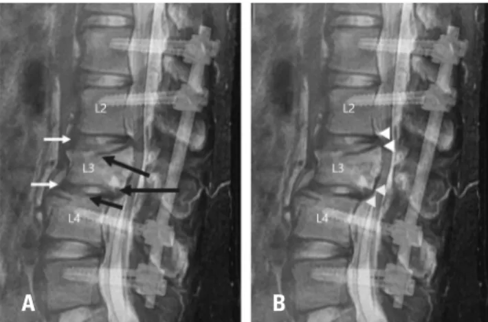
We analyzed 90 intervertebral disc levels adjacent to EF and 180 adjacent vertebral endplates using magnetic resonance imaging (MRI) at the time of injury (baseline) and at follow- up (Fig. 1). Initial radiological work-up was performed as soon as possible after the patient was admitted to the hospital.
Radiological work-up at follow-up was performed just before implant removal surgery (mean of 16.1±3.9 months after the index surgery).
Degenerative status of the intervertebral disc was classified according to the grading system of Pfirrmann et al.20) We defined DD at follow-up as Pfirrmann grade ≥III intervertebral disc.5,6) We evaluated the severity of defects in the cranial and caudal endplates of each intervertebral disc using the TEPS.15) The TEPS for a disc is the sum of the endplate
Chang-Hoon Jeon et al Volume 28 • Number 1 • March 31 2021
www.krspine.org 24
defect score of the cranial and caudal endplates. The endplate defect score ranges from 1 to 6; thus, the TEPS ranges from 2 to 12. Endplate defects are scored as follows (Grade 1, normal endplate; Grade 2, focal thinning of endplate; Grade 3, focal disc marrow contact (breakage); Grade 4, defect (depression of endplate) up to 25% of width of endplate with or without
Modic change; Grade 5, def (depression of endplate) up to 50% of width of endplate with or without Modic change;
Grade 6, complete endplate damage (irregularity and sclerosis) with or without Modic change (Fig. 2). All evaluations of intervertebral discs and endplates were performed based on reconstructed T2 sagittal images. Three experienced spine surgeons analyzed the images and classified the TEPS and DD three times, at 1-month intervals.
4. Statistical Analyses
We calculated the required sample size using statistical power analysis based on Rajasekaran et al.15) (121 cases with DD of Pfirrmann grade ≥III among 226 cases with TEPS ≥6, and 15 cases with DD of Pfirrmann grade ≥III among 139 cases with TEPS <6); At least 30 cases were required to achieve a power of 80% with a two-tailed significance level of p<0.05 The intraclass correlation coefficient (ICC) was used to calculate the intra- and interobserver reliability of the Pfirrmann grade of the intervertebral discs at follow-up, and of the TEPS at baseline. The ICC values for the intraobserver reliability were obtained using the single measure method for each observer’
s three measurements. The ICC values for the interobserver reliability were obtained using the average measures method for the last measurement round values of three observers.
Values of <0.40, 0.40–0.59, 0.60–0.74, and 0.75–1.00 were considered poor, fair, good, and excellent, respectively.21)
We assessed the associations between various factors and the development of DD using Pearson’s correlation and Spearman’s rank correlation, depending on the type of Fig. 1. This image is a combination of magnetic resonance imaging at the
time of injury and a postoperative radiograph of a patient with unstable lumbar fractures. The patient had an AO type A4 fracture with both cra- nial and caudal endplate fractures at L3 and an AO type A1 fracture with cranial endplate fracture at L4. (A) Intervertebral discs (white arrows) included in the analysis of this study. Among the segments included in fixation, only the L2-3 and L3-4 discs adjacent to the endplate fractures (black arrows) were analyzed in this study. (B) Endplates (arrows heads) included in the analysis of this study. Two adjacent endplates (white ar- rowheads, L1 caudal endplate and L3 cranial endplate) were included in the analysis as a factor related to L2-3 disc degeneration. L3-4 was analyzed separately using endplates adjacent to it (grey arrowheads, L3 caudal endplate and L4 cranial endplate).
Fig. 2. The total endplate defect score (TEPS).
Total endplate defect score=score of cranial endplate defect+score of caudal endplate defect
Score Characteristics
1 No endplate breaks or defects 2 Focal thinning of endplate 3 Focal disc marrow contacts,
Normal contour, No Modic changes
4 Defect upto 25% of width of endplate 5 Defect upto 50% of width of endplate 6 Complete endplate damage
A
1
4
2
5
3
6 B
variable. Spearman’s rank correlation coefficient ranged from -1 to 1, with 1 indicating a perfect correlation, 0 indicating no correlation, and -1 indicating a perfect inverse correlation. A correlation coefficient of R<0.3 is considered weak, while R=
0.3–0.59 is moderate and R>0.6 is strong.
Multivariate logistic regression analysis was used to identify independent risk factors for the development of DD. Variables eligible for inclusion in the multivariate models included those significant at p<0.20 in the univariate analyses. Receiver operating characteristic (ROC) curves were generated to determine the cut-off value of TEPS at baseline to predict the development of DD. The Youden index was used to determine the cut-off value. The data from this study were analyzed using SPSS statistical software (ver. 20.0; IBM, Armonk, NY, USA). In all analyses, p<0.05 was taken to indicate statistical significance.
Results
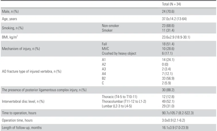
1. Demographic Data (Table 1)
Of the 34 patients, 24 were male and 7 were female. The mean age was 37.0 years. The majority of patients were non- smokers (n=23 [68.6%]). The mean BMI was 23.6 kg/m2. Fall was the most common cause of injury (n=18 [51.4%]).
Motor vehicle crash (n=10 [28.6%]) and crush injury (caused by a heavy object; n=6 [17.1%]) were the next most common injury types. The total number of damaged vertebras with endplate fracture was 58. AO type B2 was the most common fracture type (n=33, 56.9%), followed by A1 (n=14, 24.1%) and A4 (n=7, 12.1%).
2. Reliability of Pfirrmann Grade and TEPS in Thoracol- umbar Fracture
The ICC value for the intraobserver reliability of the Pfirrmann grade of DD, of the intervertebral discs at follow-
Table 1. Baseline characteristics of the patients
Total (N = 34)
Male, n (%) 24 (70.6)
Age, years 37.0±14.2 (13-64)
Smoking, n (%) Non-smoker
Smoker 23 (68.6)
11 (31.4)
BMI, kg/m2 23.6±2.9 (18.9-30.1)
Mechanism of injury, n (%) Fall
MVC
Crushed by heavy object
18 (51.4) 10 (28.6) 6 (17.1)
AO fracture type of injured vertebra, n (%)
A1 A2 A3 A4 B2 C
14 (24.1) 0 (0) 2 (3.4) 7 (12.1) 33 (56.9) 2 (5.9)
The presence of posterior ligamentous complex injury, n (%) 30 (88.2)
Intervertebral disc level, n (%) Thoracic (T4-5 to T10-11)
Thoracolumbar (T11-12 to L1-2) Lumbar (L2-3 to L4-5)
12 (12.8) 49 (52.1) 29 (31.0)
Time to operation, hours 90.7±105.7 (8.2-522.3)
Operation time, hours 3.0±0.9 (2.1-6.2)
Length of follow-up, months 16.1±3.9 (7.0-23.9)
BMI: body mass index, MVC: motor vehicle crash.
*Note: unless otherwise stated, range is indicated in parentheses.
Chang-Hoon Jeon et al Volume 28 • Number 1 • March 31 2021
www.krspine.org 26
up, was excellent [0.845, 95% confidence interval (CI): 0.709–
0.920)], and that for interobserver reliability was good (0.693, 95% CI: 0.378–0.848). The ICC value for the intraobserver reliability of the TEPS at baseline was excellent (0.946, 95% CI:
0.894–0.973). The ICC value for interobserver reliability of the TEPS at baseline was excellent (0.768, 95% CI: 0.531–0.886).
3. Development of Disc Degeneration and Endplate defect
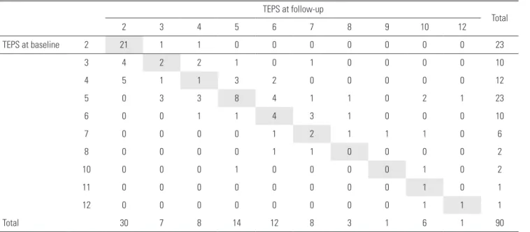
All discs were grade II at injury, which changed to grade III in 20 cases (21.2%) and grade IV in 4 cases (4.3%) at follow- up. The mean TEPS was 4.4 ± 2.2 at baseline, which changed to 4.6±2.5 at follow-up (p=0.339). Table 2 shows the changes in TEPS between baseline and follow-up.
4. Risk Factors for Disc Degeneration
Among the various demographic and radiological factors, intervertebral disc level and the TEPS were correlated with the development of DD. Spearman’s rank correlation test showed a weak correlation between intervertebral disc level and the development of DD (r=0.210, p=0.047). A strong correlation was observed between the TEPS at baseline and the development of DD (r=0.669, p<0.001).
Potential risk factors for DD, as determined by univariate analysis, included intervertebral disc level and TEPS at baseline.
The results of multivariate logistic regression analysis to identify independent risk factors for DD are shown in Table 3. The TEPS at baseline showed the strongest association with the development of DD, with an adjusted odds ratio of 3.163 after controlling for other variables in the model (95% CI: 1.792–
5.582, p<0.001). ROC curve analysis indicated that the optimal cut-off value of TEPS at baseline for the development of DD was 6, with 79.2% sensitivity, 95.5% specificity, and an area under the curve (AUC) value of 0.93 (95% CI: 0.87–0.99).
Discussion
This is the first study to demonstrate the impact of the severity of EF on the development of DD in patients with unstable thoracic and lumbar fractures treated with spinal Table 2. TEPS at baseline and follow-up
TEPS at follow-up
Total
2 3 4 5 6 7 8 9 10 12
TEPS at baseline 2 21 1 1 0 0 0 0 0 0 0 23
3 4 2 2 1 0 1 0 0 0 0 10
4 5 1 1 3 2 0 0 0 0 0 12
5 0 3 3 8 4 1 1 0 2 1 23
6 0 0 1 1 4 3 1 0 0 0 10
7 0 0 0 0 1 2 1 1 1 0 6
8 0 0 0 0 1 1 0 0 0 0 2
10 0 0 0 1 0 0 0 0 1 0 2
11 0 0 0 0 0 0 0 0 1 0 1
12 0 0 0 0 0 0 0 0 1 1 1
Total 30 7 8 14 12 8 3 1 6 1 90
TEPS: total endplate defect score.
Table 3. Multiple logistic regression model for the occurrence of disc degeneration
Risk factor Adjusted Odds Ratios
(95% Confidence Interval) p-value Intervertebral disc level
- Thoracolumbar vs. thoracic
- Lumbar vs. thoracic 1.495 (0.151-14.838) 2.610 (0.214-31.877)
0.682 0.731 0.452 TEPS at baseline 3.163 (1.792-5.582) <0.001 TEPS: total endplate defect score.
*Nagelkerke R2=0.565.
instrumentation and fusion. We analyzed the relationships between the development of DD and various baseline parameters, including the severity of EF at injury, which was graded using the TEPS. Our results showed that, among the baseline parameters, intervertebral disc level and TEPS at baseline were correlated with DD. In multivariate logistic analysis, the TEPS at baseline was the only risk factor for DD.
The cut-off value of TEPS at baseline for the development of DD was 6.
DD has been commonly observed after thoracolumbar spine fractures under spinal instrumentation.5,6) Wang et al.6) evaluated the development of DD in 26 patients (mean age, 39.6±10.3 years) who were surgically treated for thoracolumbar burst fracture. On MRI scans obtained at 23.5 months (15–36 months) follow-up, they found changes in the Pfirrmann grade in about half of the patients (from II to III in 14 cases, II to IV in 1 case, and III to IV in 4 cases). Choi et al.5) evaluated the DD in 57 surgically treated thoracolumbar fracture patients. An MRI evaluation was performed 2 years after the removal operation and revealed a DD of Pfirrmann grade>II in 43.3% of intervertebral discs adjacent to the fractured vertebrae, predominantly affecting intervertebral discs in contact with the EF. In this study, approximately 20%
of grade II discs adjacent to the endplate fracture changed to grade III disc and approximately 4% of grade II to grade IV discs with a mean follow-up of 16.1±3.9 months under spinal instrumentation and fusion.
Previous studies have confirmed that the presence of EF is related to the occurrence of DD. Wang et al.6) found new disc degeneration occurred only at level adjacent to the cranial endplate of fractured vertebra. Verlaan et al.22) treated 20 thoracolumbar fracture patients with endplate restoration using a balloon catheter and cement augmentation, and evaluated the intervertebral discs by MRI 1 month after the removal operation (12–18 months after the index surgery). They found that the change to severe DD (Pfirrmann grade ≥IV) was not significant. Choi et al.5) compared intervertebral discs contacting with and without EF under spinal instrumentation and they found a higher incidence of DD in the intervertebral discs contacting with EF (43.3% vs. 32.1%, p<0.001).
They also found that DD occurred statistically significantly more in the discs under instrumentation (7.9% vs. 32.1%, p<0.001). They postulated that fixation with pedicle screws
can reduce cyclical loading and lead to disc degeneration.
The low incidence of DD in this study compared to previous studies may be due to the short period of time when cyclic loading is limited due to a relatively short follow-up period.
However, the fact that severe DD did not occur when the endplate was sufficiently restored despite instrumentation over a similar period, as the study by Verlaan et al.22) may suggest that endplate damage acted as a more important factor in DD development than instrumentation itself.
We confirmed that the severity of EF influenced DD using the TEPS (Fig. 3, 4). The TEPS is a 6-point grading system for endplate defects in humans that was developed by Rajasekaran et al.15) As EF is a type of endplate defect, we postulated that the TEPS may also be used for grading EFs. Prior to application of the TEPS for grading of EFs, we analyzed the intra- and Fig. 3. A 21-year-old man presented to our emergency room for back pain that occurred after falling 30 meters. He was not a smoker. His body mass index was 25.7 kg/m2. He was diagnosed with an AO type B2 fracture of L1 and A1 fractures of L2 and L3. The vertebral canal was compromised, but he was neurologically intact. The patient was treated with postural and instrumental reduction, instrumentation two levels above and two levels below the L1 vertebra, and fusion of all segments within T11–
L3. (A)Magnetic resonance imaging obtained at the time of the injury showed that the intervertebral discs at T12–L1, L1–2, and L2–3 were of Pfirrmann grade II. There was severe endplate fracture and, in both inter- vertebral discs adjacent to L1, the total endplate defect score was 10 at T12–L1 (4 for the T12 caudal endplate and 6 for L1 cranial endplate), 7 at L1-2 (1 for the L1 caudal endplate and 5 for the L2 cranial endplate), and 5 at L2–3 (1 for the L2 caudal endplate and 4 for the L3 cranial endplate).
(B) At the final follow-up (23.9 months after the index operation), the endplates adjacent to the intervertebral discs of T12–L1 and L1–2 had changed to show an irregular surface, and both intervertebral discs had changed to Pfirrmann grade IV. However, the intervertebral disc of L2–3 remained at Pfirrmann grade II.
A B
Chang-Hoon Jeon et al Volume 28 • Number 1 • March 31 2021
www.krspine.org 28
interobserver reliability of the TEPS for EFs; the results were excellent. Two previous studies regarding the relationship between the TEPS and DD showed cut-off values of TEPS for the development of DD of ≥6 and ≥5.15,23) In the present study, we performed longitudinal analysis between the TEPS at injury and the development of DD at follow-up, and the cut- off value of TEPS to predict the development of DD was 6.
Concerned about secondary kyphosis or treatment failure caused by DD, Verlaan et al.22) insisted on endplate restorations in thoracolumbar burst fracture. Wang et al.10) were concerned that recurrent kyphosis may occur due to disc space collapse due to disc degeneration in the patients treated with short segment instrumentation in the thoracolumbar burst fracture.
Studies have also reported that disc lesions correlate with kyphosis after implant removal in thoracolumbar fractures.9,24) DD itself may act as a cause of pain even after posterior
fusion.7) The results of this study are still insufficient as evidence for changing the treatment direction in thoracolumbar fractures with severe EF and further research is needed. However, However, the results of this study suggest that the pain and recurrent kyphosis caused by DD must be explained to the patient in advance, and special attention is required in patients with thoracolumbar fractures with severe EF.
1. Limitations
Our study had several limitations. First, it used a retrospective design with a relatively short follow-up period. The results of this study indicated that only TEPS was a predictive factor for the development of DD; other factors, including age, BMI, intervertebral disc level,25) and length of follow-up did not have good predictive utility, possibly due to nature of the study design. Second, the patients included in this study were treated surgically with long-segment instrumentation. Instrumentation may prevent cyclic loading of the disc and promote DD.5) Our results cannot be applied to patients treated surgically with short-segment instrumentation and to non-surgical patients.
Third, this study did not include clinical outcomes or the recurrent kyphosis. Since this study included multi-segment fractures, which may act as a confounder, it was difficult to assess the clinical outcomes or the degree of kyphosis. Further studies are needed on the degree of kyphosis, pain, and disc degeneration in patients with single segment spinal fractures.
Finally, we defined the development of DD as Pfirrmann grade ≥III. However, several previous reports regarding the relationship between TEPS and DD defined DD as Pfirrmann grade ≥IV,15,22,23) while others defined DD as Pfirrmann grade
≥ III.5,6) As our study had a short follow-up, progression of DD to grade IV was very rare. Nevertheless, the change to Pfirrmann grade III was definite and reliable.
Conclusions
The severity of EF at injury was associated with the development of DD. The severity of EF at injury was the only risk factor for the development of DD. Severe EF (TEPS ≥6) at injury resulted in significant DD within a short period. DD can occur in intervertebral discs in contact with severe EF. This can be an important criterion for determining whether DD is due to trauma. In addition, since pain and recurrent kyphosis Fig. 4. A 58-year-old man presented to our emergency room due to back
pain that occurred after a motor vehicle injury. He had no medical dis- eases, was a non-smoker, and was not obese. He was diagnosed with an AO type B2 fracture of L1. He showed a lamina fracture at L1 and the posterior ligamentous complex at T12–L1 was ruptured. A huge epidural hematoma from T12–L4 was present, but he had no neurological deficits.
The patient was treated with instrumentation and fusion, two levels above and two levels below the injury. (A) A magnetic resonance imag- ing scan obtained at the time of the injury showed that the intervertebral discs above and below the fractured vertebra were of Pfirrmann grade II.
There was no severe endplate fracture. In the intervertebral disc above the fractured vertebra, the total endplate defect score (TEPS) was 5 (1 for the cranial endplate and 4 for the caudal endplate). In the intervertebral disc below the fractured vertebra, the TEPS was 3 (2 for the cranial end- plate and 1 for the caudal endplate). (B) At the final follow-up (13 months after the index operation), the endplate had healed and both interverte- bral discs were still of Pfirrmann grade II.
A B
related to DD may occur, it is necessary to explain this to the patient of thoracolumbar fracture with severe EF (TEPS ≥6) in advance and pay special attention.
Acknowledgments
We thank Textcheck (http://www.textcheck.com) for checking the English in this paper. An editor with specialist professional knowledge (MSc or PhD./MD) reviewed and corrected the English. An English language specialist then rechecked the paper. The first language of both editors is English. Reference number is ‘19050202’.
REFERENCES
1. Luoma K, Riihimäki H, Luukkonen R, et al. Low back pain in relation to lumbar disc degenera- tion. Spine. 2000;25(4):487-92. DOI: https://doi.
org/10.1097/00007632-200002150-00016.
2. de Schepper EI, Damen J, van Meurs JB, et al. The associa- tion between lumbar disc degeneration and low back pain:
the influence of age, gender, and individual radiographic features. Spine. 2010;35(5):531-6. DOI: https://doi.
org/10.1097/BRS.0b013e3181aa5b33.
3. Dudli S, Haschtmann D, Ferguson SJ. Persistent degenera- tive changes in the intervertebral disc after burst fracture in an in vitro model mimicking physiological post-traumatic conditions. European Spine Journal. 2015;24(9):1901-8.
DOI: https://doi.org/10.1002/jor.21573.
4. Kerttula LI, Serlo WS, Tervonen OA, et al. Post-traumatic findings of the spine after earlier vertebral fracture in young patients: clinical and MRI study. Spine. 2000;25(9):1104- 8. DOI: https://doi.org/10.1097/00007632-200005010- 00011.
5. Choi W, Song S, Chae S, et al. Comparison of the extent of degeneration among the normal disc, immobilized disc, and immobilized disc with an endplate fracture. Clinics in orthopedic surgery. 2017;9(2):193-9. DOI: https://doi.
org/10.4055/cios.2017.9.2.193.
6. Wang J, Zhou Y, Zhang ZF, et al. Radiological study on disc degeneration of thoracolumbar burst fractures treated by percutaneous pedicle screw fixation. European Spine Journal. 2013;22(3):489-94. DOI: https://doi.
org/10.1007/s00586-012-2462-1.
7. Barrick WT, Schofferman JA, Reynolds JB, et al. Anterior Lumbar Fusion Improves Discogenic Pain at Levels of Prior Posterolateral Fusion. Spine. 2000;25(7):853-7. DOI:
https://doi.org/10.1097/00007632-200004010-00014.
8. Speth MJ, Oner FC, Kadic MA, et al. Recurrent kyphosis after posterior stabilization of thoracolumbar fractures: 24 cases treated with a Dick internal fixator followed for 1.5- 4 years. Acta Orthopaedica Scandinavica. 1995;66(5):406- 10. DOI: https://doi.org/10.3109/17453679508995575.
9. Chen J-X, Xu D-L, Sheng S-R, et al. Risk factors of ky- phosis recurrence after implant removal in thoracolumbar burst fractures following posterior short-segment fixation.
International orthopaedics. 2016;40(6):1253-60. DOI:
https://doi.org/10.3109/17453679508995575.
10. Wang X-Y, Dai L-Y, Xu H-Z, et al. Kyphosis recur- rence after posterior short-segment fixation in thoraco- lumbar burst fractures. Journal of Neurosurgery: Spine.
2008;8(3):246-54. DOI: https://doi.org/10.3171/
spi/2008/8/3/246.
11. Möller A, Maly P, Besjakov J, et al. A vertebral frac- ture in childhood is not a risk factor for disc degeneration but for Schmorl’s nodes: a mean 40-year observational study. Spine. 2007;32(22):2487-92. DOI: https://doi.
org/10.1097/brs.0b013e3181573d6a.
12. Oner FC, van der Rijt RR, Ramos LM, et al. Changes in the disc space after fractures of the thoracolumbar spine. J Bone Joint Surg Br. 1998;80(5):833-9. DOI: https://doi.
org/10.1302/0301-620x.80b5.0800833.
13. Dudli S, Ferguson SJ, Haschtmann D. Severity and pattern of post-traumatic intervertebral disc degeneration depend on the type of injury. The Spine Journal. 2014;14(7):1256- 64. DOI: https://doi.org/10.1002/jor.21573.
14. Su Y, Ren D, Zou Y, et al. A retrospective study evaluat- ing the correlation between the severity of intervertebral disc injury and the anteroposterior type of thoracolumbar verte- bral fractures. Clinics. 2016;71(6):297-301. DOI: https://
doi.org/10.6061/clinics/2016(06)02.
15. Rajasekaran S, Venkatadass K, Babu JN, et al. Pharma- cological enhancement of disc diffusion and differentia- tion of healthy, ageing and degenerated discs. European Spine Journal. 2008;17(5):626-43. DOI: https://doi.
org/10.1007/s00586-008-0645-6.
Chang-Hoon Jeon et al Volume 28 • Number 1 • March 31 2021
www.krspine.org 30
16. Rade M, Määttä JH, Freidin MB, et al. Vertebral End- plate Defect as Initiating Factor in Intervertebral Disc Degeneration: Strong Association Between End- plate Defect and Disc Degeneration in the General Population. Spine. 2018;43(6):412-9. DOI: 10.1097/
BRS.0000000000002352.
17. Wang Y, Videman T, Battié MC. Lumbar vertebral endplate lesions: prevalence, classification, and association with age.
Spine. 2012;37(17):1432-9. DOI: https://doi.org/10.1097/
brs.0b013e31824dd20a.
18. Selby MD, Clark SR, Hall DJ, et al. Radiologic assess- ment of spinal fusion. J Am Acad Orthop Surg. 2012 Nov;20(11):694-703. DOI: 10.5435/jaaos-20-11-694.
DOI: https://doi.org/10.5435/JAAOS-20-11-694.
19. Zampolin R, Erdfarb A, Miller T. Imaging of lumbar spine fusion. Neuroimaging Clin N Am. 2014 May;24(2):269- 86. DOI: https://doi.org/10.1016/j.nic.2014.01.004.
20. Pfirrmann CW, Metzdorf A, Zanetti M, et al. Magnetic resonance classification of lumbar intervertebral disc de- generation. Spine. 2001;26(17):1873-8. DOI: https://doi.
org/10.1097/00007632-200109010-00011.
21. Lachin JM. The role of measurement reliability in clini- cal trials. Clin Trials. 2004;1(6):553-66. DOI: https://doi.
org/10.1191/1740774504cn057oa.
22. Verlaan J-J, Dhert WJ, Oner FC. Intervertebral disc viability after burst fractures of the thoracic and lumbar spine treated with pedicle screw fixation and direct end-plate restoration.
The Spine Journal. 2013;13(3):217-21. DOI: https://doi.
org/10.1007/s00586-012-2462-1.
23. Rade M, Maatta JH, Freidin MB, et al. Vertebral Endplate Defect as Initiating Factor in Intervertebral Disc Degenera- tion; Strong Association between Endplate Defect and Disc Degeneration in the General Population. Spine (Phila Pa 1976). 2017 Jul 26. DOI: 10.1097/brs.0000000000002352.
DOI: 10.1097/brs.0000000000002352.
24. Kanezaki S, Miyazaki M, Ishihara T, et al. Mag- netic resonance imaging evaluation of intervertebral disc injuries can predict kyphotic deformity after pos- terior fixation of unstable thoracolumbar spine inju- ries. Medicine. 2018;97(28):e11442-e. DOI: 10.1097/
MD.0000000000011442.
25. Dolan P, Luo J, Pollintine P, et al. Intervertebral disc de- compression following endplate damage: implications for disc degeneration depend on spinal level and age. Spine.
2013;38(17):1473-81. DOI: https://doi.org/10.1097/
brs.0b013e318290f3cc.
척추 기기 고정술과 유합술로 치료한 불안정성, 외상성 흉추 및 요추의 불안정성 골절 환자에서 추체 종판 골절의 심각도가 추간판 퇴행의 발생에 미치는 영향
전창훈 • 정남수 • 정희웅 • 이한동 아주대학교 의과대학/병원, 정형외과학교실
연구 계획: 후향적 분석
목적: 본 연구에서는 척추 기기 고정술과 유합술로 치료한 불안정성, 외상성 흉추 및 요추의 불안정성 골절 환자 코호트를 이용하여 추체종판 골절의 심 각도가 인접 추간판의 퇴행에 미치는 영향을 알아보고자 하였다.
선행 연구문헌의 요약: 종판 골절의 심각도와 추간판의 퇴행의 관계에 대해서는 알려진 바가 없다.
대상 및 방법: 본연구는 골절된 척추체에 인접한 총 90분절의 추간판과 그 추간판에 인접한 총 180개의 추체종판을 분석하였다. 본 연구는 본원에서 흉 추 및 요추의 불안정성 골절로 수술적 치료를 받은 34명의 연속적인 환자를 대상으로 하였다. 수상 당시 및 최종 추시(수상 당시로부터 평균 16.1±3.9 개월)에서의 Magnetic resonance imaging (MRI)에서 골절에 인접한 90분절의 추간판을 Pfirrmann grade를 이용하여 분석하였다. 수상 당시의 추체 종 판 골절의 심각도는 MRI에서 total endplate defect score (TEPS)를 측정하여 평가하였다. 다중 로지스틱 회귀 분석을 이용하여 추간판의 퇴행(Pfirrmann grade ≥III)에 대한 수상 당시의 위험인자를 분석하였다.
결과: 모든 추간판은 수상당시 grade II였으며, 최종 추시에서 20개의 분절(21.2%)은 grade III로 퇴행성 변화가 진행됨이 관찰되었으며, 4개의 분절(4.3%) 는 grade IV로 진행된 것이 관찰되었다. 수상당시의 TEPS가 추간판 퇴행의 발생에 가장 중요한 위험인자였다. Receiver operating characteristic curve분 석에서 TEPS의 cut-off 값은 6점이었다.
결론: 추체종판의 골절의 정도는 추간판의 퇴행의 위험인자이며, 수상 당시의 심각한 추체 종판의 손상은(TEPS ≥ 6) 추간판 퇴행에 매우 밀접한 관계가 있다.
색인 단어: 추간판 퇴행, 추체종판 손상, 추체종판 골절, Total endplate defect score, 척추 골절 약칭 제목: 추체종판 골절과 추간판 퇴행
접수일: 2020년 11월 19일 수정일: 2020년 12월 2일 게재확정일: 2021년 2월 16일 교신저자: 이한동
경기도 수원시 영통구 월드컵로 164 아주대학교 의과대학/병원 정형외과학교실
TEL: 031-219-5220 FAX: 031-219-5229 E-mail: [email protected]