경막하 지방종으로 오인될 수 있는 외상성 요천추부 아급성 경막하 출혈: 컴퓨터단층촬영의 유용성
황선철*
,§,//• 김형간* • 조성희* • 남대철*
,§,//• 이헌근
†,§,//• 박경언
†,§,//• 나재범
‡,§,//• 김동희*
,§,//경상대학교 의학전문대학원 *정형외과학교실, †마취통증의학교실, ‡영상의학교실, §임상의학연구소, //건강과학연구원
Traumatic Lumbosacral Spinal Subdural Hematoma Mimicking Subdural Lipoma: Value of Computed Tomography
Sun-Chul Hwang, M.D.*
,§,//, Hyung-Kan Kim, M.D.*, Seong-Hee Cho, M.D.*, Dae-Chul Nam, M.D.*
,§,//, Heon-Keun Lee, M.D.
†,§,//, Kyeong-Eon Park, M.D.
†,§,//, Jae-Boem Na, M.D.
‡,§,//, and Dong-Hee Kim, M.D.*
,§,//Departments of *Orthopaedic Surgery,
†Anesthesiology, and
‡Radiology,
§Resarch Institute of Clinical Medicine,
//
Institute of Health Sciences, Gyeongsang National University School of Medicine, Jinju, Korea
Traumatic lumbosacral spinal subdural hematoma due to anatomical and pathological causes is rare, compared to epidural hematoma. If the time of trauma cannot be determined, intracranial and intraspinal signal intensity according to lapse of time are not coincident, resulting in confusion in terms of differentiation. Fat suppression magnetic resonance image (MRI) and computed tomography (CT) are utilized for differentiation. The intention of this study is to report on a case where spinal subdural hematoma of unknown time of occurrence is differentiated from subdural lipoma by taking advantage of fat suppression MRI and CT in order to perform an early surgical decompression with auxiliary review of literature demonstrating good prognosis of the procedure.
Key words: lumbosacral spinal subdural hematoma, subdural lipoma, surgical decompression
두개골 내부의 손상이 동반되지 않은 단독의 외상성 요천추 경막 하 혈종은 경막하 공간이 해부학적으로 거의 무혈관 구조이므로 경막외 혈종에 비해 아주 드물게 발생한다.1) 원인으로는 출혈성 경향, 항응고제 복용, 척추 천자, 종양, 혈관 기형, 그리고 드물게 외상에 의해 발생하며, 임상적으로 드물게 발생하고 심각한 후유 증을 남길 수 있는 경우가 많기 때문에 빠른 진단과 적절한 치료 가 조기에 시행되어야 하는 질환이다.2,3) 본 저자들은 외상 후 발 생한 요천추부 아급성 경막하 혈종을 자기공명영상과 보조적 컴 퓨터단층촬영을 이용하여 경막하 지방종과 감별진단하여 조기에 수술적 감압술 시행 후, 좋은 예후를 보인 증례를 문헌 고찰과 함
께 보고하고자 한다.
증례보고
과거력상 특별한 질환이 없던 61세 남자 환자가 내원 10일 전에 넘어진 후, 발생한 요통 및 양측 하지 방사통을 주소로 내원하였 다. 항응고제 복용력은 없으며, 최근 척추 천차 및 뇌척수액 배액 술 등의 병력은 없었다. 내원 당시 시행한 이학적 검사상 요추부 타진 시 동통과 양측 대퇴부 및 하퇴부 외측 부위로 방사통이 있 었으나 감각 저하 및 근력 약화는 관찰되지 않았다. 통증은 보행 시 양측 요추 제4, 5 신경근 피부 분절을 따라 방사통이 심해지면 서 요통이 악화되는 소견을 보였으며 하지 직거상 검사상 40/40 으로 양성 소견이 나타났다. 진단 검사 의학적 소견은 정상 범위 였고, 응고 장애 및 출혈 경향은 관찰되지 않았다. 단순 방사선 사진상, 요추의 추체 골단판과 추간판 인접 부위에서 골극 형성
Copyright © 2013 by The Korean Orthopaedic Association
“This is an Open Access article distributed under the terms of the Creative Commons Attribution Non-Commercial License (http://creativecommons.org/licenses/by-nc/3.0/) which permits unrestricted non-commercial use, distribution, and reproduction in any medium, provided the original work is properly cited.”
The Journal of the Korean Orthopaedic Association Volume 48 Number 5 2013 Received June 26, 2013 Revised July 31, 2013 Accepted August 18, 2013 Correspondence to: Dong-Hee Kim, M.D.
Department of Orthopaedic Surgery, Gyeongsang National University School of Medicine, 15 Jinju-daero 816beon-gil, Jinju 660-751, Korea
TEL: +82-55-750-8669 FAX: +82-55-762-4640 E-mail: dhkim8311@gnu.ac.kr
황선철·김형간·조성희 외 5인
과 같은 퇴행성 변화 소견이 관찰되었다(Fig. 1). 외상 후 9일경 외 부 병원에서 시행한 자기공명영상 시상면에서 T1 강조 영상에 서는 균일한 고음영상, T2 강조 영상에서 균일한 중간 및 저음영 상이 제1 요추에서부터 제2 천추까지 천추관을 따라 나타났으며 지방억제 기법은 시행되지 않았다. 축면상에서 마미총 전후면에 서 고음영 영상의 척수 압박 소견이 관찰되며, 이로 인해 'inverted Mercedes sign' 소견이 보였다(Fig. 2). 그러나 자기공명영상상 경 막하 지방종과 그 음영이 비슷하여 감별진단이 되지 않았다. 그 래서 컴퓨터단층촬영을 시행하였고 축상면에서 경막 내부에 있 는 동일 밀도의 종괴 영향으로 인해 척수가 압박되고 있어 혈종 에 의한 것으로 생각되었으며, 시상면에서 경막외 후방에 있는 저밀도의 영상 소견이 경막외 지방 조직으로 생각되었다(Fig. 3).
환자의 신경학적 증상은 입원 경과 관찰하면서 족관절 족배 굴곡 및 무지 신전의 근력이 4단계로 약화되어 있었으며, 배뇨 장애가 관찰되어 불완전 마미 증후군 진단하에 본 저자는 요추 천자를 통해 액화된 혈종 흡입을 시행하였으나, 아급성 경막하 혈종으로 응고되어 혈괴(clot) 때문에 혈액은 흡인되지 않았다. 요추 천자용 바늘을 더 전진하였을 때 혈종과 희석된 뇌척수액이 약 3 ml 정도 만 흡입되었으며 신경학적 증상이 호전되지 않아 응급 수술을 시 행하였다(Fig. 4). 제4, 5 요추 척추궁 절제술 시행 후, 경막 내부에 검붉은 혈종으로 인해 압박된 마미총이 관찰되어 경막을 절개하
여 혈종을 감압하였다. 우측 제4 요추 신경근 부위에 무판막성 신 경근수질 정맥(valveless radiculomedullary vein)으로 생각되는 혈 관에서 출혈 소견이 관찰되어, 양극방식 전기 수술기(bipolar bo- vie)를 이용하여 소작 지혈 후 후외측 유합술을 시행하였다(Fig.
5). 수술 후, 방사통 및 근력은 정상으로 회복되었으며 술 후 3개 월째 촬영한 자기공명영상상 이전에 관찰되었던 요천추부 경막 하 혈종은 관찰되지 않았다(Fig. 6).
고 찰
전체 척추에 발생하는 혈종 중에 경막하 혈종은 약 3%로 알려져 Figure 1. Preoperative radiographs. (A) Posteroanterior and (B) lateral
plain radiographs showed endplate sclerotic changes and osteophytes of the lumbar spine.
Figure 2. (A) T1 weight image sagittal magnetic resonance image (MRI) demonstrating a hyperintense signal (B) T2 weight image sagittal MRI demonstrating an intermediate to low signal from L1 to S2 level suggestive of a subdural hematoma causing thecal sac compression (arrows). Axial (C) T1 and (D) T2-weighted MRI showing signal intensity, correlating with an early subacute subdural hematoma at the anterior and posterior location of the spinal canal (asterisk points: ‘inverted Mercedes sign’).
있다. 경막하 강과 달리 경막외 강은 넓고 느슨한 지방층이 차있 고 풍부한 정맥총이 있기 때문에 경막외 출혈이 잘 일어나는 반 면 경막하 혈종의 빈도는 상대적으로 낮은 것으로 알려져 있다.4) 경막하 혈종의 남녀의 비율은 1:2.1이고, 발생 연령은 평균 41세 이며, 흉추와 흉요추 이행부가 가장 많이 발생한다고 알려져 있 다.3) 시기에 따라 급성, 아급성 그리고 만성으로 분류하였는데 급 성은 갑자기 발생한 심한 요통과 방사통을 호소할 수 있으며 하 지 마비로 진행이 빠르다. 아급성은 서서히 약 2주에 걸쳐 진행을 하며 덜 국소적인 요통, 둔부 및 하지 통증을 호소하며 종종 하지 의 저림, 마비 등을 호소할 수 있다. 만성은 수개월에서 수년에 걸 쳐 천천히 진행되는 증상을 호소하고 통증은 없거나 경미하고 신
경학적 장애는 미미하며 중심성 척수 증후군을 보이는 경우도 있 다.5) Kreppel 등6)은 613명의 척수 혈종에 대해 원인 분석을 위해 메타분석을 시행한 결과, 병인을 찾을 수 없는 경우(29.7%), 항응 고요법(17.0%), 혈관 기형(9.1%), 자발적 척수 혈종(8.5%), 항응고 치료를 병행하는 환자에게 척추 및 경막외 마취를 시행하는 경우 (6.0%), 종양 출혈(5.7%), 출혈성 소인을 가지고 있는 경우(5.6%), 골절이 동반되지 않은 외상(5.4%), 골절이 동반된 외상(4.4%), 척 추 및 경막외 마취 단독인 경우(4.3%)로 보고하였다. 즉 경막하 혈 종의 원인으로 항응고제 복용, 혈관 기형, 종양, 출혈성 경향 및 척추 천차 등에 의해 발생하는 것으로 알려져 있으며 본 증례처 럼 골절이 동반되지 않은 외상에 의한 경우는 매우 드물다.2) Johnson 등7)은 'inverted mercedes sign'에 대해 언급하였으며, 자기공명영상이 경막하 혈종을 진단하기 위한 가장 좋은 영상기 법이라고 하였다. 자기공명영상상 아급성 경막하 혈종의 경우에 methemoglobin으로 인해 초기 아급성기(3-7일)에는 T1 강조 영 상에서 고음영, T2 강조 영상에서 저음영으로 나타나며, 후기 아
Figure 3. Preoperative computed tomography (A: upper doted line on C, B: lower doted line on C) different axial level view and (C) sagittal view showing differences in signal density between the hematoma (isodensity) and epidural fat (low density, arrows).
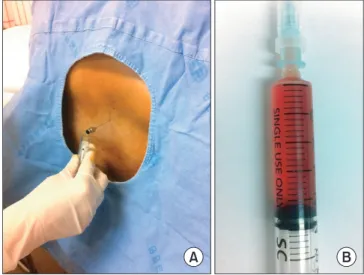
Figure 4. (A) Lumbar 4-5 level puncture. (B) Hematoma mixed with cerebrospinal fluid was aspirated by 3 ml.
Figure 5. (A) After total laminectomy of L4 and L5, a dark-reddish transparent hematoma was observed beneath the dura. (B) Bleeding was observed around the right L4 root which may be considered a valveless radiculomedullary vein, and it was coagulated with bipolar bovie.
황선철·김형간·조성희 외 5인
급성기(1-2주)에는 T1, T2 강조 영상에서 모두 고음영으로 나타 난다.8) 그러나 자발적 혈종이나 외상의 시기가 불분명한 경우, 그 리고 시간에 따른 혈종의 신호 강도가 두개강 내와 척추강 내에 서 반드시 일치하지 않기 때문에 많은 혼돈을 주게 된다. 이러한 경우 조영증강 지방억제 T1 강조 영상에서 경막외 지방보다 경 막하 혈종이 고신호 강도로 나타나기 때문에 조영증강 지방억제 T1 강조 영상의 경우, 정확한 진단을 내리는 데 도움이 된다.8) 또 한, 컴퓨터단층촬영에서도 지방 조직은 저신호 강도로 확인되며 혈종의 경우 등신호 강도로 관찰되기 때문에 혈종과 지방 조직과 의 감별이 가능하며 구획화에도 도움을 주어 자기공명영상과 함 께 컴퓨터단층촬영은 서로 보완적으로 경막하 혈종의 진단에 적 절한 진단 도구로 사용할 수 있겠다.9,10) 본 증례의 경우 약 10여 일 전에 외상의 병력이 있어 수상 시기로 보았을 때는 후기 아급성 기에 해당하였으나 자기공명영상 소견상 T1 강조 영상에서 고음 영, T2 강조 영상에서 저음영으로 나타나 초기 아급성기 경막내 혈종 소견이 관찰되었다. 지방 조직은 자기공명영상에서 초기 아 급성기 혈종과 같이 T1 강조 영상에서 고음영, T2 강조 영상에서 저음영으로 나타나 경막내 혈종과 같은 음영을 보여 경막하 지방 종과의 감별이 힘들었다. 상대적으로 두께가 얇은 경막이 잘 보 이지 않을 경우 경막외 지방 조직과도 감별이 모호해질 수 있다.9) 본 환자의 경우 경막외 지방 조직이나 경막하 지방종으로 오인될 수 있는 외상 후 아급성기 경막하 출혈 환자에 컴퓨터단층촬영을 시행하였다. 이를 통해 경막외 지방은 저신호 강도로, 경막하 공
간의 병변은 등신호 강도로 관찰되어 혈종과 지방 조직을 감별 할 수 있었다. 이를 통해 경막외 지방은 저신호 강도로, 경막하 공 간의 병변은 등신호 강도로 관찰되어 혈종과 지방 조직을 감별할 수 있었다.
외상성 척추 경막하 혈종은 드문 질환이므로 임상에서 간과할 수 있는 경우가 많고 다른 질환으로 오인될 수 있으며 심각한 후 유증을 남길 수 있기 때문에 빠른 진단과 적절한 치료가 조기에 시행되어야 하는 질환이다. 경막하 지방 조직의 경계가 불분명한 경우와 경막하 지방종을 아급성 경막하 혈종과 감별 진단을 요할 때, 신속하게 시행할 수 있는 컴퓨터단층촬영이 감별진단에 유용 할 것으로 생각한다.
REFERENCES
1. Diyora B, Sharma A, Mamidanna R, Kamat L. Chronic cer- vicothoracic spinal subdural hematoma. Neurol Med Chir (Tokyo). 2009;49:310-2.
2. Rader JP. Chronic subdural hematoma of the spinal cord: re- port of a case. N Engl J Med. 1955;253:374-6.
3. Russell NA, Benoit BG. Spinal subdural hematoma. A review.
Surg Neurol. 1983;20:133-7.
4. Hung KS, Lui CC, Wang CH, Wang CJ, Howng SL. Traumatic spinal subdural hematoma with spontaneous resolution. Spine (Phila Pa 1976). 2002;27:E534-8.
5. Mavroudakis N, Levivier M, Rodesch G. Central cord syn- drome due to a spontaneously regressive spinal subdural he- matoma. Neurology. 1990;40:1306-8.
6. Kreppel D, Antoniadis G, Seeling W. Spinal hematoma: a lit- erature survey with meta-analysis of 613 patients. Neurosurg Rev. 2003;26:1-49.
7. Johnson PJ, Hahn F, McConnell J, Graham EG, Leibrock LG.
The importance of MRI findings for the diagnosis of nontrau- matic lumbar subacute subdural haematomas. Acta Neurochir (Wien). 1991;113:186-8.
8. Braun P, Kazmi K, Nogués-Meléndez P, Mas-Estellés F, Apar- ici-Robles F. MRI findings in spinal subdural and epidural hematomas. Eur J Radiol. 2007;64:119-25.
9. Kamo M, Watanabe Y, Numaguchi Y, Saida Y. Spinal subdural hematoma mimicking epidural lipomatosis. Magn Reson Med Sci. 2012;11:197-9.
10. Post MJ, Becerra JL, Madsen PW, et al. Acute spinal subdural hematoma: MR and CT findings with pathologic correlates.
AJNR Am J Neuroradiol. 1994;15:1895-905.
Figure 6. Magnetic resonance images taken three months after surgery (A: upper doted line on C, B: lower doted line on C). T2-weight axial images demonstrate recovery of normal signal density without thecal sac compression. (C) T2-weight sagittal image showing disappearance of the subdural hematoma.
경막하 지방종으로 오인될 수 있는 외상성 요천추부 아급성 경막하 출혈: 컴퓨터단층촬영의 유용성
황선철*
,§,//• 김형간* • 조성희* • 남대철*
,§,//• 이헌근
†,§,//• 박경언
†,§,//• 나재범
‡,§,//• 김동희*
,§,//경상대학교 의학전문대학원 *정형외과학교실, †마취통증의학교실, ‡영상의학교실, §임상의학연구소, //건강과학연구원
외상성 요천추부 경막하 혈종은 해부학 및 병리학적인 원인으로 인하여 경막외 혈종에 비해 빈도가 극히 드문 것으로 알려져 있다.
외상의 시기가 불분명한 경우 시간 경과에 따른 혈종의 신호 강도가 두개강내 혈종과 척추강 내 혈종에서 일치하지 않는 경우가 많기 때문에 감별진단에 있어서 많은 혼돈을 주게 된다. 이에 지방억제 자기공명영상 및 컴퓨터단층촬영 등의 시도들이 행해지고 있다. 외 상 시기를 정확히 알 수 없는 경막하 혈종을 자기공명영상과 컴퓨터단층촬영을 이용하여 경막하 지방종과 감별진단하여 조기에 수술 적 감압술 시행 후, 좋은 예후를 보인 증례를 문헌 고찰과 함께 보고하고자 한다.
색인단어: 요천추부 경막하 혈종, 경막하 지방종, 수술적 감압술
Copyright © 2013 by The Korean Orthopaedic Association
“This is an Open Access article distributed under the terms of the Creative Commons Attribution Non-Commercial License (http://creativecommons.org/licenses/by-nc/3.0/) which permits unrestricted non-commercial use, distribution, and reproduction in any medium, provided the original work is properly cited.”