Chest radiographs are the most frequently performed radiologic examination and they account for a large por- tion of medical image data. For example, 16,110 chest
radiographs were performed, resulting in a data volume of 229.9 gigabytes accumulated at the Seoul National University Bundang Hospital in September 2009.
Irreversible (“lossy”) image compression appears to be an immediate and effective means to reduce operational costs on transmission and storage of medical image data (1-4). However, such compression techniques are not always accepted by radiologists due to concerns of arti- facts that potentially hinder diagnosis.
There have largely been two approaches in determin- ing optimal compression thresholds: the diagnostically lossless approach (5-11) and visually lossless approach
Visually Lossless Threshold: JPEG 2000 compression of Digital Chest Radiographs1
Gyoung Min Kim, M.D., Kil Joong Kim, M.S.2
1Department of Radiology, Seoul National University College of Medicine, Seoul National University Hospital
2Department of Radiation Applied Life Science, Seoul National University College of Medicine
“This study was supported by a grant from the Seoul National University Bundang Hospital (project no. 02-2007-001).”
Received April 30, 2010 ; Accepted July 7, 2010
Address reprint requests to : Kil Joong Kim, M.S., Department of Radiation Applied Life Science, Seoul National University College of Medicine, 28 Yongon-dong, Chongno-gu, Seoul 110-744, Korea.
Tel. 82-31-787-7609 Fax. 82-31-787-4011 E-mail: [email protected]
Purpose: To estimate the visual lossless threshold of Joint Photographic Experts Group (JPEG) 2000 compression digital chest radiograph images.
Materials and Methods: Fifty (n=50) selected chest radiograph images were com- pressed to 5 different levels: reversible (as negative control) and irreversible 5:1, 10:1, 15:1, and 20:1. By alternately displaying the original image and its paired compressed image on the same monitor, five radiologists independently determined if the image pairs had detectable differences. For each reader, we compared the proportion of the image pairs (the compressed image and the original image) rated to have detectable differences between reversible compression and each of the four irreversible compres- sions using the exact test for paired proportions.
Results: For each reader, the proportion of the image pairs rated to have detectable dif- ference was not significantly different between the reversible and irreversible 5:1 and 10:1 compressions. However, the proportion significantly increased with 15:1 and 20:1 irreversible compressions, versus reversible compression in all readers (p=7.4×10⁻²² - 0.027).
Conclusion: 10:1 compressed chest radiograph images can be considered visually loss- less and are therefore potentially acceptable for primary interpretation.
Index words :Data Compression
Radiology Information Systems Radiography, Thoracic
(12-14). The diagnostically lossless approach aims to preserve diagnostic accuracy in compressed images.
Although this approach addresses the diagnostic perfor- mance directly, its practicability is limited as the com- pression threshold intrinsically varies with the diagnos- tic task itself (e.g., the size of the target lesion) (12). The visually lossless approach is based on the concept that if compression artifacts are imperceptible, they should not affect the diagnosis. The latter approach has been advo- cated to be more robust and conservative and, therefore, to be more suitable for medical image compression than the former. In this study, we focus on the principle of vi- sual losslessness.
If a compressed image cannot be distinguished from the original (non-compressed) image by radiologists, there is no basis for arguing that this “visually lossless”
compression impedes diagnostic accuracy (12, 15). In other words, a visually lossless threshold (VLT) for im- age compression can be higher than a mathematically lossless threshold (reversible threshold), and lower than a diagnostically lossless threshold. Although the visually lossless criterion would likely allow a relatively lower compression level, this conservative criterion would be more readily accepted, even by radiologists skeptical to irreversible compressions (12).
The effect of image compression on chest radiographs has been studied since the early 1990s. The acceptable compression ratios were reported to be as high as be- tween 10:1 and 25:1 (7-9, 16). All published reports on studies concerned the evaluation of diagnostic perfor- mance - the diagnostic lossless threshold - typically in a receiver operating characteristic study.
There are studies on VLT of abdominal and chest computed tomography (CT) compressed with JPEG 2000. Previous studies show that VLT differed with body parts, scan parameters, and modalities (4, 14, 17- 23). To our knowledge, there is only one published re- port on VLT assessment in compressed digital chest ra- diographs (13). In that study, the chest radiographs were compressed with the JPEG algorithm, and displayed with film and CRT.
The purpose of this study was to evaluate the visually lossless threshold of irreversibly compressed chest radi- ograph adopting recent imaging trends, i.e. digital radi- ography with flat detector, JPEG 2000 compression al- gorithm, and flat panel liquid crystal displayer.
Materials and Methods
The Institutional Review Board at the Seoul National University Bundang Hospital approved the use of clini- cal images in this study. Patient confidentiality was pre- served with anonymized images therefore informed pa- tient consent was waived.
Chest Radiography
The study included 50 patients (age range, 0.6-79 years; mean age, 48.6; 26 males and 24 females) who underwent chest radiography using a commercial digital radiography system (DigitalDiagnost; Philips Medical Systems, Shelton, Conn, US) in Seoul National University Bundang Hospital in September 2009.
A chest radiologist with seven years of experience se- lected four normal chest radiographs, 40 chest radi- ographs showing specific abnormal findings, and six chest radiographs showing medical instrumentations or post-operative changes to be included in this study.
Specific abnormal findings were selected following the glossary of terms for thoracic imaging compiled by the Fleischner Society (Table 1) (19). This glossary consists of 107 terms describing anatomy (n=19), specific dis- ease entity (n=10), specific abnormal findings (n=66), and synonyms which are referred to other terms (n=12). We excluded the terms describing anatomy, specific disease entity, synonyms, and specific abnormal findings which could be seen on CT only (n=26), and se- lected 40 terms describing specific abnormal findings which could be seen on chest radiographs.
Image Compression
Each of the 50 original images had a bit depth of 16 bits/pixel packed into two bytes. The compression ratio was defined as the ratio of original image file size (16 bits/pixel) to the compressed size (bits/pixel) (20). Using a JPEG 2000 algorithm (Pegasus Imaging Co., Tampa, FL, USA), each image was compressed to five different compression ratios: reversible (as negative control) and irreversible 5:1, 10:1, 15:1, and 20:1. The encoder was set to default settings (20): single tile; 6 levels of wavelet decomposition; size of code block 64×64; size of precinct 32,768×32,768; and a single layer. The actual compression ratios achieved for the four nominal irre- versible ratios were 4.98 ± 0.01 (mean ± SD), 9.92 ± 0.04, 14.82 ± 0.10, and 19.67 ± 0.17, respectively.
Minute differences in the actual compression ratios
from the nominal compression ratios were considered inconsequential in this study.
Visual Analysis
Each compressed (and then decompressed) image was paired with its corresponding original, yielding 250 im- age pairs (50 images×5 compression ratios). Five radiol- ogists with three years of working experiences (fourth- year-residents) interpreting chest radiograph findings participated in the study. Each reader was informed of the purpose of the evaluation and a description of the study protocol. The 250 pairs of the original and com- pressed images were randomly assigned to five reading sessions, while avoiding repetition of patient in a ses- sion. The order of reading sessions was changed among readers. Reading sessions were separated by a mini- mum of one week.
To compare an image pair, we used a previously re- ported image presentation method (21-26). On a single
monitor, the reader selectively toggled between the two images in rapid fashion. The order of the original and compressed images was randomized and blinded to the readers. The reader could return to the first image as de- sired. Each reader independently determined if the sec- ond image was identical to the first image or if any de- tectable difference was present (binary response). This method is known to be extremely sensitive to image dif- ference (12), and therefore, provides a conservative standpoint on estimating the visually lossless threshold.
When making comparisons, the readers were asked to pay attention to structural detail, particularly pul- monary vascular structures, bone, soft tissues, and if any, abnormally increased pulmonary opacity, such as nodules, masses, and consolidations.
Images were displayed in a one-by-one format using a Digital Imaging and Communications in Medicine im- age viewing software (M-view version 5.4, Infinitt Healthcare, Seoul, Korea), a flat-panel monochrome
Table 1. Selected Patient Images and Their Findings
Findings Number of Images
Normal infant, child, male adult, female adult 4
Abnormal air bronchogram, air crescent, apical cap, atelectasis, bronchiectasis, bronchocele, broncholith, 40 bulla, bullous emphysema, cavity, consolidation, cyst, ground-glass opacity, honeycombing,
infarction, infiltrate, interlobular septal thickening, interstitial emphysema, juxtaphrenic peak, linear atelectasis, mass, military pattern, mycetoma, nodular pattern, nodule, oligemia, opacity, parenchymal band, parenchymal opacification, pleural plaque, pneumatocele, pneumomedi- astinum, pneumonia, pneumopericardium, pneumothorax and tension pneumothorax, pul- monary blood flow redistribution, reticular pattern, reticulonodular pattern, rounded atelecta- sis, silhouette sign
Medical endotracheal tube, central venous catheter, chest tube, pacemaker, coronary stent, 6 instrumentations spinal prosthesis
Table 2. Results of Visual Analysis of 50 Chest Radiograph Images
Compression Ratio
Reader Reversible Irreversible
5:1 10:1 15:1 20:1
1 2% (0, 6) 2% (0, 6) 12% (3, 21) 64% (50, 78) 96% (90, 100)
p = 1.000 p = 0.050 p = 4.3×10⁻¹¹ p = 5.4×10⁻²¹
2 0% (0, 0) 4% (0, 10) 6% (0, 13) 60% (46, 74) 94% (87, 100)
p = 0.495 p = 0.067 p = 5.9×10⁻¹¹ p = 4.6×10⁻²¹
3 2% (0, 6) 2% (0, 6) 6% (0, 13) 56% (42, 70) 92% (84, 100)
p = 1.000 p = 0.617 p = 2.7×10⁻⁹ p = 1.9×10⁻¹⁹
4 2% (0, 6) 2% (0, 6) 0% (0, 0) 14% (4, 24) 58% (44, 72)
p = 1.000 p = 1.000 p = 0.027 p = 10.0×10⁻¹⁰
5 0% (0, 0) 0% (0, 0) 4% (0, 10) 70% (57, 83) 96% (90, 100)
p = 0.495 p = 2.2×10⁻¹³ p = 7.4×10⁻²²
Note.─Data are the percentages of the compressed images being rated distinguishable from the corresponding original (uncompressed) images. Data in parentheses are the 95% confidence intervals of the percentages.
P-values are obtained by comparing the proportions in the irreversible 5:1, 10:1, 15:1, and 20:1 compressions with that in the reversible compression by using the exact test for paired proportions.
monitor (ME315, Totoku, Tokyo, Japan) with a matrix size of 2,048×2,560 and a diagonal display size of 20.8 inches (52.8 cm), and a matching video hardware (LV32P1, Totoku, Tokyo, Japan). The readers were en- couraged to adjust window centers and level settings.
Since reading distance would affect the readers’ sensitiv- ity to compression artifacts (13), the reading distance was limited to a range used in clinical practice. A re- search assistant had measured this range, 35-75 cm, by aiming a laser beam in front of the forehead of each reader to a ruler perpendicular to the monitor during 30 minutes of clinical work. In a similar manner, the re- search assistant monitored the reading distance during visual analysis, and instructed the readers to keep their reading distance within the range.
Further magnification was not allowed. The ambient room light was subdued. Reviewing was conducted at the readers’ convenience, without a time constraint.
Statistical Analysis
For each reader and for each compression ratio, the proportion that the image pairs rated as having de- tectable difference and the corresponding 95% confi- dence interval (27) were calculated. For each reader, the proportions in the irreversible 5:1, 10:1, 15:1, and 20:1 compressions were compared with that in the reversible compression (as negative control) using the exact test for paired proportions (28). A p-value of less than 0.05 was considered to indicate a statistically significant differ- ence. If the proportion in an irreversible compression ra- tio was statistically different compared to that in the re- versible compression, we determined the VLT to be be- low the compression ratio. Inter-observer agreements over 250 image pairs were measured using kappa statis- tics for multiple reviewers (29). StatsDirect version 2.7.2 (StatsDirect Ltd., Cheshire, UK) was used for the statisti- cal analyses.
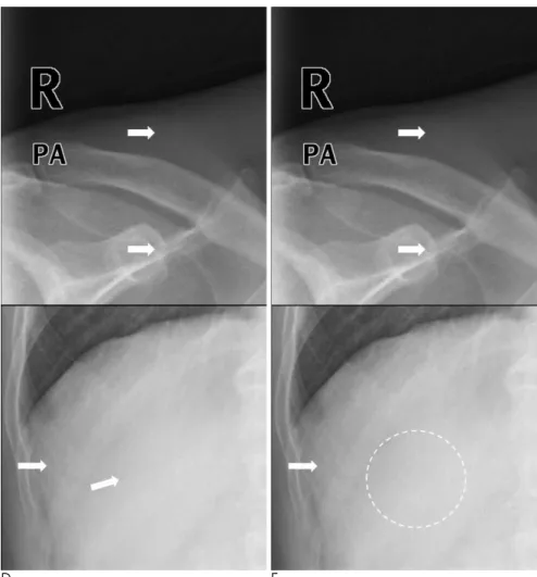
A B C
Fig. 1. JPEG 2000 compression artifacts in a chest radiograph image of a 59-year-old male with a mass in the right middle lung field.
Irreversibly 5:1 (B) and 10:1 (C) compressed images are indistinguishable from the original (A).
Results
Each reader rated 0-2% (0/50 to 1/50) of the image pairs for the reversible compression (as negative control) and 0-4% (0/50 to 2/50) of the image pairs for the irre- versible 5:1 compression having detectable differences between the compressed images and the original im- ages. 0-12% (0/50 to 6/50) of the image pairs for the 10:1 compression, 14-70% (7/50 to 35/50) of the image pairs for the 15:1 compression, and 58-96% (29/50 to 48/50) of the image pairs for the 20:1 compression had de- tectable difference between the compressed images and the original images (Table 2) (Fig. 1). Kappa statistics of the five readers’ responses was 0.686. Kappa statistics for each compression levels were also analyzed, but the kappa values were statistically not significant (p=0.607 -0.676).
Reader 5 rated none of the image pairs in the re- versible compression and irreversible 5:1 compression having detectable difference between the compressed images and the original images. Consequently, p-values
could not be calculated for comparisons between the re- versible and irreversible 5:1 compressions for reader 5.
For readers 1, 2, 3, and 4, the proportion of image pairs having detectable difference between the compressed images and the original images was not significantly dif- ferent between the reversible and irreversible 5:1 com- pressions. For irreversible 10:1 compression, the pro- portion increased in four out of five readers (reader 1, 2, 3, and 5), but the difference between reversible and irre- versible 10:1 compressions was not statistically signifi- cant for each reader.
However, the proportion significantly increased with irreversible 15:1 and 20:1 compressions, versus re- versible compression in all readers (p=0.027-p=7.4×
10-22). And we concluded VLT was considered to lie be- tween 10:1 and 15:1.
There was no significant difference in VLT between normal chest radiographs and chest radiographs with abnormal findings.
D E
Fig. 1. At a compression level of 15:1 (D), vertical linear artifact appears in the right part of the image (arrows). At a compres- sion level of 20:1 (E), the manifestation of the artifacts is apparent (arrows) and the internal texture of the soft tissue is de- graded (dashed circle). The compression artifacts are best demonstrated if the two images are alternately displayed on a sin- gle monochrome monitor calibrated ac- cording to the Digital Imaging and Communications in Medicine Gray Scale Standard Display Function.
Discussion
In our results, the overall response patterns of the five readers were similar and suggested that there was no difference in image quality between the compressed and the original images at 5:1 and 10:1 compression, while there was significant difference at 15:1 or greater compression levels. From these results, we estimate the VLT to be somewhere between 10:1 and 15:1 for chest radiograph images compressed using the JPEG 2000 al- gorithm.
Most previous studies have concerned the diagnosti- cally lossless threshold (7-9, 16). However, image com- pression artifacts can be detectable even though their presence does not affect the reader’s performance for a given diagnostic task (9, 10, 17). The presence of such perceivable artifacts possibly obscure ancillary findings (18) or induce false positive findings (13). Therefore, the threshold determined by the previous studies can ad- dress only narrowly defined diagnostic tasks (11, 12). To provide an acceptable threshold that covers a broad range of potential abnormalities with confidence, many receiver operating characteristic studies would be re- quired, which are time consuming and expensive (12).
This inefficiency of diagnostic lossless threshold and the conservativeness of VLT advocate the use of VLT in our study. Although the concepts of VLT yielded very con- servative threshold, we tried to include as diverse image findings as possible, and selected chest radiographs pos- sessing all specific abnormal findings from the glossary terms defined by the Fleischner Society.
Our image comparison method was intended to be as conservative as possible in any estimate of the visually lossless threshold. We used alternating presentation of registered images on the same monitor instead of pre- sentation in a side-by-side orientation, similar to that in Slone’s previous study, because the human visual sys- tem is naturally drawn to changes in structure or bright- ness (12). We believe this image comparison method, to- gether with the adoption of a visually lossless threshold, should result in a very conservative and, we hope, wide- ly accepted threshold for the compression level.
Therefore, the visually lossless threshold measured in this study is the minimum (baseline) of acceptable com- pression level, and should not be mistaken as an optimal compression level in practice.
In addition, we used the JPEG 2000 compression algo- rithm which is regarded as the most sensible choice in a
modern PACS (27), and brighter flat panel monitor as displaying device.
This study was conducted in the context of primary, rather than preliminary, interpretation of chest radi- ograph images, regardless of viewing tasks. Our results suggest that 10:1 JPEG 2000 compression is visually lossless for most images and is, therefore, potentially ac- ceptable for the primary interpretation of chest radi- ograph images with minimal risk negatively impacting on diagnosis, eliminating the need to maintain the origi- nal images as the diagnostic standard. Modern hospitals transmit original (non-compressed) images to worksta- tions for primary reading, and use reversible compres- sion for the image storage. Considering the large amount of image data, the practical benefits of 10:1 (as a mini- mum) irreversible compression over reversible com- pression (3.5:1 in this study) are not insignificant. This reduction in data (approximately 65%) would directly affect operational costs in transmission and storage.
The limitations of the present study are as follows.
First, the tested images did not include all potential ab- normal findings and anatomical variations.
Furthermore, the readers had to determine if the image pairs had detectable differences and did not compared all the selected abnormal findings and anatomical varia- tions of the tested images. However, we believe that our results would be reproducible even with a study sample containing other abnormalities, because our study de- sign was sensitive enough to detect perceptible com- pression artifacts, regardless of the image content.
Second, more studies are needed to further generalize our results, since the acceptable compression level can be affected by imaging parameters (30) and radiography systems. Third, loss of information is inevitable in irre- versible compression (either visually lossless or diagnos- tically lossless), and it can limit future use of the images, such as quantitative analysis. However, we focused on the adequacy of using irreversibly compressed images for primary interpretation, and such future use is out of our scope.
In conclusion, chest radiographs irreversibly com- pressed at a level of 10:1 using the JPEG 2000 algorithm are visually lossless and is therefore potentially accept- able for primary interpretation while does not impede diagnostic accuracy.
References
1. Rubin GD. Data explosion: the challenge of multidetector-row CT.
Eur J Radiol 2000;36:74-80
2. Rubin GD. 3-D imaging with MDCT. Eur J Radiol 2003;45 Suppl 1:S37-S41
3. Tamm EP, Thompson S, Venable SL, McEnery K. Impact of multi- slice CT on PACS resources. J Digit Imaging 2002;15 Suppl 1:96- 101
4. Lee KH, Lee HJ, Kim JH, Kang HS, Lee KW, Hong H, et al.
Managing the CT data explosion: initial experiences of archiving volumetric datasets in a mini-PACS. J Digit Imaging 2005;18:188- 195
5. Ko JP, Rusinek H, Naidich DP, McGuinness G, Rubinowitz AN, Leitman BS, et al. Wavelet compression of low-dose chest CT data:
effect on lung nodule detection. Radiology 2003;228:70-75 6. Ko JP, Chang J, Bomsztyk E, Babb JS, Naidich DP, Rusinek H.
Effect of CT image compression on computer-assisted lung nodule volume measurement. Radiology 2005;237:83-88
7. Ishigaki T, Sakuma S, Ikeda M, Itoh Y, Suzuki M, Iwai S. Clinical evaluation of irreversible image compression: analysis of chest imaging with computed radiography. Radiology 1990;175:739-743 8. Kido S, Ikezoe J, Kondoh H, Takeuchi N, Johkoh T, Kohno N, et al.
Detection of subtle interstitial abnormalities of the lungs on digi- tized chest radiographs: acceptable data compression ratios. AJR Am J Roentgenol 1996;167:111-115
9. MacMahon H, Doi K, Sanada S, Montner SM, Giger ML, Metz CE, et al. Data compression: effect on diagnostic accuracy in digital chest radiography. Radiology 1991;178:175-179
10. Mori T, Nakata H. Irreversible data compression in chest imaging using computed radiography: an evaluation. J Thorac Imaging 1994;9:23-30
11. Ohgiya Y, Gokan T, Nobusawa H, Hirose M, Seino N, Fujisawa H, et al. Acute cerebral infarction: effect of JPEG compression on de- tection at CT. Radiology 2003;227:124-127
12. Slone RM, Foos DH, Whiting BR, Muka E, Rubin DA, Pilgram TK, et al. Assessment of visually lossless irreversible image compres- sion: comparison of three methods by using an image-comparison workstation. Radiology 2000;215:543-553
13. Slone RM, Muka E, Pilgram TK. Irreversible JPEG compression of digital chest radiographs for primary interpretation: assessment of visually lossless threshold. Radiology 2003;228:425-429
14. Ringl H, Schernthaner RE, Kulinna-Cosentini C, Weber M, Schaefer-Prokop C, Herold CJ, et al. Lossy three-dimensional JPEG2000 compression of abdominal CT images: assessment of the visually lossless threshold and effect of compression ratio on image quality. Radiology 2007;245:467-474
15. Daly S. Application of a noise-adaptive contrast sensitivity func- tion to image data compression. Opt Eng 1990;29:977-987 16. Aberle DR, Gleeson F, Sayre JW, Brown K, Batra P, Young DA, et
al. The effect of irreversible image compression on diagnostic ac- curacy in thoracic imaging. Invest Radiol 1993;28:398-403 17. Savcenko V, Erickson BJ, Palisson PM, Persons KR, Manduca A,
Hartman TE, et al. Detection of subtle abnormalities on chest radi- ographs after irreversible compression. Radiology 1998;206:609- 616
18. Kalyanpur A, Neklesa VP, Taylor CR, Daftary AR, Brink JA.
Evaluation of JPEG and wavelet compression of body CT images for direct digital teleradiologic transmission. Radiology 2000;217:
772-779
19. Hansell DM, Bankier AA, MacMahon H, McLoud TC, Muller NL, Remy J. Fleischner Society: glossary of terms for thoracic imaging.
Radiology 2008;246:697-722
20. Kim KJ, Kim B, Choi SW, Kim YH, Hahn S, Kim TJ, et al.
Definition of compression ratio: difference between two commer- cial JPEG2000 program libraries. Telemed J E Health 2008;14:350- 354
21. Lee KH, Kim YH, Kim BH, Kim KJ, Kim TJ, Kim HJ, et al.
Irreversible JPEG 2000 compression of abdominal CT for primary interpretation: assessment of visually lossless threshold. Eur Radiol 2007;17:1529-1534
22. Woo HS, Kim KJ, Kim TJ, Hahn S, Kim B, Kim YH, et al. JPEG 2000 compression of abdominal CT: difference in tolerance be- tween thin- and thick-section images. AJR Am J Roentgenol 2007;189:535-541
23. Kim KJ, Kim B, Lee KH, Kim TJ, Mantiuk R, Kang HS, et al.
Regional difference in compression artifacts in low-dose chest CT images: effects of mathematical and perceptual factors. AJR Am J Roentgenol 2008;191:W30-37
24. Kim B, Lee KH, Kim KJ, Mantiuk R, Kim HR, Kim YH. Artifacts in slab average-intensity-projection images reformatted from JPEG 2000 compressed thin-section abdominal CT data sets. AJR Am J Roentgenol 2008;190:W342-W350
25. Kim B, Lee KH, Kim KJ, Mantiuk R, Hahn S, Kim TJ, et al.
Prediction of perceptible artifacts in JPEG 2000-compressed chest CT images using mathematical and perceptual quality metrics.
AJR Am J Roentgenol 2008;190:328-334
26. Kim TJ, Lee KH, Kim B, Kim KJ, Chun EJ, Bajpai V, et al. Regional variance of visually lossless threshold in compressed chest CT im- ages: lung versus mediastinum and chest wall. Eur J Radiol 2009;
69:483-488
27. Newcombe RG. Two-sided confidence intervals for the single pro- portion: comparison of seven methods. Stat Med 1998;17:857-872 28. Liddell FD. Simplified exact analysis of case-referent studies:
matched pairs; dichotomous exposure. J Epidemiol Community Health 1983;37:82-84
29. Fleiss JL, Cuzick J. The reliability of dichotomous judgements: un- equal numbers of judges per subjects. Appl Psychol Meas 1979;3:
537-542
30. Erickson BJ, Manduca A, Palisson P, Persons KR, Earnest F 4th, Savcenko V, et al. Wavelet compression of medical images.
Radiology 1998;206:599-607
대한영상의학회지 2010;63:371-378
JPEG 2000으로 압축한 디지털 흉부 X선 사진의 시각적 손실 없는 압축률1
1서울대학교병원 영상의학과, 서울대학교 의과대학 영상의학교실
2서울대학교 의과대학 방사선응용생명과학 협동과정
김 경 민∙김 길 중2
목적: JPEG 2000으로 압축한 디지털 흉부 X선 사진의 시각적 손실 없는 압축률을 추정한다.
대상과 방법: 50장의 디지털 흉부 X선 사진을 선택하여 다섯 수준 (가역적(대조군), 비가역적 5:1, 10:1, 15:1, 20:1)으로 압축하였다. 동일한 모니터 상에 짝지워진 원본 영상과 압축 영상을 교대로 표시하여, 5명의 영상의학과 의사가 독립적으로 압축된 영상과 원본 영상 사이에 차이가 있는지를 결정하게 하였다. 각각의 판독자에서 가역적 압축 및 4 수준의 비가역적 압축에 대하여 원본 영상과 압축 영상 사이에 차이가 있다고 판정한 분율을 짝지워진 분 율에 대한 정확 검정을 이용하여 비교하였다.
결과: 각 판독자에 대하여, 가역적 및 5:1, 10:1 압축 사이에서는 원본 영상과 압축 영상 사이에 차이가 있다고 판정 한 분율에 통계적으로 유의한 차이가 없었다. 그러나 그 분율은 15:1 및 20:1 압축 영상에서 모든 판독자에 대하여 유의하게 증가하였다 (p=0.027 - p=7.4×10⁻²²).
결론: 10:1로 압축된 흉부 X선 사진은 시각적으로 손실이 없는 것으로 간주할 수 있으며, 따라서 잠재적으로 일차 판독에 사용할 수 있을 것이다.