Printed in the Republic of Korea
http://dx.doi.org/10.5012/jkcs.2015.59.4.353 단신
(Notes)
Phenolic Compounds from the Branches of Malus sieboldii (Regel) Rehder and Their Antioxidative Activities
In Jeong Yang, Sang-Hee Byeon, Jong Seok Baik, and Nam Ho Lee*
Department of Chemistry and Cosmetics, Jeju National University, Ara-1, Jeju 690-756, Korea.
*E-mail: [email protected]
(Received May 15, 2015; Accepted June 4, 2015)
Key words: Malus sieboldii, Phenolic compounds, DPPH free radical scavenger, ABTS+ radical, Isolation, Structure determination
Malus is a genus of about 35-50 species of deciduous woody plants in the family Roseaceae that includes apple (M. pumila), a common orchard fruit. The species M. sie- boldii (Regel) Rehder is a shrub 2-6 m tall native to Japan and Korea.1 This plant has been used as an ornamental tree for its showy flowers in the spring. The extracts from the leaves of M. sieboldii have been reported to exhibit scav- enging activities on 2,2-diphenyl-1-picrylhydrazyl (DPPH) radicals2 and inhibition activities on nitric oxide production.3 However, as far as we know, no phytochemical study on M. sieboldii has yet been published.
Antioxidants exert protective effects in human health against oxidative damage associated with reactive oxygen species (ROS). ROS are chemically reactive substances that can attack lipids, proteins and nucleic acids within liv- ing organisms. ROS include free radicals (O2·−, HO·, RO·
and ROO·) and neutral molecules (1O2 and H2O2). Even though ROS generation is a normal metabolic process,4 its excessive formation can cause peroxidation in human tis- sues, ultimately leading to various diseases related with aging.5 When the skin is exposed to ultraviolet (UV) rays, ROS are generated in excess to induce aged-skin symptom such as wrinkles. In order to reduce the cumulative effects of ROS-related damage, the addition of antioxidants to food or cosmetic formulations has been considered.6 The development of novel antioxidative agents, especially from natural sources, has attracted attention in terms of their ecologically friendly properties.7
We are continuously conducting studies to find biolog- ically active natural products, especially for use in cos- metic formulations from plants in Jeju Island, a place of great biodiversity in Korea.8 Preliminary study indicated that the ethanol extract prepared from the branches of Malus sieboldii exhibited significant DPPH radical scavenging activities. Therefore, we decided to carry out a full phy- tochemical investigation of the M. sieboldii extract to
determine the bioactive metabolites. Described herein are the isolation and structural determination of the chemical constituents in the plant extract. In addition, the antioxi- dative properties of the isolates were examined using the DPPH and ABTS+ radical scavenging activity tests.
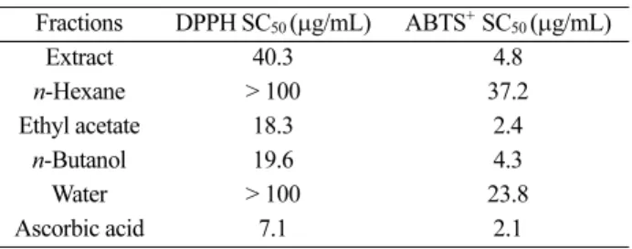
The ethanol extract (147.4 g) was prepared from the branches of M. sieboldii (1.0 kg) with a 14.7% yield. The extract suspended in water was partitioned successively to provide n-hexane, ethyl acetate (EtOAc), and n-butanol fractions. The extract and fractions were then tested for their anti-oxidative capacities using DPPH radical scav- enging assay first reported by Blois.9 Ascorbic acid show- ing an SC50 (50% radical scavenging concentration) value of 7.1 μg/mL was used as a positive control in this exper- iment.
As shown in Table 1, the ethanol extract exhibited consid- erable anti-oxidative activity with an SC50 value of 40.3 μg/mL.
Among the solvent fractions, the EtOAc soluble layer dis- played the highest potency with SC50 of 18.3 μg/mL. The n-butanol fraction also exhibited considerable activity, whereas the n-hexane and water portions showed very poor antioxidative activities. As another method to deter- mine the in vitro anti-oxidation capacities for the extracts, an assay using ABTS [2,2’-azino-bis(3-ethylbenzothiazo- line-6-sulfonic acid)] was used.10 ABTS is a chemical that can be oxidized by persulfate to yield an ABTS radical
Table 1. DPPH and ABTS+ radical scavenging activities (SC50) of the solvent fractions
Fractions DPPH SC50 (µg/mL) ABTS+ SC50 (µg/mL)
Extract 40.3 4.8
n-Hexane > 100 37.2
Ethyl acetate 18.3 2.4
n-Butanol 19.6 4.3
Water > 100 23.8
Ascorbic acid 7.1 2.1
cation (ABTS·+) featuring a characteristic blue green color in solution. In the presence of antioxidants such as phe- nolic compounds, this radical cation is converted back to its colorless non-radical form, which can be monitored by a spectrophotometer. After assaying the ethanol extract and solvent fractions from M. sieboldii, the ethanol extract, as well as EtOAc and n-butanol fractions, exhibited very strong radical scavenging activities (Table 1). The activity of the EtOAc fraction was comparable to that of the positive control, ascorbic acid.
Polyphenol compounds such as tannins and flavonoids are found in virtually all plant families. Polyphenols func- tion as natural antioxidants via the scavenging of reactive oxygen species. As the solvent fractions from M. sieboldii exhibited DPPH radical inhibition activities, the total polyphenolic contents were measured for their extract and fractions (Fig. 1). The amount of total phenols was quan- tified by colorimetric measurement using the Folin-Cio- calteu (FC) assay,11 which measures the change in color from yellow of the FC reagent to dark blue in the presence of antioxidant samples. The gallic acid equivalent (GAE) per 100 mg sample was calculated based on the calibration curve. As shown in Fig. 1, the ethanol extract was found to have 19.1 mg GAE polyphenols. Comparing the solvent fractions, the EtOAc fraction exhibited the highest poly- phenol concentration with 45.3 mg GAE.
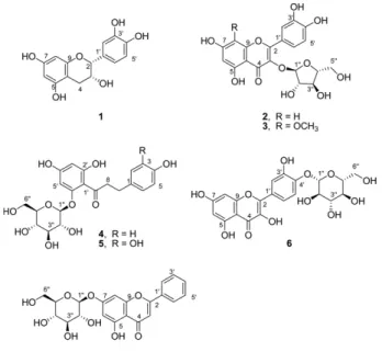
As the EtOAc-soluble layer showed the highest radical scavenging activities and polyphenol contents, it was cho- sen for further fractionation to identify the active constit- uents. The EtOAc fraction was subjected to vacuum liquid chromatography with gradients of n-hexane/EtOAc fol- lowed by EtOAc/methanol to provide 37 fractions. Each fraction was purified by repeated column chromatogra- phy with silica gel or Sephadex LH-20, leading to the iso- lation of seven compounds (1-7) (Fig. 2). The chemical structures of the isolates were characterized by spectro-
scopic data including 1D and 2D NMR spectra.
Compound 1 showed 15 signals in its 13C NMR spectrum, where twelve olefinic carbons and two oxygen-bearing sp3 carbons were easily identified. The presence of aromatic rings was also suggested by analysis of 1H and DEPT NMR signals. Diastereotopic methylene proton (H2-4) signals appeared at δ 2.73 and 2.86 with a large coupling constant (J = 16.7). Based on other spectroscopic data, compound 1 was verified as epi-catechin, a phenolic compound com- monly found in plant species.12 Compound 2 was estimated to possess 20 carbons according to 13C and DEPT NMR spectra. The data indicated that it contains 15 sp2 carbons, including one carbonyl (δ 179.9 ppm) and five oxygen- bearing sp3 carbons. In the 1H NMR spectrum, the meta- coupled protons (H-5 and H-7) appeared at δ 6.16 and 6.35 with J = 2.1. In addition, ABX spin system was observed for H-2’, H-5’ and H-6’. The signal of H-5’ (δ 6.89) showed an ortho-coupling to H-6’ (δ 7.48) with 3J = 8.3 Hz. The sig- nal of H-6’ exhibited meta- as well as ortho-coupling to H-2’
(δ 7.52) with 4J = 2.1 Hz. The 15 sp2 carbons and the cor- responding proton signals in 2 suggested a skeleton com- prised of a flavonol, quercetin. A pentose attached to the quercetin was surmised as arabinose with α-configura- tion, as determined by its coupling constant (J = 3.0 Hz) of the anomeric proton (H-1”, δ 5.45). Compound 2 was, therefore, identified as quercetin 3-O-α-arabinofuranoside.13 Compound 3 showed 18 signals, including 15 sp2 carbons and six oxygen-bearing sp3 carbons, in the 13C and DEPT spectra. The NMR study revealed that the B ring of quer- Figure 1. Total polyphenol contents for the fractions from M.
sieboldii.
Figure 2. Structures of compounds isolated from M. sieboldii.
cetin was appeared in the structure of 3. In addition, the arabinose unit shown in 2 was also detected in 3. Com- parative analysis of the spectra of 2 and 3 suggested that 3 has one additional methoxy group (δc 62.0) attached in the A ring of compound 2. The methoxy group was assigned by the observation of the heteronuclear multiple bond cor- relation (HMBC) cross peak between 8-OCH3 (δH 3.88) and C-8 (δC 129.5). Therefore, compound 3 was identified as 8-methoxyquercetin 3-O-α-arabinofuranoside. Although, the structure of flavonol glycoside 3 has been identified from a plant Dryas octopetala,14 as far as we know, its carbon NMR data has not been published. In this study, we com- pletely assigned the proton and carbon peaks for 3 using 1D and 2D NMR spectra. Compound 4 showed 19 peaks in the 13C NMR spectrum, corresponding to one carbonyl (δ 206.6, C-9), three methylene (δ 62.6, 48.7, 30.9) and five oxygenated methine carbons. Inspection of 1H NMR determined the para-substituted symmetric benzene ring with signals at δ 6.52 (2H, d, J = 8.5 Hz) and 6.90 (2H, d, J
= 8.5 Hz). In addition, meta-coupled protons at d 6.02 (H- 3’) and 5.79 (H-5’) with J = 2.3 Hz were also assigned in another benzene ring. The presence of a sugar glucose was revealed by the 1H and 13C NMR spectra. Combination of the observation data enabled compound 4 to be identified as phlorizin (phloretin 2’-O-β-D-glucopyranose).15 Com- pound 5 showed very similar 1H and 13C signals in the NMR spectra to those of compound 4. The only difference observed was that the symmetry of a benzene ring in 4 disappeared in compound 5. It was suggested that compound 5 has an additional hydroxyl group in the aromatic ring at C-3. Com- pound 5 was identified as 3-hydroxyphlorizin.16 Examination of the NMR spectra revealed compound 6 to be comprised of a skeleton of quercetin, as observed in 2. The presence of an additional sugar unit, glucose, was suggested in 6 by the observation of peaks for anomeric proton (δH 4.91) and carbon (δC 104.0), along with oxygenated methylene and methynes. The β-configuration of the glucose was estab- lished by the coupling constant (J = 7.3 Hz) of the anomeric proton. The position of the sugar was determined at C-4’
by comparison of the NMR data to the literature values.
Thus, compound 6 was identified as quercetin 4’-O-β-D- glucopyranoside.17 Inspection of the NMR data indicated that compound 7 is also a glycoside bearing a glucose unit.
In the 13C NMR spectrum, the aglycone has 13 peaks, all of which are sp2 carbons. A carbonyl peak appeared at d 183.1 (C-4). A phenyl group was identified by proton peaks at d 7.89 (2H, H-2’, H-6’), 7.44 (2H, H-3’, H-5’) and 7.51 (1H, H-4’). Meta-coupled aromatic protons were observed at δ 7.01 (1H, H-6) and 6.87 (1H, H-8) with J = 1.2 Hz. A
singlet proton was observed at δ 7.13 (1H, H-3). Based on these data, along with the 13C NMR data, the compound was determined as chrysin glycoside, chrysin 7-O-β-D-gluco- pyranoside.18
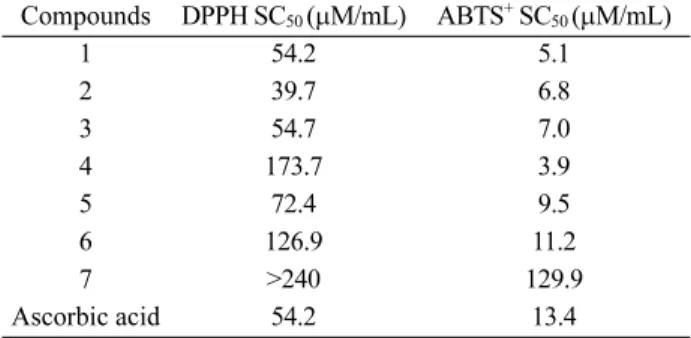
The antioxidative activities were examined for the iso- lated compounds 1-7 utilizing DPPH and ABTS radical scavenge methods. DPPH is a relatively stable radical spe- cies bearing a characteristic deep violet color in solution, and radical scavenging activity can be determined by a loss of DPPH absorbance at 515 nm. The inhibition activ- ities for the isolated compounds were measured at their concentrations of 3.0-100 μg/mL, and the results are sum- marized in Table 2. These activities were expressed as SC50, i.e., the concentration needed to reduce 50% of DPPH, and compared to ascorbic acid as a positive con- trol (SC50 54.2 μM). Among the isolates, quercetin 3-O- arabinoside (2) was determined to possess the highest free radical scavenging activity with SC50 of 39.7 μM, showing more potency than the positive control. The epi-catechin (1) and 8-methoxyquercetin glycoside (3) also exhibited strong inhibition activities with SC50 values of 54.2 and 54.7 μM, respectively. ABTS assay was used as an alter- native method to measure the antioxidative capacities for the isolated compounds and the results are summarized in Table 2. In this test, except compound 7, the remaining six compounds (1-6) were very strong scavengers against ABTS radicals. Compared to ascorbic acid (SC50 13.4 μM), isolates 1-6 had more potent activities with SC50 values below 11.2 M. Phlorizin (4) showed the most potent inhibition activity against ABTS radical, despite its relatively low activity on DPPH radical (see Table 2).
In conclusion, seven phenolic compounds were identi- fied from the ethanol extracts of M. sieboldii branches:
epi-cathechin (1), quercetin 3-O-α-arabinofuranoside (2), 8-methoxyquercetin 3-O-α-arabinofuranoside (3), phlo- rizin (4), 3-hydroxyphlorizin (5), quercetin 4’-O-β-D-glu- copyranoside (6) and chrysin 7-O-β-D-glucopyranoside
Table 2. SC50 values of DPPH and ABTS+ radical scavenging activities of compounds 1-7 isolated from M. sieboldii
Compounds DPPH SC50 (µM/mL) ABTS+ SC50 (µM/mL)
1 54.2 5.1
2 39.7 6.8
3 54.7 7.0
4 173.7 3.9
5 72.4 9.5
6 126.9 11.2
7 >240 129.9
Ascorbic acid 54.2 13.4
(7). All seven compounds were isolated for the first time from this woody plant. Among the isolates, quercetin gly- coside 2 showed the most potent DPPH free radical scav- enging activity with an SC50 value of 39.7 μM, where ascorbic acid (SC50 54.2 μM) was used as a positive control. In the study of ABTS+ radical scavenging assay, compounds 1-6 possessed more potency than ascorbic acid (SC50 13.4 μM).
Based on these results, M. sieboldii extracts are expected to be useful antioxidative agents with potential applications in food and cosmetic industries.
EXPERIMENTAL
Reagents and instruments
The chemical reagents 2,2-diphenyl-1-picrylhydrazyl (DPPH) and 2,2’-azino-bis(3-ethylbenzothiazoline-6-sul- fonic acid) (ABTS) were purchased from Aldrich. All sol- vents used were of analytical grade. A Biochrom Libra S22 UV-visible spectrophotometer was used to screen the rad- ical inhibition activities. 1H (400 MHz) and 13C (100.6 MHz) nuclear magnetic resonance (NMR) spectra were recorded on a JNM-LA 400 instrument (JEOL) with chemical shift (d) data in ppm relative to the solvent used. Merck silica gel (0.063-0.2 mm) was used for normal phased column chromatography. Silica gel 60 F254 coated on aluminum plates by Merck was used for thin layer chromatography (TLC). Gel filtration chromatography (GFC) was performed using Sephadex LH-20 (25-100 mm) from Fluka.
Plant material
The branches of M. sieboldii were collected in Novem- ber 2010 from Halla Botanical Garden in Jeju Island, Korea. A voucher specimen (sample number 317) was deposited at the herbarium of the Department of Chemistry, Jeju National University.
Extraction and isolation
The shade dried M. sieboldii branches (1.0 kg) were cut into small pieces, and extracted with 70% ethanol (20 L) two times at room temperature for 24 h. The gummy extract (147.4 g) was obtained after concentration of the filtered solution. Part of the ethanol extract (30.0 g) was suspended in water (1.0 L), and fractionated into n-hexane (1.0 g), ethyl acetate (9.4 g), n-butanol (7.0 g) and water (10.6 g) portions.
A portion (4.5 g) of the ethyl acetate (EtOAc) layer was subjected to vacuum liquid chromatography (VLC) on silica gel, using step-gradients (n-hexane/EtOAc to EtOAc/MeOH, 300 mL each) to provide 37 fractions (V1-V37). Fraction V15 was purified by silica gel column chromatography
(CC) using CHCl3-MeOH (3:1) eluent to afford compound 1 (18.8 mg). Fraction V23 (1.0 g) was column chromato- graphed over Sephadex LH-20 using CHCl3-EtOAc-MeOH (2:2:1) to give 12 subfractions (V-23-1 to V23-12), and V- 23-4 was identified to be compound 3 (24.0 mg). Fraction V-23-7 was purified over Sephadex LH-20 using CHCl3- EtOAc-MeOH-H2O (2:2:1:0.2) to give compounds 2 (1.2 mg) and 4 (8.9 mg). Compound 7 (77.1 mg) was obtained by recrystallization over MeOH from fraction V24. The MeOH- soluble V24 fraction (V24-M, 1.3 g) was subjected to VLC on silica gel using CHCl3-MeOH (100:0 to 50:50, step-gra- dients, 300 mL each) to provide five fractions (V24-M1 to V24-M5). Fraction V24-M4 was purified over Sephadex LH-20 using CHCl3-EtOAc-MeOH-H2O (2:2:1:0.2) to give compound 5 (42.0 mg). Fraction V24-M5 was recrystallized over MeOH to afford compound 6 (104.2 mg).
Compound 1: 1H NMR (400 MHz, CD3OD) δ 6.97 (1H, d, J = 1.8 Hz, H-2’), 6.80 (1H, dd, J = 8.2, 1.8 Hz, H-5’), 6.75 (1H, d, J = 8.2 Hz, H-6’), 5.94 (1H, d, J = 2.3 Hz, H- 6), 5.91 (1H, d, J = 2.3 Hz), 8.81 (1H, brs, H-2), 4.17 (1H, m, H-3), 2.86 (1H, dd, J = 16.7, 4.6 Hz, H-4), 2.73 (1H, dd, J = 16.7, 2.8 Hz, H-4); 13C NMR (100 MHz, CD3OD) δ 158.1 (C-5), 157.7 (C-7), 157.7 (C-9), 146.0 (C-3’), 145.8 (C-4’), 132.4 (C-1’), 119.5 (C-6’), 116.0 (C-5’), 115.4 (C-2’), 100.1 (C-10), 96.4 (C-8), 96.0 (C-6), 79.9 (C-2), 67.6 (C- 3), 29.4 (C-4).
Compound 2: 1H NMR (400 MHz, CD3OD) δ 7.52 (1H, d, J = 2.1 Hz, H-2’), 7.48 (1H, dd, J = 8.3, 2.1 Hz, H-6’), 6.89 (1H, d, J = 8.3 Hz, H-5’), 6.35 (1H, d, J = 2.1 Hz, H-8), 6.17 (1H, d, J = 2.1 Hz, H-6), 5.45 (1H, d, J = 3.0, H-1’’), 4.32 (1H, dd, J = 3.0, 0.9 Hz, H-2”), 3.89−3.91 (2H, m, H-3”, H-4”), 3.50 (2H, m, H-5”). 13C NMR (100 MHz, CD3OD) δ 179.9 (C-4), 167.8 (C-7), 163.1 (C-5), 159.2 (C-9), 158.8 (C-2), 150.1 (C-4’), 146.5 (C-3’), 134.8 (C-3), 123.2 (C- 6’), 123.0 (C-1’), 116.8 (C-2’), 116.5 (C-5’), 109.5 (C-1”), 105.2 (C-10), 100.5 (C-6), 95.2 (C-8), 88.1 (C-4”), 83.4 (C-2”), 78.8 (C-3”), 62.6 (C-5”).
Compound 3: 1H NMR (400 MHz, CD3OD) δ 7.59 (1H, d, J = 2.1 Hz, H-2’), 7.56 (1H, dd, J = 8.3, 2.1 Hz, H-6’), 6.94 (1H, d, J = 8.3 Hz, H-5’), 6.23 (1H, s, H-6), 5.48 (1H, d, J = 3.0 Hz, H-1’’), 4.34 (1H, dd, J = 3.0, 0.9 Hz, H-2”), 3.89−3.92 (2H, m, H-3”, H-4”), 3.88 (3H, s, 8-OCH3), 3.52 (2H, m, H-5”). 13C NMR (100 MHz, CD3OD) δ 180.0 (C-4), 160.3 (C-5), 159.0 (C-2), 158.1 (C-7), 150.6 (C-9), 150.1 (C-4’), 146.5 (C-3’), 134.9 (C-3), 129.5 (C-8), 123.3 (C-1’), 123.1 (C-6’), 116.8 (C-2’), 116.6 (C-5’), 109.6 (C-1”), 105.1 (C-10), 100.7 (C-6), 88.3 (C-2”), 88.2 (C-4”), 78.8 (C-3”), 62.6 (C-5”), 62.0 (8-OCH3).
Compound 4: 1H NMR (400 MHz, CD3OD) δ 6.90 (2H,
d, J = 8.5 Hz, H-2 & H-6), 6.52 (2H, d, J = 8.5 Hz, H-3 &
H-5), 6.02 (1H, d, J = 2.3 Hz, H-3’), 5.79 (1H, d, J = 2.3 Hz, H-5’), 5.03 (1H, d, J = 7.3 Hz, H-1”), 3.73 (1H, m, H- 8), 3.56 (1H, dd, J = 12.0, 5.8 Hz, H-8), 3.23−3.31 (6H, m, H-2”, H-3”, H-4”, H-5”, H-6”). 13C NMR (100 MHz, CD3OD) δ 206.6 (C-9), 167.7 (C-6’), 166.3 (C-4’), 162.4 (C-2’), 156.4 (C-4), 134.0 (C-1), 130.5 (C-2 & C-6), 116.2 (C-3 & C-5), 106.8 (C-1’), 102.1 (C-1”), 98.5 (C-5’), 95.7 (C-3’), 78.6 (C-3”), 78.5 (C-5”), 74.8 (C-2”), 71.2 (C-4”), 62.5 (C-6”), 48.7 (C-8), 30.9 (C-7).
Compound 5: 1H NMR (400 MHz, CD3OD) δ 6.69 (1H, d, J = 2.0 Hz, H-2), 6.62 (2H, d, J = 8.0 Hz, H-5), 6.56 (1H, d, J = 8.0, 2.0 Hz, H-6), 6.17 (1H, d, J = 2.1 Hz, H- 3’), 5.95 (1H, d, J = 2.1 Hz, H-5’), 5.05 (1H, d, J = 7.3 Hz, H-1”), 3.91 (1H, dd, J = 9.5, 2.0 Hz, H-6”), 3.57 (1H, dd, J
= 9.5, 5.3 Hz, H-6”), 3.44 (2H, m, H-8), 3.24−3.33 (4H, m, H-2”, H-3”, H-4”, H-5”). 13C NMR (100 MHz, CD3OD) δ 206.6 (C-9), 167.7 (C-6’), 166.2 (C-6’), 162.4 (C-2’), 146.1 (C-4), 144.3 (C-3), 134.8 (C-1), 120.8 (C-6), 116.7 (C-5), 116.4 (C-2), 106.8 (C-1’), 102.1 (C-1”), 98.5 (C-5’), 95.5 (C-3’), 78.6 (C-5”), 78.5 (C-3”), 74.8 (C-2”), 71.9 (C-4”), 62.5 (C-6”), 48.5 (C-8), 31.2 (C-7).
Compound 6: 1H NMR (400 MHz, pyridine-d5) δ 7.70 (1H, d, J = 2.0 Hz, H-2’), 7.62 (1H, dd, J = 8.7, 2.0 Hz, H- 6’), 7.23 (1H, d, J = 8.7 Hz, H-5’), 6.31 (1H, d, J = 1.8 Hz, H-8), 6.14 (1H, d, J = 1.8 Hz, H-6), 4.91 (1H, d, J = 7.3 Hz, H-1”), 3.94 (1H, dd, J = 12.0, 1.8 Hz, H-6”), 3.76 (1H, J = 12.0, 5.1 Hz, H-6”), 3.45−3.57 (4H, m, H-2”, H-3”, H-4”, H-5”). 13C NMR (100 MHz, pyridine-d5) δ 178.0 (C-4), 166.2 (C-7), 162.9 (C-5), 158.0 (C-9), 149.4 (C-2), 148.6 (C-3’), 147.2 (C-4’), 139.1 (C-3), 128.0 (C-1’), 120.8 (C-6’), 118.3 (C-5’), 117.7 (C-2’), 105.0 (C-10), 104.0 (C-1”), 99.8 (C-6), 94.9 (C-8), 79.7 (C-5”), 79.0 (C-3”), 75.4 (C-2”), 71.6 (C- 4”), 62.8 (C-6”).
Compound 7: 1H NMR (400 MHz, pyridine-d5) δ 7.89 (2H, d, J = 7.6 Hz, H-2’, H-6’), 7.51 (1H, t, J = 7.3 Hz, H- 4’), 7.44 (2H, dd, J = 7.6, 7.3 Hz, H-3’, H-5’), 7.13 (1H, s, H-3), 7.01 (1H, brs, H-6), 6.87 (1H, brs, H-8), 5.88 (1H, d, J = 7.3 Hz, H-1”), 4.60 (1H, dd, J = 12.0, 1.8 Hz, H-6”), 4.43 (1H, dd, J = 12.0, 6.3 Hz, H-6”), 4.38 (1H, t, J = 8.5 Hz, H-3”), 4.38 (1H, t, J = 8.5 Hz, H-4”), 4.37 (1H, m, H- 5”), 4.25 (1H, m, H-2”). 13C NMR (100 MHz, pyridine-d5) δ 183.1 (C-4), 164.5 (C-2), 164.4 (C-7), 162.7 (C-5), 158.2 (C-9), 132.4 (C-4’), 131.7 (C-1’), 129.6 (C-3’ & C-5’),
127.0 (C-2’ & C-6’), 106.9 (C-10), 106.5 (C-3), 101.9 (C- 1”), 101.1 (C-6), 95.6 (C-8), 79.4 (C-5”), 78.6 (C-3”), 75.0 (C-2”), 71.3 (C-4”), 62.5 (C-6”).
Acknowledgments. This research was financially sup- ported by the Ministry of Trade, Industry and Energy (MOTIE), Korea Institute for Advancement of Technology (KIAT), and Jeju Institute for Regional Program Evaluation through the Leading Industry Development for Economic Region.
REFERENCES
1. Lee, Y. N. Flora of Korea, Kyo Hak Publishig: Seoul, 1996; p 354.
2. Fu, F.; Lu, R. Chinese Bull. Botany, 2004, 21, 74.
3. Yang, E. J.; Yim, E. Y.; Song, G. P.; Kim, G. O.; Hyun, C.
G.. Interdisciplinary Toxicology, 2004, 2, 245.
4. Nickel, A.; Kohlhaas, M.; Maack. J. Mol. Cell Cardiol.
2014, 73, 26.
5. Denisov, E. T.; Afanas’ev, I. B. Free radicals and oxidative stress in pathophysiological processes. In Oxidation and antioxidants in organic chemistry and biology, CRC Press:
Boca Raton, 2005; pp 905-950.
6. Mukherjee, P. K.; Maity, N.; Nema, N. K.; Sakar, B. K.;
Phytomedicine, 2011, 19, 64.
7. Pietta, P. G. J. Nat. Prod. 2000, 63, 1035.
8. (a) Kim, S. S.; Hyun, C. G.; Choi, Y. H.; Lee, N. H. J Enz Inhibit Med Chem. 2013, 28, 685. (b) Kim, S. Y.; Kim, J.
E.; Bu, H. J.; Hyun, C. G.; Lee, N. H. Nat. Prod. Com- mun. 2014, 9, 1683.
9. Blois, M. S. Nature, 1958, 26, 1199.
10. Huang, D.; Boxin, O.; Prior, R. L. J. Agric. Food Chem.
2005, 53, 1841.
11. Folin, O.; Ciocalteu, V. J. Biol. Chem. 1927, 73, 627.
12. Kanwal, Q.; Hussain, I.; Siddiqui, H. L.; Javaid, A J. Serb.
Chem. Soc. 2011, 76, 375.
13. Kim, S. M.; Kang, K. S.; Jho, C. H.; Jung, Y. J.; Nho, C.
W.; Um, B. H.; Pan, C. H. Phytother. Res. 2011, 25, 1011.
14. Servettaz, O.; Laura, C. M.; Bernardi, M.; Uberti, E.; Vidari, G.; Vita-Finzi, P. J. Nat. prod. 1984, 47, 809.
15. Hilt, P.; Schieber, A.; Yildirim, C.; Arnold, G.; Klaiber, I.;
Conrad, J.; Beifuss, U.; Carle, R. J. Agric. Food Chem.
2003, 51, 2896.
16. Lu, Y.; Foo, L. Y. Food Chem. 1997, 59, 187.
17. Fossen, T.; Pedersen, A. T.; Andersen, Q. M. Phytochem, 1998, 47, 281.
18. Pereira, O. R.; Artur, S. M. S.; Domingues, M. R. M.; Car- doso, S. M. Food Chem. 2012, 131, 652.