www.krspine.org
Fistula Formation Between the Disc and Dura after Percutaneous Endoscopic Lumbar Discectomy
- A Case Report -
Hak Sun Kim, M.D., Hyoung Bok Kim, M.D., Hoon-Jae Chung, M.D., Jea Ho Yang, M.D.
J Korean Soc Spine Surg 2018 Dec;25(4):180-184.
Originally published online December 31, 2018;
https://doi.org/10.4184/jkss.2018.25.4.180
Korean Society of Spine Surgery
Asan Medical Center 88, Olympic-ro 43 Gil, Songpa-gu, Seoul, 05505, Korea Tel: +82-2-483-3413 Fax: +82-2-483-3414
©Copyright 2017 Korean Society of Spine Surgery pISSN 2093-4378 eISSN 2093-4386
The online version of this article, along with updated information and services, is located on the World Wide Web at:
http://www.krspine.org/DOIx.php?id=10.4184/jkss.2018.25.4.180
This is an Open Access article distributed under the terms of the Creative Commons Attribution Non-Commercial License (http://
creativecommons.org/licenses/by-nc/4.0) which permits unrestricted non-commercial use, distribution, and reproduction in any medium, provided the original work is properly cited.
Journal of Korean Society of
Spine Surgery
Fistula Formation Between the Disc and Dura after Percutaneous Endoscopic Lumbar Discectomy - A Case Report -
Hak Sun Kim, M.D., Hyoung Bok Kim, M.D.
*, Hoon-Jae Chung, M.D.
*, Jea Ho Yang, M.D.
Department of Orthopaedic Surgery, Gangnam Severance Hospital, Yonsei University College of Medicine, Seoul, Korea
*Department of Orthopedic Surgery, Bumin Hospital, Seoul, Korea
Study Design: Case report
Objectives: To document fistula formation between the disc and dura by an unrecognized dural tear after percutaneous endoscopic lumbar discectomy (PELD).
Summary of Literature Review: The risk of durotomy is relatively low with PELD, but cases of unrecognized durotomies have been reported. An effective diagnostic tool for such situations has not yet been identified.
Materials and Methods: A patient twice underwent transforaminal PELD under the diagnosis of a herniated lumbar disc at L4-5.
She still complained of intractable pain and motor weakness around the left lower extremity at 6 months postoperatively. Magnetic resonance imaging showed no specific findings suggestive of violation of the nerve root. However, L5 and S1 nerve root injury was noted on electromyography. An exploratory operation was planned to characterize damage to the neural structures.
Results: In the exploration, a dural tear was found at the previous operative site, along with a fistula between the disc and dura was also found at the dural tear site. The durotomy site was located on the ventrolateral side of the dura and measured approximately 5 mm.
The durotomy site was repaired with Nylon 5-0 and adhesive sealants. The patient’s preoperative symptoms diminished considerably.
Conclusions: Fistula formation between the disc and dura can be caused by an unrecognized dural tear after PELD. Discography is a reliable diagnostic tool for fistulas formed by an unrecognized durotomy.
Key Words: Percutaneous endoscopic lumbar discectomy, Dural tear, Fistula formation
Received: September 16, 2018 Revised: October 18, 2018 Accepted: November 16, 2018 Published Online: December 31, 2018 Corresponding author: Hyoung Bok Kim, M.D.
ORCID ID: Hak Sun Kim: https://orcid.org/0000-0002-8330-4688 Hyoung Bok Kim: https://orcid.org/0000-0001-6798-6386 Department of Orthopaedic Surgery, Bumin Hospital, 389, Gonghang-daero, Gangseo-gu, Seoul, 07590, Korea
TEL: +82-2-2620-0003, FAX: +82-2-2620-0100 E-mail: esshappy@daum.net
Percutaneous endoscopic lumbar discectomy (PELD) is considered a safe and minimally invasive procedure for herniated lumbar discs.1) This procedure is less likely to lead to complications, such as dural sac retraction and subsequent durotomy, than open discectomy.2-4) In contrast to the posterior interlaminar approach, the posterolateral transforaminal approach permits access to the herniated fragment without dural sac retraction.5) Hence, the transforaminal approach is associated with a lower risk of dural injury. Nonetheless, a few cases of incidental durotomy during PELD have recently been reported.6) Owing to atypical symptoms and ambiguity in the postoperative MRI, the diagnosis of unrecognized durotomy is always a challenge before revision surgery.6) We report a case in which intra-operative discography was used to establish a diagnosis of unrecognized durotomy, which occurred during a previous PELD; and fistula formation between the
intervertebral disc and the dura.
Case report
At a local clinic, a 50-year-old woman presented with left buttock pain radiating down to her left lower extremity.
Fistula Formation Between Disc and Dura Journal of Korean Society of Spine Surgery
www.krspine.org 181 MRI revealed left-sided L4-5 disc extrusion with nerve
root impingement. The patient underwent left-sided PELD with the transforaminal approach but complained of pain radiating down to her left lower extremity after surgery.
Despite conservative management, including neuroplasty and medication for about 3 months, her symptoms did not resolve.
She then underwent PELD again, but radiculopathy of the left lower extremity persisted, so she visited our spine clinic.
The patient had left lower extremity radiculopathy with a visual analogue score (VAS) of 9 and left buttock pain with a VAS score of 8 in the area of the L5 and S1 dermatome.
A straight leg raising test could not be performed due to the severe radiculopathy. The motor power of the left ankle in dorsiflexion and the great toe in extension was Grade IV.
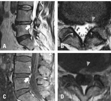
Plain radiographs showed mild disc space narrowing in the L4-5 area. On MRI, the T2-weighted image revealed hyperintense signal changes in the annulus fibrosus at the site of the previous discectomy, and the gadolinium-enhanced T1- weighted image revealed mild enhancement of the annulus fibrosus at the site of the previous discectomy. No definitive nerve root sleeve violation by the disc material was observed
(Fig. 1). Electromyography (EMG) revealed mild to moderate denervation potentials in the left L5 paraspinal muscles and moderate to severe denervation potentials in the left S1 paraspinal muscles. An exploratory surgery was planned to confirm the neural tissue damage evident on EMG.
The patient underwent a revision of the previous operative site through an open posterior approach. Intraoperative discography was performed to evaluate the status of the intervertebral disc. The injection of contrast media confirmed the presence of a fissured disc, and contrast media leakage was noted from the intervertebral disc into the dura, just like myelogram (Fig. 2). We suspected that a left ventro-lateral dural injury occurred during a previous PELD and resulted in a fistula between the intervertebral disc and the dura. Careful dissection revealed many adhesions and considerable scar tissue around the disc, dura, and nerve root. In the inferolateral region of the disc space, a thin membranous adhesion was noted between the disc and the dura. Dissection of the scar tissue revealed leakage of cerebrospinal fluid (CSF). At the site of CSF leakage, a fistula had formed between the fissured disc and the dura. Further facectectomy and extended laminectomy was done for dural repair. Durotomy site located on ventro- lateral side of dura and size was about 5mm. The durotomy site was repaired with Nylon 5-0 and adhesive sealants. After repair, there was no more CSF leakage or herniation of the nerve root. Posterior and posterolateral fusion was performed to stabilize the lumbar spine. Regular follow-ups revealed no evidence of CSF leakage. The patient’s preoperative symptoms improved with VAS 4 after 2 weeks postoperatively. After 6
Fig. 1. Magnetic resonance imaging (MRI) findings. (A, B) T2-weighted MRI images showing hyperintense signal changes in the annulus fibrosus at the previous discectomy site (arrow, sagittal view; arrowhead, axial view). (C, D) Gd-enhanced T1-weighted images showing mild enhance- ment of the annulus fibrosus at the previous discectomy site (arrow, sag- ittal view; arrowhead, axial view). Nerve root sleeve violation by the disc material was not observed.
Fig. 2. Discography findings. (A) Discography showing the fissured L4-5 disc and leakage of contrast media into the dura (arrow). (B) Intraopera- tive anteroposterior radiographs showing leakage of contrast media from the disc to the left side of the dura (arrowhead).
A
C
B
D
A B
weeks, VAS score was 2. The patient’s VAS score improved to 1 without any neurologic sequelae after 3 months.
This paper has been reviewed since IRB approval (201809- BM-003).
Discussion
Although iatrogenic durotomy is a well-known complication of open lumbar spine surgery, reports of durotomy during PELD are rare.5,7) However, the transforaminal PELD procedure has evolved over time - from indirect decompression to selective fragmentectomy and direct neural decompression - and this has led to an increase in the occurrence of incidental durotomy. Patients with fibrotic adhesion, eroded dura, or large disc herniations are at greater risk of incidental durotomy.8)
In open posterior lumbar surgery, dural repair can be carried out concurrent to surgery. In PELD, endoscopic visualization is usually ineffective, so durotomy management is usually restricted to immediate open conversion and direct repair. It is also more difficult to detect a durotomy during PELD than during open surgery.6 Indeed, the incidence of durotomy after PELD is 1.1%, but there are twice as many cases of unrecognized durotomy as recognized durotomy.6) Since the PELD operative field is usually filled with irrigation fluid, a small amount of CSF leakage due to durotomy is difficult to detect, and the positive pressure of the irrigation fluid may prevent the rootlet from being exposed during PELD.6) In contrast to open spinal surgery, the cardinal signs of incidental durotomy are conspicuously absent after PELD because there is not enough space for collection of the CSF.6,9)
MRI is a valuable tool for diagnosis of CSF leakage because enlargement of the subdural space and tethered dura can be identified on MRI. In addition, MRI provides excellent resolution of the soft tissue and fluids, and it is very sensitive to CSF accumulation.10) Unfortunately, it is difficult to find a PELD-associated durotomy on MRI because there is not enough space for collection of the CSF.
In our study, we used discography to diagnose a patient with durotomy and fistula formation, but this diagnosis occurred six months after the patient’s first surgery. Due to the lack of a precise diagnostic tool to confirm the exact cause of intractable radiculopathy after PELD, prompt management of the durotomy was delayed, and the patient had to undergo an
unnecessary posterior fusion.
In cases of intractable radiculopathy after PELD, the un- derlying pathology could be caused by the recurrence of disease or incidental durotomy. In cases suggestive of du- rotomy, we recommend discography as a reliable diagnostic tool for detection of durotomy.
REFERENCES
1. Smith JS, Ogden AT, Shafizadeh S, et al. Clinical outcomes after microendoscopic discectomy for recurrent lumbar disc herniation. J Spinal Disord Tech. 2010 Feb;23(1):30-4.
DOI: 10.1097/BSD.0b013e318193c16c.
2. Hermantin FU, Peters T, Quartararo L, et al. A prospective, randomized study comparing the results of open discectomy with those of video-assisted arthroscopic microdiscec- tomy. J Bone Joint Surg Am. 1999 Jul;81(7):958-65. DOI:
10.2106/00004623-199907000-00008.
3. Kambin P, Brager MD. Percutaneous posterolateral dis- cectomy. Anatomy and mechanism. Clin Orthop Relat Res. 1987 Oct;(223):145-54. DOI: 10.1097/00003086- 198710000-00016.
4. Jang EC, Song KS, Kang KS, et al. Comparative evaluation of percutaneous endoscopic discectomy and microdiscec- tomy using tubular retractor system at L4-5 level. J Korean Soc Spine Surg. 2009 Sep;16(3):186-93. DOI: 10.4184/
jkss.2009.16.3.186.
5. Yeung AT, Tsou PM. Posterolateral endoscopic excision for lumbar disc herniation: surgical technique, outcome, and complications in 307 consecutive cases. Spine (Phila Pa 1976). 2002 Apr;27(7):722-31. DOI: 10.1097/00007632- 200204010-00009.
6. Ahn Y, Lee HY, Lee SH, et al. Dural tears in percutane- ous endoscopic lumbar discectomy. Eur Spine J. 2011 Jan;20(1):58-64. DOI: 10.1007/s00586-010-1493-8.
7. Hasegawa K, Yamamoto N. Nerve root herniation sec- ondary to lumbar puncture in the patient with lumbar canal stenosis. A case report. Spine (Phila Pa 1976). 1999 May;24(9):915-7. DOI: 10.1097/00007632-199905010- 00015.
8. Bosacco SJ, Gardner MJ, Guille JT. Evaluation and treat- ment of dural tears in lumbar spine surgery: a review.
Clin Orthop Relat Res. 2001 Aug;(389):238-47. DOI:
Fistula Formation Between Disc and Dura Journal of Korean Society of Spine Surgery
www.krspine.org 183 10.1097/00003086-200108000-00033.
9. Khan MH, Rihn J, Steele G, et al. Postoperative man- agement protocol for incidental dural tears during de- generative lumbar spine surgery: a review of 3,183 consecutive degenerative lumbar cases. Spine (Phila Pa 1976). 2006 Oct;31(22):2609-13. DOI: 10.1097/01.
brs.0000241066.55849.41.
10. Johnsin DB, Brennnan P, Toland J, et al. Magnetic reso- nance imaging in the evaluation of cerebrospinal fluid fistu- lae. Clin Radiol. 1996 Dec;51(12):837-41. DOI: 10.1016/
S0009-9260(96)80079-1.
경피적 내시경 요추 추간판 절제술 후 발생한 추간판과 경막 사이의 누공 형성 - 증례 보고 -
김학선 • 김형복* • 정훈재* • 양재호
연세대학교 의과대학 강남세브란스병원 정형외과학교실, *서울부민병원 정형외과
연구 계획: 증례 보고
목적: 경피적 내시경하 요추 추간판 절제술 후 인식하지 못한 경막 손상에 의해 생긴 추간판과 경막 사이의 누공에 대해 보고하고자 한다.
선행 연구문헌의 요약: 경피적 내시경하 요추 추간판 절제술시 경막 손상의 위험은 낮은 것으로 알려져 있으며, 인지하지 못한 경막 손상이 보고 되고 있 으나, 이를 정확히 진단할 수 있는 도구는 아직 알려지지 않고 있다.
대상 및 방법: 50세 여자 환자는 제 4-5요추 추간판 탈출증에 대해 두 차례 경피적 내시경하 요추 추간판 절제술을 시행 받았다. 하지만, 환자는 술 후 6개 월에도 지속적으로 좌측 하지에 참을 수 없는 동통과 운동 신경 마비를 호소하였다. 이에 신경 조직 손상을 찾기 위해 수술을 계획하였다.
결과: 이전 수술 부위에서 경막 손상이 발견되었으며, 또한 경막 손상 부위에서 추간판과 경막 사이에 누공이 형성된 것을 발견 하였다. 경막 손상 부위는 경막 전측부에 위치 하였으며, 크기는 5 mm 정도 였다. 5~0 나일론과 접착 봉합제로 경막 손상 부위를 봉합하였다. 술 후 환자의 술 전 증상은 호전되었 다.
결론: 경피적 내시경하 요추 추간판 절제술 후 인지하지 못한 경막 손상은 추간판과 경막 사이의 누공 형성의 원인이 될 수 있다. 추간판 조영술은 인지하 지 못한 경막 손상에 의해 생성된 누공에 대한 신뢰할 수 있는 진단 도구이다.
색인 단어: 경피적 내시경하 요추 추간판 절제술, 경막 손상, 누공 형성 약칭 제목: 추간판과 경막 사이의 누공 형성
접수일: 2018년 9월 16일 수정일: 2018년 10월 18일 게재확정일: 2018년 11월 16일 교신저자: 김형복
서울특별시 강서구 공항대로 389 서울부민병원 정형외과
TEL: 02-2620-0003 FAX: 02-2620-0100 E-mail: esshappy@daum.net