A Case of Therapy-related Acute Lymphoblastic Leukemia with t(11;19) (q23;p13.3) and MLL/MLLT1 Gene Rearrangement
Byong-Joon Yoo, M.D.1, Myung-Hyun Nam, M.D.1, Hwa-Jung Sung, M.D.2, Chae-Seung Lim, M.D.1, Chang-Kyu Lee, M.D.1, Yun-Jung Cho, M.D.1, Kap-No Lee, M.D.1, and Soo-Young Yoon, M.D.1
Departments of Laboratory Medicine1 and Hemato-Oncology2, Korea University College of Medicine, Seoul, Korea
Therapy-related ALL (t-ALL) is a rare secondary leukemia that develops after chemotherapy and/or radiotherapy for primary malig- nancies. Chromosomal 11q23 abnormalities are the most common karyotypic alterations in t-ALL. The t(11;19)(q23;p13) aberration is extremely rare and has not been confirmed at the molecular genetic level. Here, we report a case of t-ALL with t(11;19)(q23;p13.3) and MLL-MLLT1 (alias ENL) gene rearrangement confirmed by cytogenetic analysis, multiplex reverse transcription-PCR (multiplex RT-PCR), and DNA sequencing in a patient who had undergone treatment for breast cancer. A 40-yr-old woman developed acute leukemia 15 months after undergoing 6 cycles of adjuvant chemotherapy (doxorubicin 60 mg/m2 and cyclophosphamide 600 mg/m2), radiation therapy (dose, 5,900 cGy), and anticancer endocrine therapy with tamoxifen. The complete blood cell counts and bone marrow examination showed increased blasts and the blasts showed B lineage immunophenotype (positive for CD19, CD34, and cytoplasmic CD79a). Cytogenetic analysis revealed the karyotype 47,XX,+X,t(11;19)(q23;p13.3)[4]/46,XX[16]. FISH analy- ses, multiplex RT-PCR, and DNA sequencing confirmed the MLL-MLLT1 gene rearrangement. The patient underwent induction che- motherapy with fractionated cyclophosphamide, vincristine, doxorubicin, and dexamethasone (Hyper-CVAD) and achieved com- plete remission. Subsequently, she underwent consolidation chemotherapy, but died of brain ischemia in the pons and the region of the middle cerebral artery. To our knowledge, this is the first case report of t-ALL with t(11;19)(q23;p13.3) and the MLL-MLLT1 gene rearrangement.
Key Words: MLL gene rearrangement, Secondary leukemia, Therapy-related ALL, Breast cancer
Received: July 26, 2010 Manuscript No: KJLM-10-119 Revision received: October 4, 2010
Accepted: October 20, 2010
Corresponding author: Soo-Young Yoon, M.D.
Department of Laboratory Medicine, Korea University Ansan Hospital, Gojan 1-dong, Danwon-gu, Ansan 425-707, Korea
Tel: +82-31-412-5300, Fax: +82-31-412-5314, E-mail: labmd@korea.ac.kr ISSN 1598-6535 © The Korean Society for Laboratory Medicine.
This is an Open Access article distributed under the terms of the Creative Commons Attribution Non-Commercial License (http://creativecommons.org/licenses/by-nc/3.0) which permits unrestricted non-commercial use, distribution, and reproduction in any medium, provided the original work is properly cited.
INTRODUCTION
Therapy-related acute leukemia (t-AL) is a rare second- ary leukemia that develops after chemotherapy and/or ra- diotherapy for primary malignancies. It can be broadly di- vided into 2 major groups: alkylating agent/radiotherapy- related t-AL and topoisomerase II inhibitor-related t-AL.
Patients in the alkylating agent-related t-AL subgroup fre-
quently exhibit complete or partial deletion of chromosome 5 or 7 and generally have antecedent myelodysplasia with a mean latency period of 5–7 yr [1]. In contrast, patients in the DNA topoisomerase II inhibitor-related t-AL subgroup generally develop secondary leukemia with relatively short latency periods (1–5 yr). The most common characteristic chromosomal aberrations in this group are translocations involving 11q23, i.e., the mixed lineage leukemia (MLL) gene locus [1]. Therapy-related ALL (t-ALL) is much less frequent than therapy-related AML (t-AML) and accounts for approximately 12% of all t-AL cases and 1.2–4% of adult ALL cases [2, 3].
Chromosomal 11q23 abnormalities are considered the most common karyotypic alterations with a frequency of up to 46% in t-ALL; the most common abnormality is t(4;11)(q21;q23) [2, 4].
Here, we report a case of t-ALL with t(11;19)(q23;p13.3) and the MLL-MLLT1 (alias ENL) gene rearrangement con- firmed by cytogenetic analysis, multiplex reverse transcrip-
tion-PCR (multiplex RT-PCR), and DNA sequencing in a patient who had undergone treatment for breast cancer. To our knowledge, this is the first report of t-ALL with the MLL-MLLT1 gene rearrangement.
CASE REPORT
A 40-yr-old woman underwent breast surgery for cancer of the right breast and received 6 cycles of adjuvant chemo- therapy (doxorubicin 60 mg/m2 and cyclophosphamide 600 mg/m2) and radiation therapy (dose, 5,900 cGy), followed by anticancer endocrine therapy with tamoxifen. Complete blood cell counts obtained at 15 months after chemotherapy indicated acute leukemia: white blood cell count, 3.5 × 10⁹/ L with 15% blasts; Hb level, 5.8 g/dL; and platelet count, 31.0 × 10⁹/L. Bone marrow examination showed hypercel- lular marrow with increased blasts, accounting for up to 95%
of all bone marrow nucleated cells (Fig. 1). An immuno- phenotyping study using flow cytometry revealed that the blasts were positive for CD19, CD34, and cytoplasmic CD79a and negative for CD2, CD3, CD5, CD7, CD10, CD13, CD14, CD20, CD22, CD33, CD41, CD117, and myeloperoxidase.
On the basis of the immunophenotype of the blasts, we di- agnosed the patient with B-ALL. Cytogenetic analysis of the bone marrow aspirates by using the G-banding revealed the karyotype 47,XX,+X,t(11;19)(q23;p13.3)[4]/46,XX[16] (Fig.
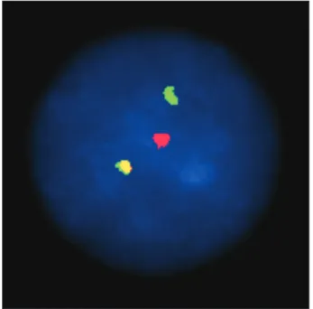
2). FISH performed using LSI MLL dual color, break apart rearrangement probes (Abbott Molecular, Des Plaines, IL, USA) showed an MLL break apart signal in 76.2% of the 500 analyzed cells (Fig. 3). The MLL-MLLT1 gene rearrange-
ment was confirmed by multiplex RT-PCR using the He- maVision kit (DNA Technology, Research Park, Aarhus, Denmark). However, 2 different amplicons (319 bp and 432 bp) were observed, and we identified the 2 amplicons by DNA sequencing. Both amplicons contained a t(11;19) (q23;p13.3)(MLL; MLLT1) translocation (Fig. 4). The patient underwent induction chemotherapy with Hyper-CVAD from March 2010. Complete remission was noted on the fol- low-up bone marrow study, and no MLL gene rearrangement was observed on follow-up FISH analysis. Subsequently, she underwent consolidation chemotherapy until June 2010;
however, she died because of brain ischemia in the pons and the region of the middle cerebral artery.
Fig. 1. Morphologic findings of B-ALL in the bone marrow. (A) Bone marrow aspirate smear shows moderate- to large-sized blasts with cytoplasmic vacuolation (Wright-Giemsa stain, ×1,000). (B) Bone marrow biopsy shows almost complete replacement of hypercellular marrow by blasts (hematoxylin and eosin stain,
×100).
A B
Fig. 2. G-banding of a bone marrow cell showing a 47,XX,+X,t(11;19) (q23;p13.3)[4]/46,XX[16] karyotype.
1
6
13
19 20 21 22 X Y
14 15 16 17 18
7 8 9 10 11 12
2 3 4 5
DISCUSSION
MLL rearrangement is one of the most frequently ob- served genetic abnormalities in patients with t-ALL. Ac- cording to a study summarizing t-ALL cases reported in the literature from 1992 to 2007, in 48 cases of t-ALL, an 11q23 abnormality involving the MLL gene locus was the pre- dominant chromosomal aberration (32/48 [67%]), followed by t(9;22) (6/48 [13%]) and a normal karyotype (4/48 [8%]) [5]. On comparing t-ALL cases with/without 11q23 abnor- malities (MLL group or non-MLL group), the distribution of primary malignancies between the 2 groups appeared to be fairly variable. Hematologic malignancies (mainly Hodg- kin’s lymphoma) were the most common primary neo- plasms in the non-MLL group (7/16 [44%]), whereas breast cancer was the most frequent primary tumor in the MLL group (12/32 [38%]). The latency period between the pri- mary malignancy and t-ALL in the non-MLL group was much longer than that in the MLL group (median, 36 vs. 19 months) [5]. A relatively short latency period, ranging from 12 months to 22 yr with a median latency of 16 months, has Fig. 3. Interphase FISH using the LSI MLL dual color, break apart rearrange-
ment probe (Abbott) set. Interphase nucleus harboring the MLL transloca- tion, 1 fusion signal, and 2 separate signals (1F1O1G).
Fig. 4. MLL1/ENL fusion transcript identified by multiplex reverse transcription-PCR. (A) Amplification products of 8 parallel multiplex reverse transcription-PCR reactions. Two bands (319 bp and 432 bp; arrow heads) are noted in lane 3. (B) Corresponding split-out reactions. Two positive bands in lane D represent the fusion transcript MLL1/ENL [t(11;19)(q23;p13.3)]. (Lanes 1 and A, 100-bp DNA ladder) (C) Part of the sequence from the 319-bp amplicon from lane D shows t(11;19) (q23;p13.3) MLL exon 9/MLLT1 exon 2. (D) Part of the sequence from the 432-bp amplicon from lane D shows t(11;19)(q23;p13.3) MLL exon 10/MLLT1 exon 2; pink, MLL exon 8; yellow, MLL exon 9; blue, MLL exon 10; green, 5’ end of MLLT1 exon 2.
A B
C
D A B C D E F
1 2 3 4 5 6 7 8 9
been reported in cases of t-ALL with MLL [4]. The primary malignancy in the present case was breast cancer, and the latency period was relatively short; this was compatible with the majority of previous findings for the MLL group. No significant differences were noted in the chemotherapy regi- men or survival between the 2 groups.
The MLL gene on chromosome 11q23 is known to have several fusion partners. The most common partner gene is AFF1 (alias AF4) on chromosome 4q21; other common partner genes include MLLT1 (alias ENL) on chromosome 19p13 and MLLT3 (alias AF9) on chromosome 9p22 [6].
The aberration t(4;11)(q21;q23) was noted in 67–89% of MLL-positive t-ALL cases, and the aberration t(1;11)(q21;
q23) was observed in 9–11% of cases [2, 4, 7, 8]. To date, only 2 cases of the aberration t(11;19)(q23;p13), i.e., involv- ing chromosome 19p13 similar to the present case, have been reported (Table 1) [9, 10].
The breakpoint on chromosome 19 is variable [11] and can occur at either p13.1 or p13.3 [12-14]. Three MLL part- ner genes on 19 p13 have been identified: ELL at 19p13.1, MLLT1 at 19p13.3, and SH3GL1 (alias EEN) on 19p13.3. A report evaluating hematologic malignancy involving chro- mosome 11q23 and chromosome 19p13 showed marked differences between t(11;19)(q23;p13.1) and t(11;19)(q23;
p13.3) [15]. Patients with t(11;19)(q23;p13.1) mostly had myeloid lineage leukemia with rare additional chromosomal changes and were predominantly adults. In contrast, patients with t(11;19)(q23;p13.3) had both myeloid and lymphoid lineage malignancy with up to 50% additional chromosomal changes and were mostly infants less than 1 yr of age. At times, the identification of the breakpoint on 19p13 by us- ing conventional G-banding may be difficult; therefore, in the case of MLL rearrangement involving 19p13, further specific banding techniques and molecular genetic testing must be performed to identify MLL abnormalities. In this patient too, the breakpoint on G-banding was unclear; there-
Table 1. Characteristics of the therapy-related ALL cases with t(11;19) abnormalities reported since 1992
Reference Age/
Gen-der Primary malignancy Previous treat-
ment Secondary
malignancy Latency period
(months) Karyotype Immunophenotype
Kobayashi et al. (1993) [10] 44/F Breast cancer Flu, Pirub B-ALL 19 46XX,t(11;19)(q23;p13) CD10-, CD19+, CD20-, HLA-DR+
Thandla et al. (1999) [9] 10/M Hepatocellular carcinoma Dox, Cis, Etop,
Carbo, Ifos T-ALL 48 t(1;13)(q4;q13),
t(11;19),+X,+5,+8,+13,+14 CD3-, CD4+, CD8+, CD2+, CD7+, CD5+, CD56+
Present case (2010) 40/F Breast cancer Dox, Ctx, RT, Tmf B-ALL 15 47,XX,+X,t(11;19)(q23;p13.3) CD10-, CD19+, CD20- , CD22-, CD34+, cCD79a+
Abbreviations: Flu, fludarabine; Pirub, pirarubicin; Dox, doxorubicin; Cis, cisplatin; Etop, etoposide; Carbo, carboplatin; Ifos, ifosfamide; Ctx, cyclophosphamide; RT, radiotherapy;
Tmf, tamoxifen.
fore, we performed multiplex RT-PCR and thereby identi- fied the MLL-MLLT1 rearrangement. Previously reported t- ALL cases with the aberration t(11;19)(q23;p13) had not been further confirmed at the molecular level, i.e., the re- gion of p13 on chromosome 19 that was involved was not clarified [9, 10].
Among patients with acute leukemia and MLL rearrange- ments, approximately 10% have been shown to have the MLL/MLLT1 rearrangement. Fu et al. [16] reported the fre- quency of the MLL/MLLT1 rearrangement to be 3.5%
(4/114) in MLL-positive de novo AML, 23% (7/30) in MLL- positive de novo B-ALL, and 100% (4/4) in MLL-positive de novo T-ALL. Therefore, the incidence of MLL/MLLT1 rear- rangement in t-ALL is significantly lower than that in de novo ALL.
The presence of the MLL/MLLT1 rearrangement appears to have certain clinical implications, such as on the progno- sis. For example, non-infants with the B-ALL and the MLL/
MLLT1 rearrangement have a favorable prognosis [17-21], whereas the prognosis of infants with B-ALL and the MLL/
MLLT1 rearrangement is controversial [16, 17]. Further studies are essential for clarifying the clinical implications of MLL/MLLT1 rearrangement in t-ALL patients.
In a majority of t-ALL cases with the MLL rearrangement, the leukemic cells typically have a pro-B immunophenotype and are positive for CD19 and the aberrant expression of CD15 and CD65 and are negative for CD10 and CD24 pat- terns [6, 8, 11]. Our patient too showed a CD19-positive and CD10-negative immunophenotype; however, we did not evaluate the CD15 and CD24 immunophenotypes.
In summary, to the best of our knowledge, this is the first case of t-ALL with the aberration t(11;19)(q23;p13.3) and the MLL/MLLT1 rearrangement that was confirmed by molecular genetic testing; moreover, this case exhibited fea- tures of a CD10-negative pro-B immunophenotype similar to other cases with the MLL rearrangement.
Authors’ Disclosures of Potential Conflicts of Interest No potential conflict of interest relevant to this article was reported.
REFERENCES
1. Zhang Y, Poetsch M, Weber-Matthiesen K, Rohde K, Winkemann M, Haferlach T, et al. Secondary acute leukaemias with 11q23 re- arrangement: clinical, cytogenetic, FISH and FICTION studies. Br J Haematol 1996;92:673-80.
2. Ishizawa S, Slovak ML, Popplewell L, Bedell V, Wrede JE, Carter NH, et al. High frequency of pro-B acute lymphoblastic leukemia in adults with secondary leukemia with 11q23 abnormalities. Leu- kemia 2003;17:1091-5.
3. Pagano L, Pulsoni A, Mele L, Leone G. Clinical and epidemiologi- cal features of acute lymphoblastic leukemia following a previous malignancy. Leuk Lymphoma 2000;39:465-75.
4. Secker-Walker LM, Moorman AV, Bain BJ, Mehta AB. Secondary acute leukemia and myelodysplastic syndrome with 11q23 abnor- malities. EU Concerted Action 11q23 Workshop. Leukemia 1998;
12:840-4.
5. Chen W, Wang E, Lu Y, Gaal KK, Huang Q. Therapy-related acute lymphoblastic leukemia without 11q23 abnormality: report of six cases and a literature review. Am J Clin Pathol 2010;133:75-82.
6. Hrusák O and Porwit-MacDonald A. Antigen expression patterns reflecting genotype of acute leukemias. Leukemia 2002;16:1233- 7. Andersen MK, Christiansen DH, Jensen BA, Ernst P, Hauge G, 58.
Pedersen-Bjergaard J. Therapy-related acute lymphoblastic leukae- mia with MLL rearrangements following DNA topoisomerase II inhibitors, an increasing problem: report on two new cases and re- view of the literature since 1992. Br J Haematol 2001;114:539-43.
8. Cho J, Hur M, Moon HW, Yun YM, Lee CH, Lee HG. A case of therapy-related ALL with MLL gene rearrangement following treatment of breast cancer. Korean J Lab Med 2010;30:255-9.
9. Thandla S, Alashari M, Green DM, Aplan PD. Therapy-related T cell lymphoblastic lymphoma with t(11;19)(q23;p13) and MLL gene rearrangement. Leukemia 1999;13:2116-8.
10. Kobayashi Y, Yang J, Shindo E, Tojo A, Tani K, Ozawa K, et al.
HRX gene rearrangement in acute lymphoblastic leukemia after adjuvant chemotherapy of breast cancer. Blood 1993;82:3220-1.
Kowarz E, et al. The MLL recombinome of adult CD10-negative B-cell precursor acute lymphoblastic leukemia: results from the GMALL study group. Blood 2009;113:4011-5.
12. Huret JL, Brizard A, Slater R, Charrin C, Bertheas MF, Guilhot F, et al. Cytogenetic heterogeneity in t(11;19) acute leukemia: clini- cal, hematological and cytogenetic analyses of 48 patients--up- dated published cases and 16 new observations. Leukemia 1993;
7:152-60.
13. Mitani K, Sato Y, Kobayashi Y, Shibasaki Y, Kasuga M, Inaba T, et al. Heterogeneity in the breakpoints of chromosome 19 among acute leukemic patients with the t(11;19)(q23;p13) translocation.
Am J Hematol 1989;31:253-7.
14. Hudson MM, Raimondi SC, Behm FG, Pui CH. Childhood acute leukemia with t(11;19) (q23;p13). Leukemia 1991;5:1064-8.
15. Moorman AV, Hagemeijer A, Charrin C, Rieder H, Secker-Walker LM. The translocations, t(11;19)(q23;p13.1) and t(11;19)(q23;p13.3):
a cytogenetic and clinical profile of 53 patients. European 11q23 Workshop participants. Leukemia 1998;12:805-10.
16. Fu JF, Liang DC, Shih LY. Analysis of acute leukemias with MLL/
ENL fusion transcripts: identification of two novel breakpoints in ENL. Am J Clin Pathol 2007;127:24-30.
17. Rubnitz JE, Camitta BM, Mahmoud H, Raimondi SC, Carroll AJ, Borowitz MJ, et al. Childhood acute lymphoblastic leukemia with the MLL-ENL fusion and t(11;19)(q23;p13.3) translocation. J Clin Oncol 1999;17:191-6.
18. Pui CH, Behm FG, Downing JR, Hancock ML, Shurtleff SA, Ri- beiro RC, et al. 11q23/MLL rearrangement confers a poor progno- sis in infants with acute lymphoblastic leukemia. J Clin Oncol 1994;12:909-15.
19. Rubnitz JE, Link MP, Shuster JJ, Carroll AJ, Hakami N, Frankel LS, et al. Frequency and prognostic significance of HRX rearrange- ments in infant acute lymphoblastic leukemia: a Pediatric Oncol- ogy Group study. Blood 1994;84:570-3.
20. Schoch C, Schnittger S, Klaus M, Kern W, Hiddemann W, Hafer- lach T. AML with 11q23/MLL abnormalities as defined by the WHO classification: incidence, partner chromosomes, FAB sub- type, age distribution, and prognostic impact in an unselected se- ries of 1897 cytogenetically analyzed AML cases. Blood 2003;102:
2395-402.
21. Pui CH, Chessells JM, Camitta B, Baruchel A, Biondi A, Boyett JM, et al. Clinical heterogeneity in childhood acute lymphoblastic leukemia with 11q23 rearrangements. Leukemia 2003;17:700-6.
![Fig. 2. G-banding of a bone marrow cell showing a 47,XX,+X,t(11;19) (q23;p13.3)[4]/46,XX[16] karyotype.1613192021 22 X Y14151617 187891011 122345](https://thumb-ap.123doks.com/thumbv2/123dokinfo/5190697.114264/2.918.106.818.739.1037/fig-banding-bone-marrow-cell-showing-xx-karyotype.webp)