J Korean Soc Surg Hand 2013;18(2):49-58.
http://dx.doi.org/10.12790/jkssh.2013.18.2.49
THE HAND
INTRODUCTION
Scaphoid nonunions present a challenging problem because of the geometry of the fracture and vascular pat- tern of the scaphoid. Natural history of the scaphoid nonunion was well known. A gradual onset of arthritic changes were noted beginning between 5 and 10 years after nonunion. The initial abnormalities were confined
to the scaphoid and consisted of cyst formation, fracture site resorption, and sclerosis at the fracture margins.
Subsequently, pointing of the radial styloid and narrow- ing of the radioscaphoid articulation became evident.
And then, progressive degeneration ensured, with fur- ther periscaphoid involvement.
The simple or displaced nonunion can be treated by open reduction and internal fixation with or without
Scaphoid Nonunion: Herbert Screw Fixation through Dorsal Approach
Sang Hyun Lee1, Jong Seok Oh2, Chang Hyo Seo1, Yong Jin Kim2
1Department of Orthopaedic Surgery, Pusan National University School of Medicine, Busan, Korea
2Centum Institute for Hand and Microsurgery, West Busan Centum Hospital, Busan, Korea
Received:April 24, 2013 Revised:June 9, 2013 Accepted:June 10, 2013
Correspondence to:Yong Jin Kim
Centum Institute for Hand and Microsurgery, West Busan Centum Hospital,
226 Saebyeok-ro, Sasang-gu, Busan 617-060, Korea
TEL:+82-51-329-3000 FAX:+82-51-329-3100 E-mail:[email protected]
Purpose:To evaluate the clinical and radiographic outcomes of scaphoid nonunion patients who had treated by open reduction and internal fixation with Herbert screw through dorsal approach.
Methods:We reviewed prospectively a series of 102 consecutive patients with scaphoid nonunion (Mack-Lichtman stage I, II, III). All patients were managed with open reduction with dorsal approach and internal fixation with a Herbert screw and additional K-wires. Exclusion criteria included conservative treat- ment, percutaneous fixation, scaphoid nonunion advanced collapse wrist.
There were 94 male and 8 female with an average age of 28 years (range, 13-65 years). The mean follow period was 35 months (range, 12-96 months).
Postoperative radiographs were reviewed to assess the fracture union, carpal alignment, and screw position. Functional results were evaluated by modified Mayo wrist score.
Results: Ninety-eight of 102 patients (96.1%) showed radiographic union at an average time of 12.7 weeks. Modified Mayo wrist score was 87.5 points in an average. Ninety-two of 102 patinets (91.3%) showed more than good results.
There was no major complications. There was no statistically significant differ- ence between the preoperative and postoperative radiolunate angle, scapholu- nate angle, or height to length scaphoid ratio.
Conclusion: Herbert screw fixation through dorsal approach was a reliable method for patients of scaphoid nonuinion to achieve bony union with high functional scores and without major complications.
Keywords: Scaphoid, Nonunion, Dorsal approach, Herbert screw
This is an Open Access article distributed under the terms of the Creative Commons Attribution Non-Commercial License (http://creativecommons.org/ licenses/by- nc/3.0/) which permits unrestricted noncommercial use, distribution, and reproduction in any medium, provided the original work is properly cited.
bone graft. When considering the appropriate way to treat scaphoid nonunion, we can use either volar or dor- sal approach. Volar approach is most commonly used approach. The great advantages of the volar approach is that it provides excellent visualization of the entire volar surface of the bone. However, in a recent cadaveric study, Chan and McAdams1found that the dorsal approach permits screw placement closer to the central axis compared with the volar approach. Central place- ment of the screw is advantagenous biomechanically, with greater stiffness and load to failure.
So we reviewed prospectively the radiographic and functional outcomes in a consecutive series of scaphoid nonunion patients who had Herbert screw fixation through dorsal approach.
MATERIALS AND METHODS
We reviewed prospectively a series of 102 consecutive patients with scaphoid nonunion (Mack-Lichtman stage I, II, III) and followed more than 1 year during last 10 years. All patients were managed with open reduction with dorsal approach and internal fixation with a Herbert screw and additional K-wires. Styloidectomy was also performed for 6 patients because of radioscaphoid degeneration (Table 1). The study group included 94 male and 8 female patients with an average age of 28 years (range, 13-65 years). Sites of nonunion included distal 1/3 (6 cases, 5.8%), waist (88 cases, 86.4%), and proximal 1/3 (8 cases, 7.8%). The mean follow up time for this group was 35 months (range, 12-96 months).
Exclusion criteria included conservative treatment, per- cutaneous fixation, scaphoid nonunion advanced col- lapse (SNAC) wrist.
1. Assessment of patient outcomes
Posteroanterior (PA), lateral and 30 pronation PA X-rays were used to assess the radiologic consolidation.
Consolidation was accepted when there was disappear- ance of the nonunion line, evidence of bone trabeculae crossing the nonunion line, absence of the gap at the fracture site, absence of the lucency around the implant and no signs of internal fixation failure2,3. Radiologic study included change of carpal alignment, progress of arthritic change.
Wrist range of motion and grip strength were mea- sured in both limbs at the final follow-up. Range of motion was checked and grip strength parameters were converted into a percentage of the contralateral side for each patient. Grip strength of the nondominant hand was multiplied by 1.07 before comparing it to that of the dominant hand. Pain and tenderness at the final follow- up visit and return to previous occupation also were obtained. A modified Mayo wrist score4was used for functional assessment. We compared the preoperative and postoperative radiolunate angle, scapholunate angle, and height to length ratio of the scaphoid, as described by Bain et al.5Scaphoid length was measured preoperatively and postoperatively to rule out any short- ening following screw compression, which might prove detrimental to normal carpal mechanics. Central versus eccentric screw position, as based on previously estab- lished criteria by Trumble et al.6, was assessed on antero- posterior, lateral, and dedicated scaphoid views.
A paired sample t test was used to compare. A differ- ence was considered to be statistically significant when p<0.05.
2. Surgical technique
The patient is placed in a supine position with the affect- Table 1.Method of treatment
Nonunion I 41 231 - - 44
Nonunion II 20 17 15 - 52
Nonunion III - - - 6 6
Total 61 20 15 6 102
Scaphoid Open reduction and internal fixation
Only Bone graft (radius) Bone graft (iliac) Bone graft radial styloidectomy Total
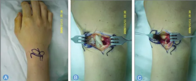
ed hand on the C-arm. The procedure is carried out under regional anesthetic block. The upper arm tourni- quet is inflated. The forearm is pronated and a longitudi- nally curved skin incision that is approximately 3 cm in length is placed beginning at the proximal aspect of Lister’s tubercle. The extensor retinaculum between sec- ond and third extensor compartment is incised. By plac- ing retractors, the extensor carpi radialis longus tendon is retracted radially and the extensor carpi radialis brevis and the extensor pollicis longus tendons are retracted ulnarly. A longitudinal capsulotomy is performed along the long axis of the incision. Now we can expose the articular surface of the radius, radioscaphoid joint, scapholunate interval and scaphoid nonunion site (Fig.
1). Reduction of the nonunion site is attempted. To help the reduction, the carpus is distracted manually via longitudinal traction on the index and long fingers and then manual reduction is facilitated. When a satisfactory reduction has been achieved, provisional fixation is obtained with two 0.045 K-wires which were inserted eccentrically from the dorsal ridge of the radius to slightly volar to the central axis of the scaphoid axis. Using the K- wires as lever-arm, a scaphoid is volar flexed. Guide K- wire is then inserted in the central axis of the scaphoid under the C-arm control. To determine the appropriate
screw length, an identical second guide wire is applied to the proximal cortex of the scaphoid. The difference between the two wires is measured and the screw length is determined. After remove the guidewire, drilling and tapering for the fixation of the Herbert screw was done.
And then Herbert screw fixation is inserted with a free hand technique. Final position of the Herbert screw is checked with C-arm. Two K-wires which were fixed eccentrically is retracted volarly but not removed for additional stability. The end of the K-wires were left on the volar skin. These K-wires are removed after 8 weeks operation at the time of cast off (Fig. 2).
In case of cyst formation, we can do additional bone graft through dorsal window after fixation of the scaphoid.
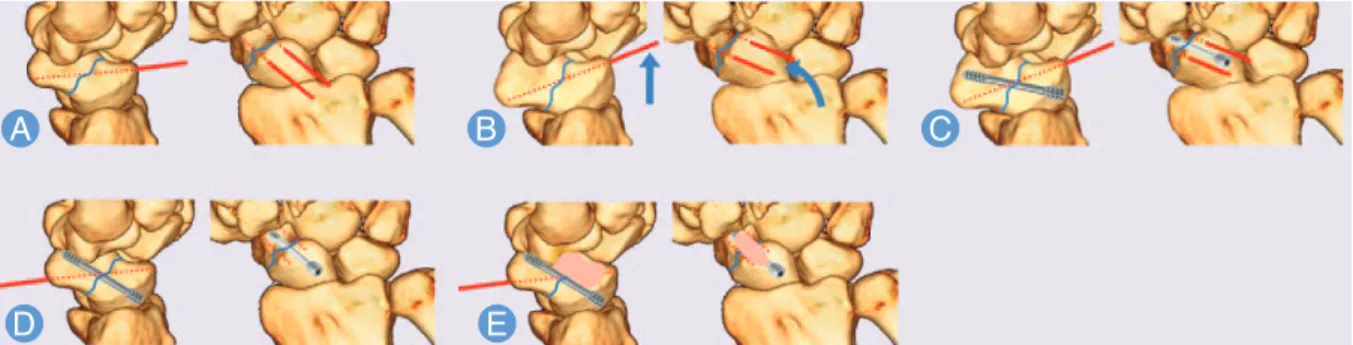
When scaphoid nonunion is accompanied with scle- rotic margin or humpback deformity, 0.045 K-wire are inserted perpendicularly to the central axis of the scaphoid into the proximal and distal scaphoid frag- ments. These two K-wires can be used to assist reduction and handling of the scaphoid. Decortication of the scle- rotic margin with electric saw until fresh bone is exposed is made. Reduction of the nonunion site is attempted with assist of the joystick K-wires. After confirm the reduction with the C-arm. Insertion of the iliac bone graft
Fig. 1. (A) Skin incition.(B)The extensor retinaculum between second and third extensor compartment is incised.(C)By placing retractors, the extensor carpi radialis longus tendon is retracted radially and the extensor carpi radialis brevis and the extensor pollicis longus tendons are retracted ulnarly. A longitudinal capsulotomy is performed along the long axis of the incision.
into the gap. Provisional fixation with two K-wires which were inserted eccentrically as the same procedure as simple nonunion. Using the K-wires as a lever-arm, the scaphoid is dorsiflexed. Herbert screw fixation is done in the central position with a free hand technique. Two pro- visionally fixed K-wires retracted volarly but left for addi- tional stability (Fig. 3).
Postoperatively, the patient is immobilized in a short arm plaster splint and is advised to elevate the hand the first days for control of swelling. At 2 weeks, the patient returns for suture removal and short arm thumb spica cast. At 8 weeks postoperation, cast and K-wires were removed. Range of motion exercises and occupational
theraphy are begun. Fracture healing is assessed at 2, 4, 8, 12, and 16 weeks postoperatively with routine plain radiographs. The duration of follow-up depends on radi- ographic fracture healing and clinical evaluation.
RESULTS
Ninety-eight of 102 patients (96.1%) in the scaphoid nonunion progressed to radiographic union without complication (Table 2). The average time to union was 12.7 weeks (range, 8-26 weeks). Overall, the final range of motion of the wrist averaged 61° (range, 42°-70°) of volar flexion, 47° (range, 35°-60°) of dorsiflexion, 16° (range, Fig. 2. (A)When a satisfactory reduction has been achieved, provisional fixation is obtained with two 0.045 K-wires which were inserted eccentrically from the dorsal ridge of the radius to slightly volar to the central axis of the scaphoid axis.(B, C)Using the K-wires as lever-arm, a scaphoid is volar flexed for the screw which is then inserted in the central axis.(D)Two K-wires which were fixed eccentrically is retracted volarly but not removed for additional stability.(E)In case of cyst formation, we can do addi- tional bone graft through dorsal window after fixation of the scaphoid.
Fig. 3. (A) 0.045 K-wire are inserted perpendicularly to the central axis of the scaphoid into the proximal and distal scaphoid fragments. Reduction of the nonunion site is attempted with assist of the joystick K-wires.(B)After insertion of the iliac bone graft into the gap, provisional fixation with two K-wires which were inserted eccentrically as the same proce- dure as simple nonunion.(C)Using the K-wires as a lever-arm, the scaphoid is volar flexed.(D)Herbert screw fixation is done in the central position with a free hand technique.
10°-20°) of radial deviation, and 27° (range, 22°-30°) of ulnar deviation. In 92 of 102 patients (90%), grip strength parameters were converted into a percentage of the con- tralateral side were over 85%. 9 patients (9%) had 70% to 84% grip strength parameter compared with contralater- al side and 1 patient (1%) had 50% to 69% grip strength parameter compared with contralateral side. The mean grip strength was 38 kg at the final follow-up evaluation, which was approximately 91% of the uninvolved wrist.
Paired t-test analysis showed no significant difference (p>0.05) in grip strength between the injured and unin- jured extremity at the final follow-up evaluation.
Eighty-nine patients (87%) were pain free and asymp- tomatic at final follow-up. Thirteen patient (13%) had mild pain with vigorous activities or pain only with weather changes. No patient had moderate pain with activities of daily living or pain at rest. Ninety-five employed patients (93%) eventually were able to return to their preinjury employment without restrictions and 7 employed patients (7%) returned to their employment with mild restrictions.
According to the more stringent modified Mayo wrist score system, 102 the functional results were excellent for 60 patients (58.9%), good for 32 patients (32.4%), fair for 8 patients (7.8%), and poor for 2 patient (1.9%) (Table 3).
Twelve patients that performed styloidectomy and 3 patients that performed ligament repair had satisfactory
results similarly. No statistically significant difference was found between the preoperative and postoperative radiolunateangle, scapholunate angle, or height to length scaphoid ratio, as measured by either indepen- dent reader (p>0.05). There were no perioperative com- plications. None of the patients showed radiographic signs of arthrosis during the study period. There were no complications related to Herbert screw (i.e., migration or loosening). None of the patients showed stiffness of the fingers or thumb, nor did they develop complex regional pain syndrome. Screw position relative to the central axis of the scaphoid as defined by Trumble et al.6 was evaluated on postoperative AP, lateral, and dedicated scaphoid radiographs. 98 of 102 patients (96%) showed a central screw position on the AP and lateral views. 92 of 102 percent (90%) of patients showed a central screw position on the dedicated scaphoid view.
DISCUSSION
Due to anatomical properties including tenuous vascular supply, joint fluid dilution, and the inability to form cal- lus, as well as biomechanical properties, such as high shear stress and displacement of fragments, delayed unions and nonunions are not uncommon. Delayed treatment and an inadequate period of fixation are also responsible for scaphoid nonunion7. It is known that the nonunion rate of scaphoid fracture is 5% to 10% with non-surgical treatment2,8. Established nonunion, if left untreated, will progress to osteoarthritis and impair the function of the wrist joint7,8,9. Therefore, in most instances, nonunions of the scaphoid are managed by surgery. However, the treatment of scaphoid nonunions is trouble-some, with reported failure rates between 25%
and 45%10,11. The key points of successful surgery for scaphoid nonunions include achieving union of the frac- ture, correcting the deformities, restoring anatomical alignment, and recovering the function of the wrist12.
There is more than one way to do the surgery(open volar approach or open dorsal approach, percutaneous approach). The location and type of fracture determine which approach allows the best screw placement13,14. In Table 3.Modified Mayo wrist score
Excellent 33 27 - 60 (58.9)
Good 10 20 2 32 (32.4)
Fair 1 5 2 8 (7.8)
Poor - - 2 2 (1.9)
Total 44 52 6 102 (100)
Mean modified Mayo score: 87.5 points.
Score/Stage Stage I Stage II Stage III Total (%) Table 2.Radiologic union rate
Stage I 44/44 (100)
Stage II 50/52 (96.1)
Stage III 4/6 (66.6)
Total 98/102 (96.1)
Mack-Lichtman stage No. of union (%)
nondisplaced waist nonunions perpendicular to the long axis of the scaphoid, the approach depends on the surgeon’s preference. Proximal pole nonunions are more easily managed with a dorsal approach, and distal nonunions with a volar approach. Open techniques for fixation are generally indicated for scaphoid nonunions15. The volar approach is typically used for distal third or mid-waist nonunions of the scaphoid. This approach is particularly useful to correct a humpback deformity of the scaphoid. The important dorsal blood supply is left undisturbed, and a good view of the volar surface of the scaphoid is facilitated. But volar approaches may jeopardize the already tenuous blood supply to the proximal pole, resulting in avascular necrosis. Volar open approaches necessitate division of the important volar radiocarpal ligaments. Garcia-Elias et al.16reported a significant increase in scapholunate and lunocapitate angles following a volar approach for fixation of scaphoid fractures when the radiocapitate and radiolunate liga- ments were divided. Most importantly, the disadvantage is that the screw may be placed slightly oblique to a frac- ture line in the mid-waist portion of the scaphoid. If a volar approach is used, central screw placement is hin- dered by the volar aspect of the trapezium. Investigators have attempted to identify optimal guidelines for screw placement, including manipulation and partial excision of the trapezium17,18. The additional dissection may cause attendant problems including vascular compromise, scaphotrapezial instability, and degenerative changes19,20.
Although there are articles showing excellent results with a dorsal percutaneous approach for nondisplaced waist nonunions21, recent reports highlight the potential risks of this approach. The anatomical structures at risk (e.g., the posterior interosseous nerve, extensor digito- rum communis to the index, and extensor indicis pro- prius)22and complications (e.g., hardware problems, nonunion, extensor pollicis longus rupture, and screw malpositioning) might be more common than previously assumed23,24. The disadvantage of the dorsal approach is that, as the wrist is hyperflexed, the unstable scaphoid fracture may displace to create a humpback deformity.
A open dorsal approach is most useful for proximal
pole fractures and preserves the volar carpal ligaments.
In addition, exposure of the scapholunate ligament is facilitated to address any injury there. This approach allows more central placement of the screw down the long axis of the scaphoid and avoids any possibility of damaging the important anterior stabilizing radioscaphocapitate and radioscapholunate liga- ments25,26, allowing earlier postoperative wrist mobiliza- tion. In addition, central placement of the screw in the proximal pole fragment, as described by Trumble et al.27, is more predictable using a dorsal approach, allowing for more rigid fixation. These findings are in concordance with Chan and McAdams1, who showed more accurate and reliable central axis screw placement with the dorsal versus the volar approach in a cadaveric model. A study showed central screw placement to be stronger and to have greater stiffness and increased load to failure com- pared with eccentrically placed screws. Thus, a centrally located screw reduces the likelihood of screw failure28. The main advantage of our approach is the ability to reliably and safely insert the screw along the central scaphoid axis. Using two K-wires that is inserted more dorsally to the central axis as lever-arm, it is possible for guidewire to insert into the central axis of scaphoid easily.
Generally, the disadvantage of the dorsal approach is that, as the wrist is hyperflexed, the unstable scaphoid fracture may displace to create a humpback deformity and put the extensor tendons with several anatomic structures at risk22. Because of K-wire levering technique, we can fix the screw without hyperflexion of wrist.
Consequently, 98 of 102 patients (96%) showed a central screw position on the AP and lateral views without com- plications. 92 of 102 percent (90%) of patients showed a central screw position on the dedicated scaphoid view.
Although the association of central screw position with higher rates of union has not been accepted definitely, our results showed higher union rates with higher central position of screw. Ninety-eight of 102 patients (96.1%) in the scaphoid nonunion progressed to radiographic union and the average time to union was 12.7 weeks. In addition, our results showed favorable range of motion and restoration of grip strength. Patients returned to
their employment with no restrictions and according to the modified Mayo wrist score system, most patients were above good grade.
Screw fixation provides more successful union rates than K-wire fixation but which of the screw types pro- vides better compression remains debatable29-31. Most previous studies suggest that the Acutrak screw gener- ates greater compression. But a study shows that there is no significant difference between the Acutrak and can- nulated Herbert screws with respect to functional out- come, consolidation rate and time to consolidation in the treatment of scaphoid nonunion. It seems that differ- ence in compressive force between these screws does not influence the outcome32. Apart from selection of the screw type, bone grafting and rigid fixation into the cen- tral axis are required to achieve union.
The usual treatment for scaphoid nonunion is conven- tional bone grafting, with or without bony fixation.
Satisfactory bone union rates ranging from 80% to 90%
have been reported by some authors33; but failure rates as high as 65% have been described by others10,34. Most of these failures have been related to fractures of the proxi- mal pole or avascular necrosis of the proximal fragment.
We treated that vascularised bone grafting is mandatory whenever there is a non-vascularised sclerotic fragment.
On the other hand, in patients with well vascularised fragments, conventional non-vascularised bone graft from the distal radius or ilium can be used. Iliac crest has been considered the gold standard for corticocancellous grafting in unstable scaphoid nonunions because of the quality of osteoprogenitor cells obtained and the bio- mechanical properties of the graft35,36.
In arthroscopic evaluation of displaced and nondis- placed scaphoid waist fractures, they found that 71% had associated acute scapholunate (SL)-ligament injuries and 24% had a complete SL ligament rupture37. The over- all number of ligament injuries is high compared to find- ings in previous studies involving arthroscopic examina- tion, where incidences of between 14% to 50% have been reported38,39. A high incidence of SL ligament injuries found in scaphoid nonunions has raised the possibility of an association between the 2 injuries40. We believe that
the combination of these 2 lesions is clinically important because it might lead to an increased risk of developing scaphoid nonunion. We recommend dorsal approach to assess and treat associated intrinsic ligament injuries at the same incision of screw fixation.
Styloidectomies have been unsatisfactory as isolated procedures for SNAC arthritis, but have enjoyed success when combined with bone grafts, with or without inter- nal fixation, for stage I SNAC arthritis. Using limited dor- sal approach, styloidectomy can perform for mild radioscaphoid degeneration (stage III) at the same time.
CONCLUSION
We found Herbert screw fixation using a dorsal approach was a reliable method for patients to achieve bony union with high functional scores and without major complica- tions. Furthermore, the dorsal approach enable to visual- ize any associated scapholunate ligament injury, have a radial styloidectomy for patient with arthritis, and har- vest cancellous bone graft or vascularized pedicled bone graft from distal radius. With the dorsal approach, distal nonunions of scaphoid are more easily managed in addi- tion to proximal pole and waist fractures.
REFERENCES
1. Chan KW, McAdams TR. Central screw placement in percutaneous screw scaphoid fixation: a cadaveric comparison of proximal and distal techniques. J Hand Surg Am. 2004;29:74-9.
2. Braga-Silva J, Peruchi FM, Moschen GM, Gehlen D, Padoin AV. A comparison of the use of distal radius vas- cularised bone graft and non-vascularised iliac crest bone graft in the treatment of non-union of scaphoid fractures. J Hand Surg Eur Vol. 2008;33:636-40.
3. Dias JJ. Definition of union after acute fracture and surgery for fracture nonunion of the scaphoid. J Hand Surg Br. 2001;26:321-5.
4. Cooney WP, Dobyns JH, Linscheid RL. Fractures of the scaphoid: a rational approach to management. Clin Orthop Relat Res. 1980;(149):90-7.
5. Bain GI, Bennett JD, Richards RS, Slethaug GP, Roth JH.
Longitudinal computed tomography of the scaphoid: a
new technique. Skeletal Radiol. 1995;24:271-3.
6. Trumble TE, Clarke T, Kreder HJ. Non-union of the scaphoid. Treatment with cannulated screws compared with treatment with Herbert screws. J Bone Joint Surg Am. 1996;78:1829-37.
7. Inoue G, Sakuma M. The natural history of scaphoid non-union. Radiographical and clinical analysis in 102 cases. Arch Orthop Trauma Surg. 1996;115:1-4.
8. Lindstrom G, Nystrom A. Natural history of scaphoid non-union, with special reference to "asymptomatic"
cases. J Hand Surg Br. 1992;17:697-700.
9. Proctor MT. Non-union of the scaphoid: early and late management. Injury. 1994;25:15-20.
10. Barton NJ. Experience with scaphoid grafting. J Hand Surg Br. 1997;22:153-60.
11. Schuind F, Haentjens P, Van Innis F, Vander Maren C, Garcia-Elias M, Sennwald G. Prognostic factors in the treatment of carpal scaphoid nonunions. J Hand Surg Am. 1999;24:761-76.
12. Herbert TJ. The fractured scaphoid. St. Louis, MO:
Quality Medical Publishing; 1990. 31.
13. Haisman JM, Rohde RS, Weiland AJ; American Academy of Orthopaedic Surgeons. Acute fractures of the scaphoid. J Bone Joint Surg Am. 2006;88:2750-8.
14. Soubeyrand M, Biau D, Mansour C, Mahjoub S, Molina V, Gagey O. Comparison of percutaneous dorsal versus volar fixation of scaphoid waist fractures using a com- puter model in cadavers. J Hand Surg Am. 2009;34:
1838-44.
15. Geissler WB, Slade JF. Fractures of the carpal bones. In:
Wolfe SW, Hotchkiss RN, Peterson WC, Kozin SH, editors.
Green’s operative hand surgery. 6th ed. Philadelphia, PA: Elsevier; 2010. 639-707.
16. Garcia-Elias M, Vall A, Salo JM, Lluch AL. Carpal align- ment after different surgical approaches to the scaphoid: a comparative study. J Hand Surg Am. 1988;
13:604-12.
17. Levitz S, Ring D. Retrograde (volar) scaphoid screw insertion-a quantitative computed tomographic analy- sis. J Hand Surg Am. 2005;30:543-8.
18. Leventhal EL, Wolfe SW, Walsh EF, Crisco JJ. A compu- tational approach to the "optimal" screw axis location and orientation in the scaphoid bone. J Hand Surg Am.
2009;34:677-84.
19. Kehoe NJ, Hackney RG, Barton NJ. Incidence of osteoarthritis in the scapho-trapezial joint after Herbert
screw fixation of the scaphoid. J Hand Surg Br. 2003;28:
496-9.
20. Nicholl JE, Buckland-Wright JC. Degenerative changes at the scaphotrapezial joint following Herbert screw insertion: a radiographic study comparing patients with scaphoid fracture and primary hand arthritis. J Hand Surg Br. 2000;25:422-6.
21. Saint-Cyr M, Oni G, Wong C, Sen MK, LaJoie AS, Gupta A. Dorsal percutaneous cannulated screw fixation for delayed union and nonunion of the scaphoid. Plast Reconstr Surg. 2011;128:467-73.
22. Adamany DC, Mikola EA, Fraser BJ. Percutaneous fixation of the scaphoid through a dorsal approach: an anatomic study. J Hand Surg Am. 2008;33:327-31.
23. Bushnell BD, McWilliams AD, Messer TM. Complications in dorsal percutaneous cannulated screw fixation of nondisplaced scaphoid waist fractures. J Hand Surg Am. 2007;32:827-33.
24. Weinberg AM, Pichler W, Grechenig S, Tesch NP, Heidari N, Grechenig W. The percutaneous antegrade scaphoid fracture fixation: a safe method? Injury. 2009;
40:642-4.
25. Haddad FS, Goddard NJ. Acute percutaneous scaphoid fixation. A pilot study. J Bone Joint Surg Br. 1998;80:95-9.
26. Ledoux P, Chahidi N, Moermans JP, Kinnen L.
Percutaneous Herbert screw osteosynthesis of the scaphoid bone. Acta Orthop Belg. 1995;61:43-7.
27. Trumble TE, Salas P, Barthel T, Robert KQ 3rd.
Management of scaphoid nonunions. J Am Acad Orthop Surg. 2003;11:380-91.
28. McCallister WV, Knight J, Kaliappan R, Trumble TE.
Central placement of the screw in simulated fractures of the scaphoid waist: a biomechanical study. J Bone Joint Surg Am. 2003;85:72-7.
29. Adla DN, Kitsis C, Miles AW. Compression forces gener- ated by Mini bone screws: a comparative study done on bone model. Injury. 2005;36:65-70.
30. Beadel GP, Ferreira L, Johnson JA, King GJ. Interfrag- mentary compression across a simulated scaphoid fracture: analysis of 3 screws. J Hand Surg Am. 2004;29:
273-8.
31. Hausmann JT, Mayr W, Unger E, Benesch T, Vecsei V, Gabler C. Interfragmentary compression forces of scaphoid screws in a sawbone cylinder model. Injury.
2007;38:763-8.
32. Gereli A, Nalbantoglu U, Sener IU, Kocaoglu B,
Turkmen M. Comparison of headless screws used in the treatment of proximal nonunion of scaphoid bone. Int Orthop. 2011;35:1031-5.
33. Cooney WP 3rd, Dobyns JH, Linscheid RL. Nonunion of the scaphoid: analysis of the results from bone grafting.
J Hand Surg Am. 1980;5:343-54.
34. Green DP. The effect of avascular necrosis on Russe bone grafting for scaphoid nonunion. J Hand Surg Am.
1985;10:597-605.
35. Fernandez DL. A technique for anterior wedge-shaped grafts for scaphoid nonunions with carpal instability. J Hand Surg Am. 1984;9:733-7.
36. Tambe AD, Cutler L, Murali SR, Trail IA, Stanley JK. In scaphoid non-union, does the source of graft affect out- come? Iliac crest versus distal end of radius bone graft. J
Hand Surg Br. 2006;31:47-51.
37. Jorgsholm P, Thomsen NO, Bjorkman A, Besjakov J, Abrahamsson SO. The incidence of intrinsic and extrin- sic ligament injuries in scaphoid waist fractures. J Hand Surg Am. 2010;35:368-74.
38. Wong TC, Yip TH, Wu WC. Carpal ligament injuries with acute scaphoid fractures: a combined wrist injury.
J Hand Surg Br. 2005;30:415-8.
39. Schadel-Hopfner M, Junge A, Bohringer G. Scapholunate ligament injury occurring with scaphoid fracture: a rare coincidence? J Hand Surg Br. 2005;30:137-42.
40. Monsivais JJ, Nitz PA, Scully TJ. The role of carpal insta- bility in scaphoid nonunion: casual or causal? J Hand Surg Br. 1986;11:201-6.
배측도달법을 이용하여 Herbert 나사못으로 내고정을 시행한 주상골 불유합의 치료
이상현
1∙오종석
2∙서창효
1∙김용진
21부산대학교 의학전문대학원 정형외과학교실, 2서부산 센텀병원 수부 및 미세재건수술센터
목적:주상골 불유합 환자에서 배측도달법을 이용하여 Herbert 나사못으로 내고정을 시행한 후 임상적, 방사선학적 결
과를 알아보고자 하였다.
방법:주상골 불유합(Mack-Lichtman stage I, II, III)으로 배측도달법을 이용하여 Herbert 나사못과 추가적인 K-강선 으로 내고정을 시행받은 환자 102명을 대상으로 전향적으로 연구하였다. 보존적 치료, 경피적 나사 고정술을 받거나 주 상골 불유합 진행성 붕괴(scaphoid nonunion advanced collapse)가 있었던 예는 제외하였다. 남자가 94예 여자가 8예 였으며, 평균 연령은 28세(범위, 13-65세), 평균 추시 기간은 35개월(범위, 12-96개월)이었다. 수술 후 방사선 검사를 통 해 골유합, 수근골 정렬, 나사못의 위치를 평가하였다. 기능적 결과는 modified Mayo wrist score를 통해 평가하였다.
결과: 102예 중 98예에서 방사선학적으로 골유합을 얻었으며 평균 기간은 12.7주였다. Modified Mayo wrist score를 이용한 기능적 평가에서 평균 87.5점으로 102예 중 92예에서 우수 이상의 결과를 보였으며 주요 합병증은 발생하지 않았 다. 수근골 정렬은 수술 전 후 유의한 차이가 없었다.
결론:배측도달법을 이용한 Herbert 나사못 내고정술은 높은 골유합률 뿐 아니라 기능적으로도 우수한 결과를 보여 주상
골 불유합 환자에서 유용한 방법으로 생각된다.
색인단어:주상골, 불유합, 배측도달법, Herbert 나사못
접수일2013년 4월 24일수정일2013년 6월 9일 게재확정일2013년 6월 10일
교신저자김용진
부산광역시 사상구 새벽로 226
서부산 센텀병원 수부 및 미세재건수술센터 TEL051-329-3000 FAX051-329-3100 [email protected]