A rare case of choledochal cyst with pancreas divisum:
case presentation and literature review
Adrián Ransom-Rodríguez1,2, Ruben Blachman-Braun3, Emilio Sánchez-García Ramos1,2, Jesús Varela-Prieto1, Erick Rosas-Lezama1, and Miguel Ángel Mercado1
1Department of Hepatopancreatobiliary Surgery, Instituto Nacional de Ciencias Médicas y Nutrición Salvador Zubirán, Mexico City, 2Facultad Mexicana de Medicina de la Universidad La Salle, Mexico City, 3Facultad de
Ciencias de la Salud, Universidad Anáhuac México Norte, Edo. de Mexico, Mexico
Choledochal cysts are rare congenital malformations of the bile duct characterized by dilatations of the intrahepatic and/or extrahepatic portion of the biliary tree, they are associated to an anomalous arrangement of the pan- creaticobiliary duct. Pancreas divisum results from a fusion failure of the pancreatic buds. The coexistence of pancreas divisum and choledochal cyst in adults has been reported in less than 10 well documented cases. This article presents a case of a 42-year-old Peruvian man with intermittent episodes of abdominal pain, initially diagnosed with chol- edocholithiasis, who underwent open cholecystectomy. During surgery, a diagnosis of choledochal cyst and pancreas divisum was made, and therefore a hepaticoduodenostomy was performed. The patient was referred to our hospital due to persistence of abdominal pain. After admission, a papillectomy was achieved without further complications. A cyst resection and dismantling of hepaticoduodenostomy with Roux-en-Y was performed 8 years later. During the sub- sequent 18-month follow-up, the patient remains asymptomatic. (Ann Hepatobiliary Pancreat Surg 2017;21:52-56) Key Words: Bile duct cysts; Choledochal cyst; Pancreas divisum; Pancreatitis
Received: September 22, 2016; Revised: November 13, 2016; Accepted: November 17, 2016 Corresponding author: Adrián Ransom-Rodríguez
Department of Hepatopancreatobiliary Surgery, Instituto Nacional de Ciencias Médicas y Nutrición Salvador Zubirán, Vasco de Quiroga No.
15. Tlalpan, 14080 Mexico City, Mexico
Tel: +52-55-54870900, E-mail: adrianrsm.md@gmail.com
Copyright Ⓒ 2017 by The Korean Association of Hepato-Biliary-Pancreatic Surgery
This is an Open Access article distributed under the terms of the Creative Commons Attribution Non-Commercial License (http://creativecommons.org/
licenses/by-nc/4.0) which permits unrestricted non-commercial use, distribution, and reproduction in any medium, provided the original work is properly cited.
Annals of Hepato-Biliary-Pancreatic Surgery ∙ pISSN: 2508-5778ㆍeISSN: 2508-5859
INTRODUCTION
Choledochal cysts are rare congenital malformations of the bile duct characterized by abnormal dilatations of the intrahepatic and/or extrahepatic portion of the biliary tree.1 The worldwide prevalence of choledochal cyst rang- es from 1:13,000 to 1:2,000,000,2 with majority of cases reported in Southeast Asian countries (e.g., Japan), where the prevalence could be as high as 1:1,000, when com- pared to Western countries where the prevalence is 1:100,000 to 150,000. Also, choledochal cyst predom- inantly affects females (3-4:1). A notable observation is that Asians immigrants to Western countries have reported lesser prevalence of choledochal cyst than in their native countries.3
From the anatomical perspective, choledochal cyst is associated with an anomalous arrangement of the pan-
creaticobiliary duct.1 Therefore, patients have a higher lifetime risk for developing acute pancreatitis, cholangitis and biliary tract malignancy (e.g., cholangiocarcinoma).1-4 Nonetheless, patients with choledochal cyst have an ex- cellent prognosis with early diagnosis and surgical re- section with reconstruction, before life-threatening com- plications arise. On the other hand, pancreas divisum is the most frequent congenital pancreatic variation; this anomaly results from a fusion failure of the ventral and dorsal pancreatic buds.5
The coexistence of choledochal cyst and pancreas divi- sum in adults is extremely rare. This article aims to pres- ent the first case of a Latin America patient with chol- edochal cyst and pancreas divisum. In addition, a liter- ature review and discussion of the clinical features of this entity is provided.
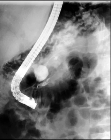
Fig. 1. Endoscopic retrograde cholangiopancreatogram.
Injection of iodine contrast agent into the major duodenal papilla reveals its amputation. Opacification of the principal and secondary pancreatic duct with an independent drainage is identified. A saccular image was also observed on the com- mon bile duct.
CASE
A 42-year-old Peruvian man hospitalized in Peru, with recurrent abdominal pain and diagnosis of chronic lithiasic cholecystitis, underwent open cholecystectomy. During the surgical procedure, choledochal cyst and a partial ob- struction of the choledochal duct was found. Thus, a hep- aticoduodenostomy was performed without removing the cyst. No complications were reported during or after the surgery.
Seven months after the surgical procedure, he presented with recurrent abdominal pain with belt-like radiation, which was exacerbated with cholecystokinetics, and asso- ciated with chronic diarrhea and steatorrhea. He was then referred to our hospital. At admission, his laboratory tests revealed elevated serum amylase (299 U/L) and serum li- pase (597 U/L), with remaining liver function tests within normal range. This led to the diagnosis of chronic pancreatitis. During subsequent endoscopic retrograde cholangiopancreatography (ERCP) procedure, pancreas di- visum and amputation of the major pancreatic duct (Wirsung) was found (Fig. 1). To confirm the diagnosis, the patient underwent an abdominal computed tomog- raphy scan, where a diverticular formation on the pancre- atic head was reported. The patient then underwent an ex-
ploratory laparotomy, with the operative finding of pan- creas divisum, congenital choledochal cyst of 20 mm in diameter and hypoplasia of the distal part of the major pancreatic duct and the major duodenal papilla, which emptied into the hypoplastic portion with flow disruption of the ventral pancreas. With histopathology findings of a major duodenal papilla with chronic inflammation and fibrosis with chronic nonspecific duodenal inflammation, a transduodenal papillectomy was performed. The post- operative course was uneventful.
The patient again presented abdominal pain 8 years lat- er, having the same clinical characteristics associated with fever, asthenia, adynamia and involuntary weight loss of 5 kg since the last 3 months. Laboratory studies showed elevated serum amylase and lipase, diagnosing recurrent acute pancreatitis with associated cholangitis. The patient was hospitalized, seven days later he was discharged with- out complications with normal serum lipase levels, but persistent elevated amylase levels (152 U/L).
However, 7 years later he presented acute cholangitis with following laboratory reports: total bilirubin, direct bi- lirubin and indirect bilirubin within normal levels, and elevated levels of alkaline phosphatase 171 U/L, aspartate aminotransferase 69 U/L and alanine aminotransferase 67 U/L. The patient then underwent a percutaneous transhepatic cholangiography (PTC), where a hepaticoduodenostomy stenosis was identified. Subsequently, a balloon dilatation and placement of an internal-external biliary catheter was performed. Next, he underwent choledochal cyst resection and dismantling of the hepaticoduodenostomy with Roux-en-Y hepaticojejunostomy, with partial resection of the IV B hepatic segment. The patient was discharged 5 days later, asymptomatic and without any complications.
The patient remained asymptomatic for 12 years, after which he presented with acute cholangitis. A percutaneous biliary drainage was performed, revealing a leak to the ab- dominal cavity. Subsequently, a magnetic resonance imag- ing showed loss of proximal left branch continuity, a T1-hypointense pericholangitic lesion consistent with in- flammatory activity, as well as pneumobilia on the left hepatic lobule, leading to the diagnosis of left hepatic duct stenosis. Subsequently, the patient underwent a hep- atojejunostomy of the left duct. Four days after surgery, the patient was discharged with all laboratory levels with- in normal limits. Currently, the patient remains asympto-
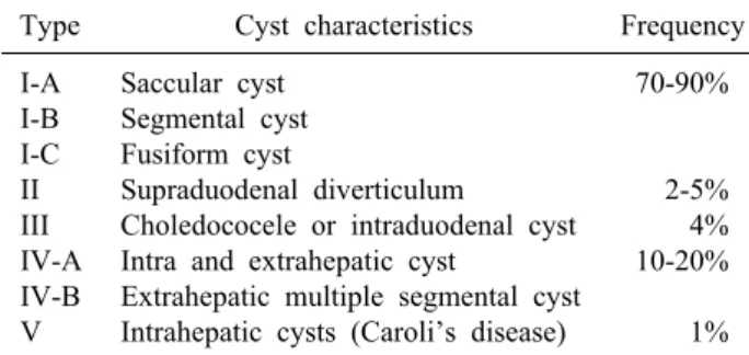
Table 1. Todani classification for choledochal cyst15
Type Cyst characteristics Frequency
I-A I-B I-C II III IV-A IV-B V
Saccular cyst Segmental cyst Fusiform cyst
Supraduodenal diverticulum
Choledococele or intraduodenal cyst Intra and extrahepatic cyst
Extrahepatic multiple segmental cyst Intrahepatic cysts (Caroli’s disease)
70-90%
2-5%
4%
10-20%
1%
matic 18 months after the surgery.
DISCUSSION
Choledochal cyst association with pancreas divisum in adults is a rare condition, and in our knowledge less than 8 well-documented cases have been reported.2,5-9 In this patient, the choledochal cysts was categorized as type I according to the Todani et al. classification, due to the cyst localization on the extrahepatic biliary tree (Table 1).
This variant is the most frequent choledochal cyst type, while pancreas divisum is the most common congenital pancreatic variant, being found in 10% of necropsies.10 In the latter cases, the ventral duct drains the ventral pan- creas through the major papilla and the rest of the pancre- atic tissue drains via the dorsal duct through the minor papilla.5 When a choledochal cyst type I coexists with a sufficiently wide common pancreatic duct, the bile and pancreatic juice drainage may not have any repercussions.
However, if the duct is narrow, there is accumulation of fluids, and pancreatitis with secondary cholangitis can be presented. In a recent study, Bertin et al.11 demonstrated that patients with pancreas divisum are more likely to have genetic predisposition to chronic pancreatitis involv- ing defects in CFTR, SPINK1, and PRSS1. Choledochal cyst increases the risk of developing cholangitis, acute pancreatitis and cholangiocarcinoma, and hence cyst ex- cision is recommended. Nevertheless, successful liver transplants have been performed in cases of bile duct ma- lignancies secondary to choledochal cyst.3,12,13 A delayed diagnosis can result in biliary cirrhosis and portal hyper- tension, which is the most common long-term complica- tion of this pathology.14
According to Todani et al.,15 the most common clinical presentation of choledochal cyst is intermittent recurrent
episodes of upper abdominal pain. The classic triad of the choledochal cyst includes abdominal pain, intermittent jaundice and palpable right upper quadrant abdominal mass; however, these are found in the minority of cases.16 Patients with this condition usually present acute pan- creatitis with hyperamylasemia, as in the current case report. However, if the physician does not suspect chol- edochal cyst and further evaluation is not done, the diag- nosis might be easily overlooked.
ERCP is considered the gold standard for the diagnosis of pancreas divisum due to its high diagnostic accuracy.
However, cannulation of the minor papilla is difficult dur- ing ERCP, and the technique presents a high rate of complications. Due to these disadvantages, the use of non- invasive procedures, such as magnetic resonance chol- angiopancreatography (MRCP), multidetector computed tomography (MDCT) and endoscopic ultrasonography, are well-accepted as diagnostic tools in the assessment of bile tree duct anatomy.17 MRCP enables a noninvasive diag- nosis of pancreas divisum without the use of contrast ma- terial, and avoids the risk of ERCP induced acute pancreatitis.18 MDCT may also identify this anomaly, and its diagnostic accuracy can be improved with the secretin stimulation test.17,18 In a series of 45 patients with pan- creas divisum, Kushnir et al.19 found that endoscopic ul- trasonography has more sensitivity for establishing the di- agnosis of pancreas divisum, than MDCT or MPCP.
In cases of suspected biliary pathology, the initial imag- ing study should be abdominal ultrasonography.16 Nevertheless, ultrasound is less accurate for the diagnosis of bile duct cysts in adults, where the cause of bile duct dilatation may differ (e.g., malignancy or other non-malig- nant entities).20 Therefore, for a precise and accurate de- limitation of the biliary tree, a cholangiography is necessary.16 In order to achieve this goal, complementary diagnostic assessments include computed tomography, MRCP and ERCP. Some patients may also present an anomalous pancreaticobiliary junction that can be diag- nosed by MRCP or ERCP.21 MRCP is emerging as a highly sensitive, safe and preoperative noninvasive diag- nostic technique for choledochal cyst detection. Hence, in clinical practice, MRCP is recommended before ERCP, in patients suspected of having choledochal cyst on ultra- sonography screening.20 Although choledochal cyst is not a frequent condition, the surgeon should always consider
the possibility of choledochal cyst during a surgical ex- ploration procedure in patients with biliary tract-related symptoms.16
Current guidelines recommend that patients with pan- creatic-type abdominal pain and pancreas divisum should undergo smoking cessation before considering any endo- scopic or surgical intervention, due to an increased risk of postsurgical complications in smokers. The majority of patients with pancreas divisum do not exhibit sympto- matic chronic pancreatitis and should not be considered candidates for surgical treatment.22 ERCP has an im- portant therapeutic role in the endoscopic treatment of pa- tients with symptomatic pancreas divisum. There are no prospective randomized controlled trials comparing endo- scopic and surgical therapy; nonetheless, retrospective studies could not establish a difference between the pooled overall response rates between both treatments.17
Recurrent pancreatitis, cholangitis or malignancy are complications associated with choledochal cyst; hence, an early diagnosis and definitive surgical management needs to be performed. Currently, the total resection of the ex- trahepatic bile duct with hepaticojejunostomy is the treat- ment choice for choledochal cyst. Early diagnosis and sur- gical treatment provide a good prognosis, with few com- plications in most cases. Although choledochal cyst shows an excellent long-term prognosis with early resection, sur- gical complications increase with age.23 Nevertheless, the risk of biliary malignancy remains elevated for 15 years or more after choledochal cyst excision.24 Therefore, long-term surveillance is essential, especially if there was persistent dilatation of the intrahepatic bile duct or biliary stones.1 The follow-up should consist of regular abdomi- nal ultrasonography and liver function test evaluation.23,24 Management of choledochal cysts depends on the type of cyst. Type I, II and IV-A treatment consist of complete extrahepatic bile duct cyst excision down to the level of communication with the pancreatic duct, cholecystectomy, and restoration of bilioenteric continuity. Hepaticoduode- nostomy and Roux-en-Y hepaticojejunostomy reconstruc- tion after choledochal cyst resection are both reported in literature, but Roux-en-Y hepaticojejunostomy is the pre- ferred technique. Hepaticoduodenostomy is associated with increased rates of gastric cancer, biliary cancer, post- operative reflux and gastritis.24 Recently, there is a report of a successful cyst excision and Roux-en-Y hep-
aticojejunostomy via laparoscopy, with faster recovery, less intestinal adhesions and aesthetic damage, and facilitation of surgery due to magnification of the operative field.23
Hackert et al.8 reported a similar case that was success- fully treated with bile duct resection, papillectomy, hep- aticojejunostomy and jejunal reinsertion of the uncinate pancreatic duct. However, there is a lack of evidence about the efficacy and safety of this procedure.
In conclusion, the association of pancreas divisum with choledochal cyst is a rare condition that should be sus- pected in adult patients with recurrent acute cholangitis and/or pancreatitis, without an identified cause.
Additionally, imaging studies should be used for diag- nosis, considering that MRCP and endoscopic ultra- sonography have a higher sensitivity compared with the current gold standard (ERCP). When choledochal cyst and pancreas divisum coexist, early diagnosis and resection are the main objectives due to the increased risk of chol- angiocarcinoma and other complications. In addition, a long follow-up duration should be considered after surgi- cal resection of the cyst. Although pancreas divisum with choledochal cyst could have a favorable prognosis, more studies are required for determining the best therapeutic approach and overall prognosis of this group of patients.
ACKNOWLEDGEMENTS
We acknowledge Manuel Campuzano Fernández and Iván Ransom Rodríguez who provided writing assistance.
REFERENCES
1. Ono S, Fumino S, Shimadera S, Iwai N. Long-term outcomes after hepaticojejunostomy for choledochal cyst: a 10- to 27-year follow-up. J Pediatr Surg 2010;45:376-378.
2. Garrido A, León R, López J, Márquez JL. An exceptional case of choledochocele and pancreas divisum in an elderly man.
Gastroenterol Hepatol 2012;35:8-11.
3. Dhupar R, Gulack B, Geller DA, Marsh JW, Gamblin TC. The changing presentation of choledochal cyst disease: an incidental diagnosis. HPB Surg 2009;2009:103739.
4. Reungwetwattana T, Dy GK. Targeted therapies in development for non-small cell lung cancer. J Carcinog 2013;12:22.
5. Arulprakash S, Balamurali R, Pugazhendhi T, Kumar SJ.
Pancreas divisum and choledochal cyst. Indian J Med Sci 2009;
63:198-201.
6. Li L, Yamataka A, Segawa O, Miyano T. Coexistence of pan- creas divisum and septate common channel in a child with chol- edochal cyst. J Pediatr Gastroenterol Nutr 2001;32:602-604.
7. Alonso-Lej F, Rever WB Jr, Pessagno DJ. Congenital chol-
edochal cyst, with a report of 2, and an analysis of 94, cases.
Int Abstr Surg 1959;108:1-30.
8. Hackert T, Hartwig W, Werner J. Symptoms and surgical man- agement of a distal choledochal cyst in a patient with pancreas divisum: case report and review of the literature. Case Rep Gastroenterol 2007;1:90-95.
9. Bechtler M, Eickhoff A, Willis S, Riemann JF. Choledochal cyst type IA with drainage through the ventral duct in pancreas divisum. Endoscopy 2009;41 Suppl 2:E71-E72.
10. Pilcher JT. Disease of the pancreas: its cause and nature. Ann Surg 1911;54:430-431.
11. Bertin C, Pelletier AL, Vullierme MP, Bienvenu T, Rebours V, Hentic O, et al. Pancreas divisum is not a cause of pancreatitis by itself but acts as a partner of genetic mutations. Am J Gastroenterol 2012;107:311-317.
12. Wiseman K, Buczkowski AK, Chung SW, Francoeur J, Schaeffer D, Scudamore CH. Epidemiology, presentation, diag- nosis, and outcomes of choledochal cysts in adults in an urban environment. Am J Surg 2005;189:527-531.
13. Ammori JB, Mulholland MW. Adult type I choledochal cyst resection. J Gastrointest Surg 2009;13:363-367.
14. Jensen KK, Sohaey R. Antenatal sonographic diagnosis of chol- edochal cyst: case report and imaging review. J Clin Ultrasound 2015;43:581-583.
15. Todani T, Watanabe Y, Narusue M, Tabuchi K, Okajima K.
Congenital bile duct cysts: classification, operative procedures, and review of thirty-seven cases including cancer arising from choledochal cyst. Am J Surg 1977;134:263-269.
16. Gadelhak N, Shehta A, Hamed H. Diagnosis and management
of choledochal cyst: 20 years of single center experience. World J Gastroenterol 2014;20:7061-7066.
17. Bor R, Madácsy L, Fábián A, Szepes A, Szepes Z. Endoscopic retrograde pancreatography: when should we do it? World J Gastrointest Endosc 2015;7:1023-1031.
18. Türkvatan A, Erden A, Türkoğlu MA, Yener Ö. Congenital var- iants and anomalies of the pancreas and pancreatic duct: imaging by magnetic resonance cholangiopancreaticography and multi- detector computed tomography. Korean J Radiol 2013;14:905-913.
19. Kushnir VM, Wani SB, Fowler K, Menias C, Varma R, Narra V, et al. Sensitivity of endoscopic ultrasound, multidetector com- puted tomography, and magnetic resonance cholangiopancreatog- raphy in the diagnosis of pancreas divisum: a tertiary center experience. Pancreas 2013;42:436-441.
20. Khandelwal C, Anand U, Kumar B, Priyadarshi RN. Diagnosis and management of choledochal cysts. Indian J Surg 2012;74:
401-406.
21. Ozturk E, Sivrioglu AK, Geceer G. Education and Imaging.
Hepatobiliary and pancreatic: choledochal cyst with involvement of the cystic duct. J Gastroenterol Hepatol 2014;29:4.
22. Nath BD, Freedman SD, Moser AJ. The Frey procedure is a treatment for chronic pancreatitis, not pancreas divisum. JAMA Surg 2013;148:1062.
23. Singham J, Yoshida EM, Scudamore CH. Choledochal cysts.
Part 3 of 3: management. Can J Surg 2010;53:51-56.
24. Soares KC, Arnaoutakis DJ, Kamel I, Rastegar N, Anders R, Maithel S, et al. Choledochal cysts: presentation, clinical differ- entiation, and management. J Am Coll Surg 2014;219:1167-1180.